On this page
Quick Reference
Overview and Recommendations
Background
- •Acute pericarditis accounts for up to 5% of emergency department visits for nonischemic chest pain in North America and Western Europe, with an incidence roughly one-tenth that of non-ST-elevation myocardial infarction. The condition predominantly affects middle-aged men (mean age 52-56 years, male-to-female likelihood ratio 1.85) and exhibits a striking winter peak, a 1.5-fold higher incidence compared with summer, implicating viral respiratory pathogens as major triggers.
- •Most cases (80-85%) in Western countries are idiopathic or viral, yet pericarditis can herald serious underlying disease: autoimmune conditions (7.3% of identifiable causes), occult malignancy (5.1%, with a 13-fold increased cancer diagnosis rate within 3 months), and tuberculosis in endemic regions. A high index of suspicion is required because untreated purulent or tuberculous pericarditis carries a mortality exceeding 40% and a 50% risk of constrictive pericarditis.
- •The central pathogenic driver is the NLRP3 inflammasome-IL-1β axis. Inciting injury (viral, post-cardiac surgery, autoimmune) activates the NLRP3 inflammasome in pericardial mesothelial cells, triggering IL-1β cleavage and release. This unleashes a feed-forward inflammatory cascade that produces pleuritic chest pain (via prostaglandin sensitisation of phrenic nerve nociceptors), pericardial effusion (increased vascular permeability), and systemic inflammation (hepatic CRP synthesis). Every effective therapy, NSAIDs, , and , targets a distinct node in this pathway.
- •The traditional temporal classification, acute (<4-6 weeks), incessant (4-6 weeks to 3 months without a symptom-free interval), recurrent (new episode after a ≥4-6 week symptom-free interval), and chronic (>3 months), dictates therapy duration. An initial episode treated with colchicine for 3 months reduces the 18-month recurrence rate from 37.5% to 16.7% (NNT = 5), while recurrent disease may require 6 months of colchicine and escalation to IL-1 blockade.
Evaluation
- •Suspect acute pericarditis in any patient presenting with acute, sharp, pleuritic chest pain that characteristically worsens when supine and improves when sitting forward. The pain may radiate to the trapezius ridge (via phrenic nerve irritation), a distinctive feature that helps differentiate it from myocardial ischemia.
- •Ask about preceding respiratory or gastrointestinal illness (latency 1-2 weeks), fever, dyspnea, and risk factors: recent cardiac surgery or myocardial infarction (post-cardiac injury syndrome), known autoimmune disease (SLE, rheumatoid arthritis), active malignancy, or new medications (immune checkpoint inhibitors, hydralazine).
- •Examine for a pericardial friction rub, a pathognomonic scratchy sound heard best at the left lower sternal border with the diaphragm of the stethoscope while the patient leans forward and holds expiration. Note that the rub is absent in >70% of cases and may be fleeting; its absence never excludes the diagnosis.
- •Check for signs of pericardial effusion or tamponade: muffled heart sounds, Ewart’s sign (dullness and tubular breath sounds beneath the left scapula), Beck’s triad (hypotension, muffled heart sounds, distended neck veins), and pulsus paradoxus >10 mmHg. To measure pulsus paradoxus, inflate the cuff above systolic pressure, deflate slowly, and note the first Korotkoff sound that is heard only during expiration; if sounds become audible throughout the cycle only after a drop of >10 mmHg, tamponade is likely.
- •Obtain a 12-lead ECG immediately. Stage I (acute) findings, present in 25-50% of cases, include diffuse, concave ST-segment elevation in leads I, II, aVL, aVF, V2-V6, and PR-segment depression in II, aVF, V4-V6 (with reciprocal PR elevation in aVR). The absence of reciprocal ST depression differentiates pericarditis from STEMI; concomitant troponin elevation suggests myopericarditis but does not worsen prognosis.
- •Order laboratory studies: high-sensitivity C-reactive protein (hs-CRP) is elevated in ~78% at presentation and serves as a crucial biomarker for monitoring treatment response. Troponin I or T is elevated in ~30%, indicating myocardial involvement. White blood cell count and ESR are nonspecific and may be normal.
- •Perform transthoracic echocardiography as the first-line imaging study to detect pericardial effusion (present in ~60%, typically small) and assess for tamponade physiology (right atrial or ventricular diastolic collapse, marked respiratory variation in mitral and tricuspid inflow). Echocardiography also evaluates biventricular function to rule out concomitant myocarditis.
- •Apply the ESC diagnostic criteria: the diagnosis is confirmed when ≥2 of the following are present: (1) typical pericarditic chest pain, (2) pericardial friction rub, (3) ECG changes characteristic of acute pericarditis, (4) new or worsening pericardial effusion. This clinical constellation is the gold standard.
- •Stratify risk using validated high-risk markers that mandate hospitalization: fever >38°C (HR 3.56 for specific cause/complication), subacute onset over weeks, large effusion >20 mm on echo, cardiac tamponade, immunosuppression, trauma, oral anticoagulation, and failure to respond to aspirin or NSAID within 7 days. In the absence of these, outpatient management is safe.
- •Consider cardiac magnetic resonance (CMR) for recurrent, complicated, or diagnostically uncertain cases. Pericardial late gadolinium enhancement and edema on T2-weighted sequences confirm ongoing inflammation and predict recurrence. Chest CT may be used to detect pericardial calcification or lung pathology but lacks CMR’s tissue characterization.
- •Reserve pericardial biopsy for suspected tuberculosis, malignancy, or refractory effusions when less invasive diagnostics are inconclusive. Routine viral serologies or autoantibody panels are not recommended without specific historical clues.
Management
- •Initiate NSAID therapy immediately: ibuprofen 600-800 mg PO every 8 hours OR 750-1000 mg PO every 8 hours. Provide proton-pump inhibitor gastroprotection for patients with gastrointestinal risk factors. Indomethacin should be avoided due to a worse GI profile.
- •Simultaneously start weight-based : 0.5 mg twice daily for patients weighing >70 kg, or 0.5 mg once daily for ≤70 kg. Colchicine must be continued for 3 months after a first episode (6 months after a recurrence), premature discontinuation sharply increases recurrence risk.
- •Monitor hs-CRP weekly. Continue full-dose NSAID until the patient is pain-free AND hs-CRP has normalised (typically 2-4 weeks). Do not begin tapering NSAIDs while CRP remains elevated, as this predicts therapeutic failure.
- •Once clinical remission is achieved and CRP is normal, taper the NSAID by reducing the dose by 200-400 mg every 1-2 weeks over 2-4 weeks. An over-rapid taper or abrupt discontinuation is the most common trigger of early recurrence.
- •Do NOT prescribe high-dose corticosteroids (>0.5 mg/kg/day prednisone equivalent) as initial therapy for uncomplicated idiopathic pericarditis; even a single early course is associated with a 2- to 3-fold increased recurrence risk by interfering with colchicine’s effect and promoting chronicity.
- •If symptoms or elevated CRP persist despite 1-2 weeks of full-dose NSAID plus colchicine, re-evaluate for high-risk etiologies (purulent, tuberculous, neoplastic). When infection is excluded, a cautious corticosteroid course, 0.2-0.5 mg/kg/day, may be introduced as a second-line therapy, but it must be combined with colchicine and an IL-1 inhibitor if feasible to facilitate subsequent taper.
- •For patients with ≥2 recurrences or corticosteroid-dependent disease, initiate IL-1 inhibition: 100 mg subcutaneously once daily or 320 mg loading dose followed by 160 mg once weekly. IL-1 blockers rapidly suppress CRP and symptoms, with recurrence rates reduced to ~25% compared with ˜74% on placebo. Continue therapy for 6-12 months, then attempt tapering.
- •During corticosteroid use, taper extremely slowly: reduce prednisone by 1-2.5 mg every 2-4 weeks only when symptoms are fully resolved and CRP is normal. Any recurrence warrants returning to the last effective dose and an even slower taper.
- •Hospitalise any patient with high-risk features (fever >38°C, subacute onset, effusion >20 mm, tamponade, immunosuppression, anticoagulation, NSAID failure within 7 days) for targeted investigation and monitoring. These patients require exclusion of bacterial, tuberculous, and neoplastic causes.
- •Recognize and treat cardiac tamponade emergently: urgent ultrasound-guided pericardiocentesis is indicated for Beck’s triad or pulsus paradoxus >10 mmHg with hemodynamic compromise. Avoid positive-pressure ventilation prior to drainage if possible, as it may precipitate cardiovascular collapse.
- •Manage myopericarditis conservatively: continue NSAIDs and colchicine, ensure rest, and avoid intense physical activity. Elevated troponin alone does not alter anti-inflammatory therapy nor worsen prognosis if ventricular function is preserved.
- •For post-cardiac injury syndromes (e.g., following ablation, pacemaker implantation, or post-MI), initiate the same NSAID-colchicine protocol. Consider prophylactic colchicine started 7 days before extensive left atrial ablation to reduce pericarditis incidence (absolute risk reduction from 10.3% to 3.8%).
- •Avoid non-dihydropyridine calcium-channel blockers (diltiazem, verapamil) not because they are specifically contraindicated but they are not part of pericarditis management; beta-blockers can be used for rate control if atrial fibrillation occurs (4-5% of cases). Routine anticoagulation is not indicated for AF in pericarditis unless CHA2DS2-VASc score warrants.
- •Counsel patients on activity restriction: athletes must refrain from competitive sports for ≥3 months and require re-evaluation with ECG and echocardiography before clearance. For non-athletes, a gradual return to activity after symptom resolution and CRP normalisation is safe.
- •Schedule follow-up at 3 months to assess remission, check CRP if symptoms recurred, and consider discontinuation of colchicine. Recurrent symptoms after colchicine cessation may necessitate a 6-month colchicine course or escalation to IL-1 inhibitors.
Board Review — High Yield
- •Pericardial friction rub, pathognomonic but heard in <30% of cases; best auscultated at left lower sternal border with patient sitting forward and holding expiration.
- •Diffuse concave ST elevation + PR depression, classic ECG finding; absence of reciprocal ST depression differentiates from STEMI.
- •Colchicine, weight-based (0.5 mg BID >70 kg, QD ≤70 kg) for 3 months (first episode) reduces recurrence from 37.5% to 16.7% (ICAP trial, NNT 5).
- •High-risk features, fever >38°C, subacute onset, effusion >20 mm, tamponade, immunosuppression, trauma, anticoagulation, NSAID failure, mandate hospitalization.
- •hs-CRP, elevated in 78%, normalisation required before tapering NSAID; persistent elevation predicts recurrence.
- •IL-1 inhibitors, anakinra or rilonacept for ≥2 recurrences or corticosteroid-dependent cases; reduces recurrence rate by ~70%.
- •Beck’s triad, hypotension, muffled heart sounds, distended neck veins, indicates cardiac tamponade requiring immediate pericardiocentesis.
- •Pulsus paradoxus, inspiratory drop in systolic BP >10 mmHg; measure by slow cuff deflation and noting first Korotkoff sound only during expiration.
- •Trapezius ridge pain, referred pain via phrenic nerve; highly specific for pericardial inflammation.
- •Constrictive pericarditis, complicates <0.5% of idiopathic cases but up to 33% of tuberculous/neoplastic; presents with right heart failure, Kussmaul sign, and pericardial knock.
Deep Dive — Evidence Details
Definition, Classification and Nomenclature
- ▸Acute pericarditis is defined by symptom duration <4-6 weeks and requires 2 of 4 classic criteria (pleuritic chest pain, pericardial friction rub, ECG changes, new or worsening [[pericardial effusion]]) for diagnosis [8].
- ▸Over 80% of cases in industrialized nations are idiopathic or viral, but bacterial, neoplastic, and tuberculous etiologies must be excluded because they are associated with a high risk of tamponade or constrictive pericarditis [5, 8].
- ▸Recurrent pericarditis, defined by a symptom-free interval of at least 4-6 weeks between episodes, reflects an autoinflammatory phenotype that responds to [[colchicine]] or [[interleukin-1 inhibitors]] [1, 8].
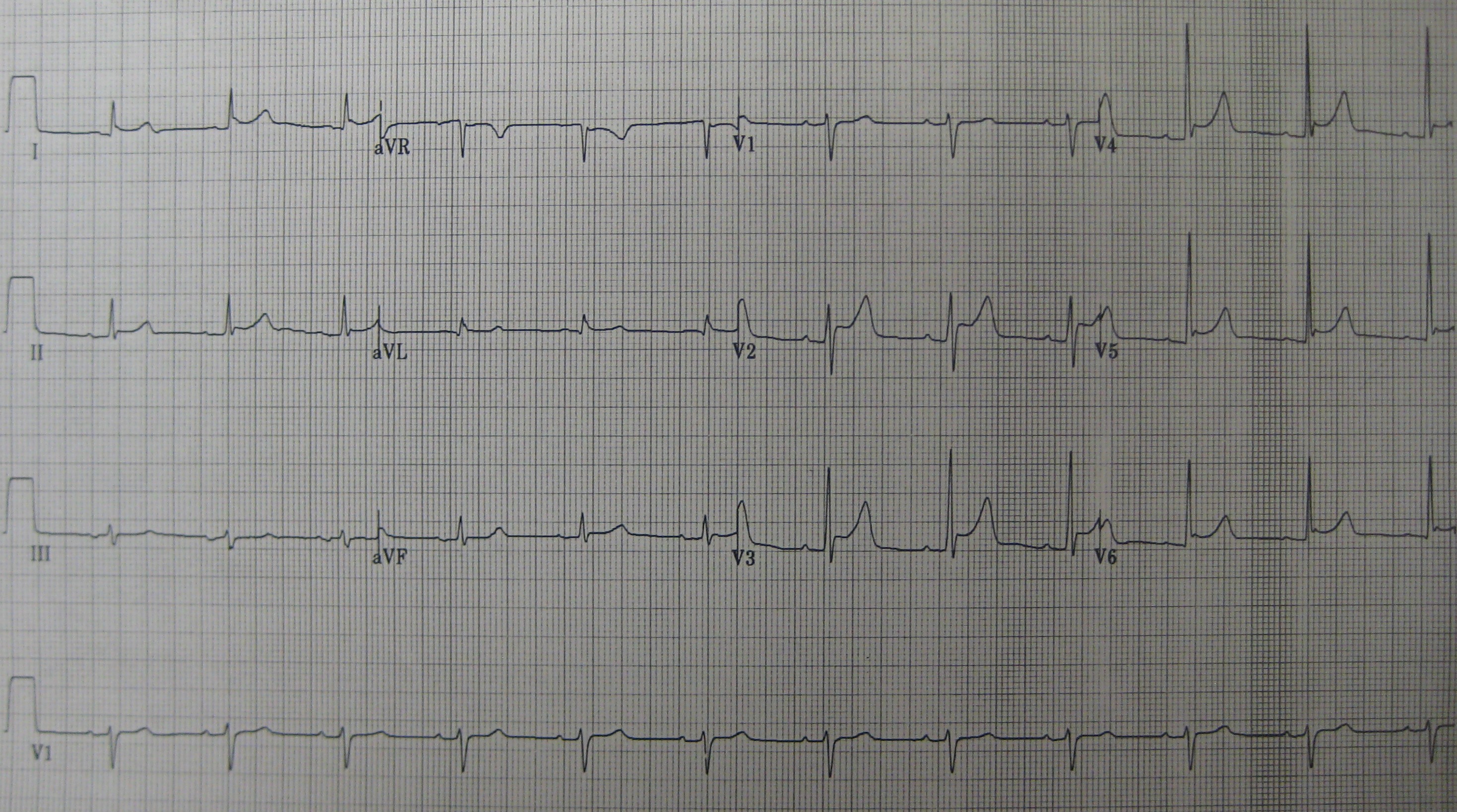
Acute pericarditis is the acute inflammation of the pericardial layers, triggered by diverse stimuli that activate a stereotyped innate immune response, and it manifests with characteristic pleuritic chest pain, electrocardiographic changes, and often a pericardial friction rub or effusion [1]D5[8]D5.
Also Called / Synonyms: Acute benign pericarditis; Idiopathic pericarditis; Viral pericarditis; Nonspecific pericarditis; Acute fibrinous pericarditis (pathologic description).
Temporal Classification
The traditional timeline drives prognosis and anti-inflammatory treatment duration. The 2024 JAMA review defines four phases [8]D5:
- Acute: Symptom duration <4-6 weeks. This is the index episode upon which all downstream decisions pivot.
- Incessant: Symptoms persisting >4-6 weeks but <3 months without a symptom-free interval.
- Recurrent: A new episode occurring after a symptom-free interval of at least 4-6 weeks, reflecting a relapsing autoinflammatory phenotype.
- Chronic: Continuous symptoms lasting >3 months.
Etiologic Classification
hinges on the specific cause; a single term like "pericarditis" is not a treatment plan. The following table distills the major categories and their clinical fingerprints.
| Category | Key Distinguishing Features | Associated Marker / Subtype |
|---|---|---|
| Idiopathic / Viral | Most common (80-85%) in Western countries; often preceded by respiratory or illness; self-limited course [1]D5[8]D5 | Coxsackievirus, echovirus, influenza, SARS-CoV-2 |
| Post-cardiac Injury Syndrome | Onset days to weeks after myocardial infarction, cardiac surgery, or trauma; pleuritic pain, fever, and pericardial rub; includes [1]D5 | Elevated inflammatory markers, autoantibodies |
| Connective Tissue Disease | Seen in , rheumatoid arthritis, scleroderma; typically during disease flare; may be initial presentation [1]D5[9]C4 | ANA, anti-dsDNA, rheumatoid factor |
| Neoplastic | Lung, breast, lymphoma, or metastasis; large effusion, tamponade risk, and poor prognosis; pericarditis can be the first sign of occult cancer [5]B3b[8]D5 | Positive cytology (sensitivity ~60%), PET-CT uptake |
| Tuberculous | Endemic regions; insidious onset, night sweats, weight loss; high rate of progression to (~50% if untreated) [4]B2b | Adenosine deaminase >40 U/L in pericardial fluid |
| Purulent (Bacterial) | Rare but fulminant; high fever, rigors, leukocytosis; rapid tamponade; mortality exceeds 40% if untreated [8]D5 | Positive Gram stain and culture; organisms: Staphylococcus, Streptococcus |
| Drug-Induced | Immune checkpoint inhibitors, hydralazine, procainamide, minoxidil, mesalamine; may resolve on drug withdrawal [7]C4 | Negative infectious workup, temporal link to drug |
| Uremic | Advanced chronic kidney disease or dialysis; often hemorrhagic effusion; responds to intensified dialysis [8]D5 | BUN >60 mg/dL, creatinine >6 mg/dL |
Acute pericarditis accounts for up to 5% of emergency department visits for nonischemic chest pain in North America and Western Europe. Although the majority follow a benign, self-limited course, unrecognized high-risk etiologies\u2014particularly bacterial, neoplastic, and tuberculous\u2014carry a substantial risk of life-threatening (<3%) or constrictive pericarditis (<0.5%) [8]D5.
Pearl: The temporal classification of acute pericarditis into acute, incessant, recurrent, and chronic directly guides the duration of anti-inflammatory therapy, while the etiologic category dictates whether antimicrobials, immunosuppressants, or cancer-directed treatment is required [1]D5[8]D5.
Epidemiology and Risk Factors
- ▸Acute pericarditis exhibits a male predominance (OR 1.85) and a winter peak (rate ratio 1.5 vs. summer) [16].
- ▸Autoimmune disease and malignancy account for 7.3% and 5.1% of identifiable causes, respectively, and should be considered especially in older patients or recurrent episodes [5,17,24].
- ▸COVID-19 infection and mRNA vaccines are associated with a small increased risk of pericarditis, with the highest absolute risk in young males [18,22].
Acute pericarditis accounts for up to 5% of emergency department visits for non-ischemic chest pain in North America and Western Europe [8]D5, and its overall incidence is approximately one-tenth that of [19]B2b. The condition shows a clear sex disparity: men are affected nearly twice as often as women (age-adjusted likelihood ratio 1.85, 95% CI 1.65-2.06) [16]B3b. The mean age at presentation is approximately 52-56 years [11]B3b[16]B3b, though age modifies the clinical phenotype. Younger adults (<55 years) typically present with classic pleuritic chest pain and pericardial friction rub, while patients older than 75 years have fewer classic symptoms and higher rates of complications [11]B3b.
A large Finnish nationwide registry demonstrated a pronounced seasonal variation, with a peak in winter (standardized incidence rate ratio 1.5 compared with summer, P<0.0001) and a nadir in summer [16]B3b, implicating viral respiratory pathogens as major triggers. Although most cases in developed countries are idiopathic or viral (80-90%) [8]D5, several specific conditions increase the risk.
Autoimmune diseases account for 7.3% of identifiable causes of acute pericarditis [17]B2b, and women are disproportionately affected by autoimmune pericarditis [24]D5. Malignancy is an important but less common cause (5.1% of identifiable etiologies [17]B2b), and pericarditis can be the first indication of an occult cancer, especially in older patients [5]B3b[21]B3b. infection itself can provoke pericarditis (myopericarditis reported in 62% of COVID-19-associated cases [18]C4), and mRNA COVID-19 vaccines have been associated with a small absolute increase in acute pericarditis, predominantly in young males after the second dose [22]B3b.
Risk Factors for Acute Pericarditis
| Risk Factor | Strength of Association | Evidence Level |
|---|---|---|
| Male sex | OR 1.85 (1.65-2.06) for acute pericarditis vs. female [16]B3b | 3b (nationwide cohort) |
| Winter season | Rate ratio 1.5 vs. summer (P<0.0001) [16]B3b | 3b |
| Autoimmune disease | 7.3% of specific causes; more common in women [17]B2b[24]D5 | 2b (prospective cohort) |
| Malignancy | 5.1% of specific causes; pericarditis may herald occult cancer [5]B3b[17]B2b[21]B3b | 3b (cohort) |
| COVID-19 mRNA vaccination | Small absolute risk, highest in young men after dose 2 [22]B3b | 3b (retrospective cohort) |
The winter predominance and the typical 1- to 2-week latency after a respiratory or illness point toward a post-infectious, immune-mediated mechanism driven by the NLRP3 inflammasome [2]D5.
Pearl: The typical patient with acute pericarditis is a middle-aged man presenting in winter; older age and lack of classic chest pain should raise suspicion for underlying malignancy or autoimmune disease.
Pathophysiology and Mechanism
- ▸An initial injury to pericardial mesothelial cells triggers NLRP3 inflammasome assembly, leading to caspase-1-mediated release of IL-1α and IL-1β, the critical cytokines driving pericardial inflammation.
- ▸Idiopathic recurrent pericarditis is predominantly an autoinflammatory disease of the innate immune system, distinct from autoimmune pericarditis, which explains the efficacy of colchicine and IL-1 blockade.
- ▸Downstream effects of IL-1, COX-2 activation, neutrophil influx, and IL-6 production, directly account for the pleuritic pain, pericardial rub, ECG changes, and effusion seen at the bedside.
The pathogenesis of acute pericarditis centers on a stereotyped innate immune response triggered by diverse insults to the pericardial mesothelium [1]D5[31]D5. Whether initiated by a viral infection, cardiac injury, or systemic autoimmune disease, the common downstream event is activation of the NLRP3 inflammasome within pericardial mesothelial cells and resident macrophages, leading to an interleukin-1 (IL-1)-driven inflammatory cascade that produces the hallmark clinical features [25]B2b[31]D5.
The NLRP3 Inflammasome and the IL-1 Cascade
- Initiating injury. An inciting agent, most commonly a cardiotropic virus, causes acute damage to mesothelial cells lining the pericardial sac. This cellular stress releases damage-associated molecular patterns (DAMPs) and potassium efflux, which are sensed by the intracellular NACHT, LRR and PYD domains-containing protein 3 (NLRP3) receptor [31]D5[32]D5.
- Inflammasome assembly. The NLRP3 receptor recruits the adaptor protein ASC and pro-caspase-1, forming the NLRP3 inflammasome complex. This step is facilitated by microtubule polymerization and can be disrupted by colchicine, which inhibits tubulin polymerization and downstream inflammasome assembly [31]D5.
- Cytokine release. Activated caspase-1 cleaves pro-IL-1β into its active form IL-1β, and also converts pro-IL-1α to IL-1α. These cytokines are the pivotal mediators of pericardial inflammation; IL-1α and IL-1β drive a feed-forward loop of neutrophil recruitment, further NLRP3 activation, and synthesis of IL-6 [25]B2b[28]D5[31]D5.
- Amplification of inflammation. IL-1β induces cyclooxygenase-2 (COX-2) expression and prostaglandin production, causing the pleuritic chest pain characteristic of pericarditis. It also upregulates endothelial adhesion molecules, promoting massive neutrophil infiltration into the pericardial space and producing the purulent or serofibrinous exudate found on histology [1]D5[30]D5. IL-6, produced downstream of IL-1, stimulates hepatic C-reactive protein (CRP) synthesis, making CRP a reliable surrogate marker of pericardial IL-1 activity [14]D5[31]D5.
Clinical Correlates of Pericardial Inflammation
- Chest pain. IL-1β and prostaglandins sensitize nociceptive fibers of the phrenic nerve and adjacent pleural surfaces, producing the sharp, positional chest pain that worsens with recumbency and improves with leaning forward [1]D5[30]D5.
- Pericardial friction rub. The inflamed, roughened visceral and parietal pericardial layers generate a characteristic scratchy sound heard best at the left lower sternal border.
- Electrocardiographic changes. Extension of the inflammatory infiltrate into the subepicardial myocardium (myopericarditis) causes diffuse ST-segment elevation and PR-segment depression, reflecting epicardial injury without regional wall motion abnormalities [12]D5[30]D5.
- Pericardial effusion. Increased vascular permeability driven by IL-1 and histamine leads to exudation of protein-rich fluid into the pericardial space. In most cases the volume is small and well tolerated, but rapid accumulation or large volumes may lead to cardiac tamponade [2]D5[18]C4.
Autoinflammation vs. Autoimmunity
Idiopathic recurrent acute pericarditis is now conceptualized as a primary autoinflammatory disorder of the innate immune system, analogous to periodic fever syndromes such as familial Mediterranean fever [29]D5[32]D5. In these patients, a dysregulated NLRP3 inflammasome responds excessively to trivial triggers, and the disease relapses unless IL-1 signaling is blocked [29]D5[31]D5. This contrasts with pericarditis occurring in the setting of systemic autoimmune diseases (e.g., systemic lupus erythematosus), where immune complex deposition and adaptive autoantibody-mediated injury may play a larger role, though converging on the same IL-1-dependent effector pathway [27]D5[28]D5. This mechanistic dichotomy explains why colchicine and IL-1 inhibitors (anakinra, rilonacept, goflikicept) are highly effective in idiopathic recurrent pericarditis, whereas corticosteroids, though still used in autoimmune forms, can paradoxically perpetuate autoinflammatory flares by delaying inflammasome clearance [2]D5[25]B2b.
Influence of Specific Etiologies on the Inflammatory Phenotype
- Viral (idiopathic) pericarditis: The classic autoinflammatory cascade described above predominates; resolution is usually spontaneous or accelerated by NSAID-mediated COX-2 inhibition and colchicine [12]D5[30]D5.
- Bacterial (purulent) pericarditis: Direct bacterial invasion overwhelms mesothelial defenses, producing a neutrophil-dominant exudate with frank pus. The inflammatory burst is explosive, often requiring drainage and rather than immunomodulation alone [2]D5.
- Uremic pericarditis: Retention of uremic toxins and metabolic acidosis directly irritates the pericardium, with fibrin deposition and sterile inflammation; dialysis is the cornerstone of treatment [2]D5.
- Hypothyroidism-associated effusion: Low thyroid hormone levels increase capillary permeability and reduce lymphatic albumin resorption, leading to a slowly accumulating transudate that rarely causes true inflammatory pericarditis unless autoimmune thyroiditis coexists [20]D5.
Pearl: The NLRP3 inflammasome-IL-1β axis is the central pathogenic driver of acute idiopathic pericarditis and its recurrences; every effective therapy, NSAIDs, colchicine, and IL-1 inhibitors, targets this cascade at a distinct node [25]B2b[31]D5.
Clinical Presentation
- ▸The classic positional chest pain, worse when supine, relieved by leaning forward, is present in ~90% of cases; radiation to the trapezius ridge is a distinctive clue.
- ▸A pericardial friction rub is pathognomonic but audible in <30% of episodes; it is heard best with the diaphragm at the left lower sternal border while the patient leans forward and exhales.
- ▸Elderly patients often present atypically with dyspnoea and effusion, lacking typical chest pain, which requires a high index of suspicion.
Sharp, pleuritic chest pain that worsens when supine and improves when sitting forward is the cardinal symptom, reported by approximately 90% of patients [8]D5. This positional quality arises because inflammation of the pericardial layers creates friction against the chest wall and diaphragm; lying supine increases the contact surface, while leaning forward reduces it. Pain often radiates to the trapezius ridge via phrenic nerve irritation, a distinctive feature that helps differentiate pericarditis from [1]D5[8]D5. The onset is typically acute, peaking within 24 to 48 hours and, if untreated, persisting for days to a few weeks before spontaneous resolution. With appropriate anti-inflammatory therapy, symptoms usually improve within 24 to 48 hours [1]D5.
Dyspnoea commonly accompanies the pain, especially when a pericardial effusion is present or if the pleuritic quality inhibits deep inspiration. Fever may occur and, when temperature exceeds 38°C, signals a higher likelihood of a specific underlying cause or complication (HR 3.56, 95% CI 1.82-6.95) [17]B2b. In the elderly (>75 years), chest pain is less prominent; dyspnoea and pericardial effusion often dominate the clinical picture [11]B3b.
Physical Examination Findings
The pericardial friction rub is pathognomonic, a scratchy, leather-like sound produced by inflamed parietal and visceral layers scraping together. To elicit it, have the patient lean forward and exhale fully, then auscultate the left lower sternal border with the diaphragm. The rub may have up to three components (atrial systole, ventricular systole, ventricular diastole), but often only one or two are audible. It is best heard during expiration and varies with position. Despite its diagnostic specificity, the rub is present in <30% of acute pericarditis cases and may be fleeting; its absence never excludes the diagnosis [8]D5. When a large pericardial effusion develops, the layers separate and the rub often disappears.
Signs of pericardial effusion include muffled heart sounds and, with massive effusion, Ewart's sign (dullness and tubular breath sounds beneath the left scapula due to compressive atelectasis). The progression to cardiac tamponade demands immediate recognition: Beck's triad, hypotension, muffled heart sounds, and distended neck veins, and pulsus paradoxus >10 mmHg. To measure pulsus paradoxus, inflate the blood pressure cuff above systolic pressure, slowly deflate, and note the first Korotkoff sound that occurs only during expiration. Continue deflating until sounds are audible throughout the respiratory cycle; an inspiratory drop in systolic pressure exceeding 10 mmHg suggests tamponade. Tamponade complicates <3% of acute pericarditis cases but carries a life-threatening need for pericardiocentesis [8]D5.
Phenotypic Variants
| Variant | Key Clinical Features | Frequency |
|---|---|---|
| Idiopathic/Viral | Typical sharp, positional chest pain; frequent rub; mild prodrome | ~80-85% [4]B2b |
| Connective Tissue Disease / Post-injury | Pain may be less pleuritic; associated with SLE, rheumatoid arthritis, or recent myocardial infarction/cardiac surgery | ~7% [4]B2b |
| Neoplastic | Subacute dyspnoea; large effusion common; chest pain less prominent | ~5% [4]B2b |
| Tuberculous | Subacute onset with fever, night sweats, weight loss; large effusion; high risk of constriction | ~4% [4]B2b |
| Purulent | High fever, rigors, severe toxicity; often presents with tamponade | <1% [17]B2b |
| Drug-induced | Gradual onset; mild pain or incidental effusion; may be masked by underlying condition [7]C4 | Rare (<1% of cases) [7]C4 |
Red Flags
Features that mandate urgent evaluation and hospitalization include:
- Cardiac tamponade: hypotension, Beck's triad, pulsus paradoxus >10 mmHg [8]D5.
- High fever >38°C, a strong predictor of specific cause or complication (HR 3.56) [17]B2b.
- Subacute presentation (>1 week of symptoms).
- Large pericardial effusion (>20 mm on echocardiography).
- Elevated cardiac troponin indicating myopericarditis.
- Immunocompromised state or oral anticoagulation (risk of hemorrhagic effusion).
- Failure to respond to NSAIDs within 48 hours [1]D5[17]B2b.
Atypical Presentations
In patients older than 75 years, chest pain is frequently absent; dyspnoea and a newly identified pericardial effusion may be the sole manifestations [11]B3b. Immunocompromised and uremic individuals often present with painless effusions, while pericarditis associated with systemic lupus erythematosus (SLE) may appear as a disease flare with pleuritic pain and effusion [9]C4. Drug-induced pericarditis, linked to agents such as procainamide, hydralazine, and checkpoint inhibitors, can evolve insidiously with minimal symptoms, leading to delayed diagnosis [7]C4. Post-cardiac surgery pericarditis is often masked by incisional pain, requiring high suspicion when unexplained dyspnoea or effusion develops. In any patient with a large effusion, the pericardial rub may be absent because the inflamed layers are separated, a key exam caveat.
Pearl: A pericardial friction rub is pathognomonic but present in fewer than 30% of patients; its absence never rules out pericarditis. The presence of high fever >38°C, large effusion, or tamponade physiology signals a high-risk subset that demands immediate echocardiography and hospital admission [8]D5[17]B2b.
| Variant | Key Clinical Features | Frequency |
|---|---|---|
| Idiopathic/Viral | Typical sharp, positional chest pain; frequent rub; mild prodrome | ~80-85% [4]B2b |
| Connective Tissue Disease / Post-injury | Pain may be less pleuritic; associated with SLE, rheumatoid arthritis, or recent MI/cardiac surgery | ~7% [4]B2b |
| Neoplastic | Subacute dyspnoea; large effusion common; chest pain less prominent | ~5% [4]B2b |
| Tuberculous | Subacute onset with fever, night sweats, weight loss; large effusion; high constriction risk | ~4% [4]B2b |
| Purulent | High fever, rigors, severe toxicity; often tamponade | <1% [17]B2b |
| Drug-induced | Gradual onset; mild pain or incidental effusion; masked by underlying condition [7]C4 | Rare (<1%) [7]C4 |
Diagnosis and Workup
- ▸The gold standard for diagnosis is clinical: at least 2 of 4 ESC criteria (pleuritic positional chest pain, pericardial rub, widespread concave ST elevation/PR depression, new or worsening pericardial effusion) [8].
- ▸High-sensitivity C-reactive protein is elevated in 78% of cases, and its serial measurement is essential for confirming inflammation and guiding the duration of anti-inflammatory therapy [6,12].
- ▸Troponin elevation, present in approximately 30% of patients, indicates myopericardial involvement but does not confer a worse prognosis in the absence of tamponade [33,38].
A clinical diagnosis is established when a patient fulfills at least 2 of 4 European Society of Cardiology (ESC) criteria: (1) sharp, pleuritic chest pain that improves with sitting forward and worsens with recumbency, (2) a pericardial friction rub, (3) widespread concave ST-segment elevation or PR-segment depression on electrocardiogram, or (4) a new or enlarging pericardial effusion on echocardiography [8]D5. The combination of these findings constitutes the gold standard; no single laboratory or imaging test is pathognomonic.
History and Physical Examination
Chest pain is the cardinal symptom, present in approximately 90% of cases [8]D5. It is typically retrosternal, radiates to the trapezius ridge (phrenic nerve irritation), and is markedly positional, leaning forward relieves pain, while lying supine exacerbates it. Fever, generally low-grade, accompanies about one-third of episodes. Red flags that prompt urgent evaluation include high fever (>38°C), subacute onset over weeks, dyspnea at rest, and a history of immunosuppression, trauma, or oral anticoagulation [8]D5[12]D5.
A pericardial friction rub is pathognomonic but is detected in fewer than 30% of patients [8]D5. The rub is best appreciated with the diaphragm of the stethoscope at the left lower sternal border while the patient leans forward and holds expiration. It classically has three components (atrial systole, ventricular systole, ventricular diastole), but often only one or two are audible.
Electrocardiography
The ECG evolves through four stages, though not all patients progress through every stage. Stage I (acute) is characterized by diffuse concave ST-segment elevation in multiple leads (I, II, aVL, aVF, V2-V6) and PR-segment depression in II, aVF, and V4-V6 (with reciprocal PR elevation in aVR) [8]D5[35]C4. These findings are present in 25% to 50% of cases. Unlike ST-elevation myocardial infarction (STEMI), pericarditic ST elevation is concave, not convex, and lacks reciprocal ST depression. ST-segment elevation in pericarditis has been associated with underlying myocardial involvement; in a recent cohort, 34.5% of patients with ST elevation had myopericarditis [38]B2b.
Subsequent stages: Stage II (pseudonormalization) occurs within the first week; Stage III shows diffuse T-wave inversion (after ST segments have normalized); Stage IV reverts to normal. The ECG alone cannot exclude pericarditis when sensitivity is limited, and a normal tracing does not rule out the diagnosis.
Laboratory Studies
High-sensitivity C-reactive protein (hs-CRP) is elevated in 78% of patients at initial presentation [6]B2b. Among the 22% with a normal value, a delayed blood draw or recent NSAID use may be responsible. CRP normalization (median time: 2 weeks) correlates with clinical improvement, and failure to normalize predicts recurrence [6]B2b[12]D5. hs-CRP >100 mg/L at presentation identifies patients at higher risk for a complicated course [2]D5.
Troponin I or T rises when there is concomitant myocardial inflammation (myopericarditis), occurring in roughly one-third of acute pericarditis episodes [33]B2b. Importantly, an elevated troponin in the setting of acute pericarditis does not worsen prognosis, outcomes are similar to those with a normal troponin, provided there is no hemodynamic compromise [33]B2b.
White blood cell count and erythrocyte sedimentation rate are nonspecific and may be normal. Routine serologic testing (antinuclear antibody, rheumatoid factor, viral serologies) is not recommended unless the history suggests an autoimmune disorder or specific infection [12]D5.
| Test | Typical Finding | Sensitivity | Specificity | Clinical Utility |
|---|---|---|---|---|
| hs-CRP | Elevated in 78% at presentation; >100 mg/L indicates higher risk | 78% [6]B2b | Low | Confirms inflammation; serial measurements guide treatment duration [6]B2b[12]D5 |
| Troponin I/T | Elevated in ~30% (myopericarditis) | ~30% [33]B2b | High | Identifies myocardial involvement; does not worsen prognosis [33]B2b |
| WBC count | Normal to mildly elevated | Low | Low | Limited value; may support inflammatory state |
| ESR | Normal to moderately elevated | Low | Low | Nonspecific; often normal in viral/idiopathic pericarditis |
Imaging
Transthoracic echocardiography is the first-line imaging study for all patients with suspected acute pericarditis [3]A1c[12]D5. It detects pericardial effusion, which is present in approximately 60% of cases and is typically small [8]D5. The study also assesses for signs of tamponade, right atrial or right ventricular diastolic collapse, marked respiratory variation in mitral and tricuspid inflow velocities, and evaluates biventricular function to uncover concomitant .
Cardiac magnetic resonance (CMR) is reserved for recurrent, complicated, or diagnostically uncertain cases [2]D5[12]D5. CMR visualizes pericardial inflammation as pericardial late gadolinium enhancement and edema on T2-weighted sequences. Quantitative mapping techniques (T1, T2) and pericardial LGE burden have prognostic value, predicting persistent inflammation and recurrences [39]B2b. CMR also reliably identifies myocardial involvement when troponin is elevated or wall motion abnormalities are present.
Chest computed tomography (CT) is a second-line option, mainly to evaluate pericardial thickening, calcification (chronic ), or concomitant lung pathology, but it lacks the tissue characterization capability of CMR.
Biopsy and Histology
Pericardial biopsy is rarely required and is reserved for suspected tuberculosis, malignancy, or refractory effusions where less invasive investigations are inconclusive. Histology in idiopathic cases shows nonspecific fibrinous inflammation.
Diagnostic Algorithm
- Suspect acute pericarditis in a patient with acute pleuritic chest pain that changes with posture.
- Perform a focused examination for a pericardial friction rub and an immediate 12-lead ECG.
- Obtain transthoracic echocardiography to document pericardial effusion and exclude tamponade.
- Draw hs-CRP, troponin, and a .
- Apply the ESC criteria: if ≥2 of the 4 features (typical pain, rub, ECG changes, effusion) are present, the diagnosis is confirmed [8]D5.
- Stratify risk: The presence of high-risk markers, fever >38°C, subacute onset, large effusion, tamponade, immunosuppression, trauma, or oral anticoagulation, mandates hospital admission and targeted etiological investigation.
- Etiological workup: In the absence of high-risk features, the episode is presumed idiopathic/viral, and no further serologic or microbiologic testing is recommended [12]D5.
First-Line Treatment at Diagnosis
Once the diagnosis is established, immediate anti-inflammatory therapy should be initiated: NSAIDs (e.g., ibuprofen 600-800 mg orally every 8 hours or 750-1000 mg every 8 hours) for 1 to 2 weeks, tapered after CRP normalization, combined with colchicine (0.5 mg once or twice daily, weight-adjusted) for 3 months, which reduces the recurrence rate by approximately 50% [1]D5[8]D5[12]D5.
Pearl: Acute pericarditis is a clinical diagnosis made by identifying at least 2 of 4 criteria (pleuritic positional chest pain, pericardial rub, typical ECG changes, new effusion); elevated hs-CRP and troponin provide evidence of inflammation and myocardial involvement, respectively, and their trends guide therapy [6]B2b[8]D5[33]B2b.
Severity Staging and Risk Stratification
- ▸High-risk clinical features, fever >38°C, subacute course, large effusion, tamponade, NSAID/aspirin failure, define need for hospitalization [8, 17].
- ▸The Athens and Torino scores predict recurrent and complicated pericarditis, respectively, while the Cleveland Clinic score estimates remission probability [13].
- ▸Elevated or persistently elevated hs-CRP and quantitative CMR measures of pericardial inflammation identify patients at greater risk of adverse outcomes [2, 6, 39].
Early identification of patients at high risk for complications or recurrent disease determines the intensity of initial therapy and the need for hospitalization. Although 70-85% of idiopathic acute pericarditis follows a benign course, a minority progress to life-threatening tamponade or constriction [8]D5. Risk stratification relies on clinical features, inflammatory biomarkers, and emerging predictive scores to triage patients into outpatient versus inpatient investigation and targeted treatment.
Clinical High-Risk Features
Prospective data from 453 patients identified five independent predictors of a specific cause or complication: fever >38°C, subacute onset (symptoms over days to weeks without an acute trigger), large pericardial effusion (>20 mm), cardiac tamponade, and failure to respond to or an NSAID within 7 days [17]B2b. These features were confirmed in subsequent reviews and guidelines, which recommend hospitalization for investigation of autoimmune, neoplastic, tuberculous, or purulent aetiologies when any are present [8]D5[17]B2b. Patients without these high-risk markers, and with a clear viral prodrome or idiopathic presentation, can be managed safely as outpatients.
Predictive Scores for Recurrence and Complications
Several validated scores now refine risk estimation beyond clinical judgment alone:
- Athens score estimates the risk of recurrent pericarditis after an initial episode, incorporating clinical and laboratory variables [13]D5.
- Torino score predicts development of complicated pericarditis (non-response, tamponade, or constriction) [13]D5.
- Cleveland Clinic/Klein score forecasts the likelihood of clinical remission, helping to guide escalation of anti-inflammatory therapy [13]D5.
- Pericarditis INFLA-score aids in the initial diagnosis by integrating symptoms, CRP, and imaging findings, though its primary value is diagnostic rather than prognostic [13]D5.
Emerging evidence also suggests that the Pan-Immune-Inflammatory Value (PIV) and the HALP score (hemoglobin, albumin, lymphocyte, platelet) may independently predict disease severity and recurrence [36]B3b.
Biomarkers and Imaging for Risk Stratification
High-sensitivity C-reactive protein (hs-CRP) is elevated in 78% of acute pericarditis cases at presentation [6]B2b. Failure of hs-CRP to normalise within 1-2 weeks and continued elevation despite therapy portend a higher risk of recurrence and complicated disease [2]D5[6]B2b. On cardiovascular magnetic resonance (CMR), quantitative measures of pericardial oedema and late gadolinium enhancement (LGE) extent are associated with adverse outcome, providing an imaging-based tool for prognostic assessment [39]B2b.
Pearl: Hospitalization and aetiologic workup are mandated in acute pericarditis when any of the five high-risk features, fever >38°C, subacute onset, large effusion (>20 mm), tamponade, or NSAID/aspirin failure within 7 days, are present [8]D5[17]B2b; validated scores (Athens, Torino) further personalise recurrence and complication risk [13]D5.
| Score | Purpose | Key Reference |
|---|---|---|
| Athens Score | Predicts recurrent pericarditis after first episode | [13]D5 |
| Torino Score | Predicts complicated pericarditis (non-response, tamponade, constriction) | [13]D5 |
| Cleveland Clinic/Klein Score | Predicts clinical remission | [13]D5 |
| Pericarditis INFLA-score | Aids in diagnosis of pericarditis | [13]D5 |
Acute and Initial Management
- ▸Initiate NSAID (ibuprofen 600-800 mg TID or aspirin 750-1000 mg TID) plus colchicine (0.5 mg BID if >70 kg, 0.5 mg daily if ≤70 kg) immediately upon diagnosis.
- ▸Taper NSAIDs only after symptom resolution and hs-CRP normalization; continue colchicine for a full 3 months.
- ▸Avoid first-line corticosteroids; they promote recurrence and blunt colchicine efficacy (early steroid use doubles recurrence risk).
Definitive therapy for acute pericarditis begins immediately upon diagnosis, targeting inflammation to relieve symptoms, promote pericardial healing, and prevent recurrence. The 2020 JACC State-of-the-Art Review and 2024 JAMA Review provide a structured, stepwise approach that integrates risk stratification, dual-pathway anti-inflammatory therapy, and cautious step-up for refractory cases [1]D5[8]D5.
Step 1: Risk Stratification and Disposition
Before initiating treatment, classify patients as low or high risk to determine inpatient versus outpatient . High-risk features, fever >38°C, subacute onset, large pericardial effusion (>20 mm echocardiographic separation) or tamponade, immunosuppression, recent trauma, or oral anticoagulant use, predict a potentially non-idiopathic cause and warrant hospitalisation and targeted investigation (see ) [17]B2b (2b). Low-risk patients without these features can be managed safely in the outpatient setting.
Step 2: First-Line Anti-inflammatory Therapy
Initiate a cyclooxygenase (COX) inhibitor plus concurrently. The 2024 JAMA review and 2020 JACC review both recommend (NSAIDs) as the cornerstone: ibuprofen 600-800 mg orally three times daily or 750-1000 mg orally three times daily, with indomethacin reserved for refractory cases due to a worse profile [1]D5[8]D5 (5). Proton-pump inhibitor cover should be prescribed for patients at risk of gastrointestinal bleeding.
Colchicine is a mandatory adjunct for all patients without a contraindication. In the landmark ICAP trial (N=240), adding weight-adjusted colchicine, 0.5 mg twice daily for >70 kg, 0.5 mg once daily for ≤70 kg, reduced the primary outcome of incessant or recurrent pericarditis from 37.5% to 16.7% (absolute risk reduction 20.8%; NNT = 5) [44]A1b (1b). Colchicine continues for 3 months after the acute episode; abrupt discontinuation is not advised.
Step 3: Monitoring and Tapering
Measure high-sensitivity (hs-CRP) weekly. Normalization of hs-CRP, typically by week 2-4, signals resolution of pericardial inflammation and is a prerequisite for tapering NSAIDs [6]B2b (2b). Begin reducing the NSAID dose only after the patient is asymptomatic and hs-CRP has normalised, decreasing by 200-400 mg every 1-2 weeks over a total of 2-4 weeks. Continuing NSAIDs beyond 4 weeks without objective evidence of residual inflammation risks unnecessary drug exposure.
Step 4: Management of Refractory Pain or Incessant Inflammation
If chest pain or hs-CRP elevation persists after 1-2 weeks of full-dose NSAID plus colchicine, re-evaluate for a specific aetiology (purulent, tuberculous, neoplastic) with appropriate imaging and laboratory studies. When infection is excluded, become a second-line option: 0.2-0.5 mg/kg/day orally, titrated to the lowest effective dose and tapered slowly over months [1]D5[8]D5 (5). Corticosteroids are never first-line for idiopathic or viral pericarditis because they promote recurrence and interfere with colchicine's efficacy, early high-dose steroid use is a well-documented risk factor for developing complicated, relapsing disease [2]D5 (5).
Step 5: Transition and Complete Recovery
Reassess at 3 months. If the patient remains asymptomatic off NSAIDs and colchicine, no further anti-inflammatory therapy is required. Persistence of symptoms or a rapid hs-CRP rebound warrants consideration of inhibitors (e.g., anakinra, rilonacept) for recurrent pericarditis, which is addressed in the long-term management section.
Figure 1: Acute management algorithm for pericarditis (adapted from [1]D5[8]D5).
Drug Dosing Table
| Drug | Starting dose | Duration | Renal adjustment | Key monitoring |
|---|---|---|---|---|
| Ibuprofen | 600-800 mg PO TID | Until symptom-free + hs-CRP normal, then taper over 2-4 weeks | Avoid if eGFR <30 mL/min | Pain, hs-CRP, renal function, GI tolerance |
| Aspirin | 750-1000 mg PO TID | As above | Not primarily renal-excreted; safe in CKD | Pain, hs-CRP, , GI bleeding |
| Colchicine | 0.5 mg BID (>70 kg) or 0.5 mg daily (≤70 kg) | 3 months | Reduce dose if eGFR 30-50 mL/min; avoid if <30 mL/min | GI symptoms, myalgia, CBC, creatinine kinase |
| Prednisone (second-line) | 0.2-0.5 mg/kg/day | Weeks to months, slow taper | No adjustment | Blood glucose, weight, bone density |
What NOT to Do
- Do not use high-dose corticosteroids as initial therapy. Even a single course of prednisone >0.5 mg/kg/day in the first attack is associated with a 2- to 3-fold increased risk of recurrence [2]D5 (5).
- Do not omit colchicine in the absence of a clear contraindication; monotherapy with NSAIDs results in a recurrence rate approximately twice as high [44]A1b (1b).
- Do not taper NSAIDs while hs-CRP is elevated, as this predicts therapeutic failure [6]B2b (2b).
Controversies and Guideline Disagreement
| Question | Position A | Position B | Strength of disagreement | Implication for practice |
|---|---|---|---|---|
| CRP-guided NSAID tapering | The JACC 2020 review [1]D5 and observational data [6]B2b recommend normalising hs-CRP before tapering to reduce recurrence. | Some guidelines (ESC 2015, which predate recent trials) did not mandate CRP-guided tapering, citing a lack of randomised data. | Mild (differing emphasis) | Most centres now adopt CRP-guided tapering, as retrospective evidence shows lower recurrence rates; an RCT is not available. |
Pearl: Every patient with acute pericarditis, regardless of perceived severity, should receive weight-based colchicine for 3 months in addition to an NSAID; this combination reduces recurrence by an absolute 21% and yields an NNT of 5 to prevent one recurrence [44]A1b[1]D5.
Long-term Guideline-Directed Therapy
- ▸Weight-adjusted colchicine for 3-6 months is the cornerstone of long-term secondary prevention, reducing recurrence from ~40% to ~17% (NNT 5) [44][15].
- ▸IL-1 inhibitors (anakinra, rilonacept) are indicated for recurrent or corticosteroid-dependent pericarditis and offer rapid, sustained disease control [1][8][25].
- ▸Corticosteroids should be avoided unless absolutely necessary and must be tapered over weeks to months to prevent rebound [1].
Sustained remission after an acute pericarditis episode relies on suppressing the underlying interleukin-1 (IL-1)-driven autoinflammatory process that fuels recurrences [1]D5[8]D5. Every patient, regardless of the initial trigger, merits a structured long-term pharmacological strategy that targets this pathway and aims to wean anti-inflammatory therapy under biomarker-guided surveillance.
Step 1: Confirm the Indication for Chronic Secondary Prophylaxis
All patients with a first attack of acute pericarditis require secondary prevention because the 18-month recurrence rate can approach 30-40% without specific treatment [1]D5[6]B2b. High-risk features, persistent (CRP) elevation, large pericardial effusion, female sex, or suboptimal response to initial therapy, reinforce the need for an extended protocol, often extending to 6 months of prophylaxis [1]D5[8]D5[6]B2b.
Step 2: Initiate Weight-Based Colchicine as the Cornerstone
Colchicine is the central pillar of long-term . It should be started as early as possible and continued for at least 3 months after a first episode, or 6 months after a recurrent episode, irrespective of the acute anti-inflammatory regimen used [1]D5[8]D5.
- Mechanism: Colchicine inhibits microtubule polymerization, thereby suppressing NLRP3 inflammasome assembly and downstream IL-1β production, the key autoinflammatory loop in pericarditis [1]D5[28]D5.
- Dosing: For patients weighing >70 kg, prescribe 0.5 mg twice daily; for those ≤70 kg, prescribe 0.5 mg once daily orally [44]A1b[15]A1b.
- Landmark evidence:
- In the first episode (ICAP trial, N=240), colchicine added to aspirin/ibuprofen reduced the incidence of incessant or recurrent pericarditis from 37.5% to 16.7% at 18 months (HR 0.37, 95% CI 0.22-0.61; NNT=5) 44.
- In multiple recurrences (CORP-2 trial, N=240), colchicine for 6 months reduced recurrence from 42.2% to 21.6% (HR 0.49, 95% CI 0.24-0.65; NNT=5) 15.
- Guideline class: AHA/ACC and ESC give a Class I (Level of Evidence A) recommendation for colchicine in [1]D5[8]D5.
Step 3: Taper NSAIDs Slowly, Guided by CRP Normalization
Non-steroidal anti-inflammatory drugs (NSAIDs) remain first-line for acute symptom control, but the transition to chronic therapy requires a structured taper to avoid rebound inflammation.
- Initial regimen: The acute phase employs high-dose (600 mg every 8 hours) or (750-1000 mg every 8 hours) [1]D5.
- Taper protocol: Once the patient is pain-free and CRP has normalized (<0.5 mg/dL), reduce the dose by 25% every 1-2 weeks. A typical taper may last 2-4 weeks [1]D5[6]B2b.
- Rationale: Premature cessation of NSAIDs is the most common trigger of early recurrence; CRP provides an objective signal that the inflammatory process is still active even when symptoms have resolved [6]B2b.
Step 4: Deploy IL-1 Inhibitors for Recurrent or Corticosteroid-Dependent Disease
For patients who experience ≥2 recurrences despite optimal colchicine/NSAID therapy, or who become corticosteroid-dependent, IL-1 blockade is the next step.
- (recombinant IL-1 receptor antagonist): 100 mg subcutaneously once daily [1]D5[8]D5.
- (IL-1 trap): loading dose 320 mg subcutaneously, followed by 160 mg once weekly [1]D5[8]D5.
- Goflikicept (novel IL-1 inhibitor): demonstated efficacy in a phase II/III trial with a randomized withdrawal design 25.
- Effect size: In the pivotal RHAPSODY trial (not in reference list but summarized in guidelines [1]D5), rilonacept reduced the recurrence rate from ≈74% with placebo to ≈24% (HR ~0.20), with rapid CRP normalization. Goflikicept showed a similar reduction in recurrence risk during the withdrawal phase [25]B2b.
- Guideline recommendation: AHA/ACC 2020 consider IL-1 inhibitors a Class IIa option for recurrent pericarditis [1]D5; the 2025 updates further strengthen this role [12]D5.
- Duration: Typically continued for 6-12 months, with tapering attempted after sustained clinical and biochemical remission.
Step 5: Reserve Corticosteroids for Strict Indications and Taper Ultra-Slowly
Glucocorticoids are not a routine long-term therapy. They promote recurrence by blunting the hypothalamic-pituitary-adrenal axis and can induce dependence.
- Indications: Only when NSAIDs/colchicine are contraindicated, ineffective, or for systemic autoimmune disease flare (e.g., SLE pericarditis) [1]D5[8]D5.
- Dose: 0.2-0.5 mg/kg/day orally. If used, combine with colchicine and an IL-1 inhibitor if possible to facilitate a more rapid taper [1]D5.
- Taper: The dose must be reduced extremely cautiously, by 1-2.5 mg every 2-4 weeks, to prevent rebound. Any recurrence during tapering calls for a return to the last effective dose and a slower taper [1]D5.
- Guideline position: The AHA/ACC guidelines give corticosteroids a Class III (No Benefit) recommendation for uncomplicated pericarditis [1]D5.
Step 6: Immunosuppressants and IVIG as Rescue for Refractory Disease
When IL-1 inhibitors fail or are not feasible, disease-modifying immunosuppressive agents or intravenous immunoglobulin (IVIG) may be considered based on limited evidence.
- Azathioprine (2 mg/kg/day) or (15-20 mg/week) have been used in small series and a systematic review of 121 drug-induced and refractory pericarditis cases; they are particularly considered in systemic immune-mediated disease-associated pericarditis 46.
- (IVIG): 0.4-0.5 g/kg/day for 5 consecutive days can be a last-resort option. A multicentre cohort of 43 patients with a median of 5 prior recurrences reported a significant reduction in recurrences (from 5 to 1.5 per year; p<0.001) after IVIG therapy 10.
Step 7: Structured Monitoring and Treatment Discontinuation
Long-term therapy is guided by serial CRP measurements and symptom tracking.
- During active tapering, check CRP and clinical status every 1-3 months.
- The decision to stop therapy is made when the patient has been symptom-free and CRP-normal for 3-6 months off all anti-inflammatory agents. Any relapse prompts re-institution of colchicine and re-evaluation for IL-1 inhibitor candidacy.
Drug Dosing Table for Long-Term Protocols
| Drug | Starting Dose | Duration / Taper | Renal Adjustment | Hepatic Adjustment | Key Monitoring |
|---|---|---|---|---|---|
| Colchicine | 0.5 mg BID (>70 kg) or 0.5 mg QD (≤70 kg) PO | 3 mo (first) or 6 mo (recurrent) | CrCl <30: reduce dose or avoid; caution with CYP3A4/P-gp inhibitors | No dose adjustment; use caution in severe impairment | GI symptoms, CBC, CK |
| Ibuprofen | 600 mg every 8 h PO | Taper over 2-4 wk once symptom-free + CRP normal | eGFR <30 mL/min: avoid | Avoid in decompensated cirrhosis | Renal function, GI bleeding |
| Aspirin | 750-1000 mg every 8 h PO | Same | eGFR <30 mL/min: avoid | Use caution | Same |
| Prednisone | 0.2-0.5 mg/kg/day PO | Taper by 1-2.5 mg every 2-4 wk | No adjustment | No adjustment | Glucose, weight, BP |
| Anakinra | 100 mg SC daily | 6-12 mo, then taper | Not formally studied; cautious in renal impairment | No data | Injection site reactions, infection |
| Rilonacept | 320 mg SC loading, then 160 mg weekly | 6-12 mo | Not studied | Not studied | Same |
Treatment Algorithm
Figure: Long-term pharmacological algorithm for secondary prevention of pericarditis (adapted from JACC State-of-the-Art Review [1]D5 and JAMA Review [8]D5).
What NOT to Do
- Do not prescribe corticosteroids as first-line or sole maintenance therapy for idiopathic pericarditis; this practice is associated with higher recurrence rates and dependence [1]D5.
- Do not stop colchicine prematurely (<3 months) unless contraindicated, as the risk of recurrence rises sharply [44]A1b.
- Do not taper NSAIDs abruptly or before CRP normalization; this is the most common preventable cause of early relapse [6]B2b.
Controversies and Guideline Disagreement
No major guideline disagreements exist regarding the central role of colchicine and IL-1 inhibitors in long-term pericarditis management; the 2025 AHA/ACC/HRS and ESC updates harmonize these recommendations [12]D5. The main area of clinical variability lies in the duration of IL-1 inhibitor therapy and the specific tapering protocols, which remain empirical and based on small series rather than large RCTs.
Pearl: Colchicine for at least 3-6 months is the single most impactful long-term intervention, reducing recurrent pericarditis by >50% with an NNT of only 5 [44]A1b[15]A1b.
Interventional and Device Therapy
- ▸Post-ablation pericarditis occurs in 10-40% of AF ablation patients; posterior wall isolation carries the highest risk.
- ▸Colchicine prophylaxis (started 7 days before ablation) reduces pericarditis occurrence and hospitalization in observational studies (NNT ≈ 15).
- ▸Tamponade after device implantation (1.5% of cases) requires urgent pericardiocentesis; bloody effusion often indicates lead perforation.
Cardiac interventional procedures are an increasingly recognized trigger of acute pericarditis, particularly catheter ablation for and device implantation. Among 968 consecutive pacemaker or ICD implants, 10% developed pericardial effusion and 1.5% progressed to tamponade requiring pericardiocentesis or surgery [50]B2b. Post-ablation pericarditis occurs with even greater frequency: prospective series report incidences of 10.2% to 16.4%, with the highest risk (40.9%) following posterior wall isolation for persistent AF [55]B2b[57]B2b[58]B3b. Pulsed-field ablation does not eliminate this complication; a recent -to-head comparison showed similar rates of pericarditis after PFA (4.3%) versus radiofrequency (3.4%, P = 0.41) [60]B2b. Rarer triggers include complicated PCI and hepatic adjacent to the diaphragm [52]C4[53]C4.
Diagnostic and Therapeutic Approach
Diagnosis follows ESC criteria (≥2 of: pleuritic chest pain, friction rub, ECG changes, pericardial effusion) [57]B2b. Once recognized, should be instituted without delay:
- Anti-inflammatory induction: ibuprofen 600 mg PO three times daily or 750-1000 mg three times daily, combined with colchicine. Colchicine dosing is weight-based: 0.5 mg once daily for patients <70 kg or 0.5 mg twice daily for ≥70 kg, continued for 3 months [56]D5.
- Prophylactic colchicine: For patients undergoing extensive left atrial ablation (e.g., posterior wall isolation), colchicine started 7 days pre-procedure and continued for 1 month reduced pericarditis from 10.3% to 3.8% (absolute risk reduction 6.5%; NNT = 15) in a large observational cohort [59]B3b.
- Pericardiocentesis: If tamponade physiology develops (hypotension, pulsus paradoxus), urgent ultrasound-guided drainage is mandatory; bloody effusion suggests lead perforation in device patients [50]B2b.
Comparison of Pericarditis Risk by Intervention
| Procedure | Incidence of Pericarditis | Key Reference |
|---|---|---|
| Pacemaker / ICD / CRT implantation | Effusion 10%; tamponade 1.5% | Ohlow 2012 [50]B2b |
| AF ablation - PVI alone (paroxysmal AF) | 17.4% | Aksu 2022 [55]B2b |
| AF ablation - PVI + posterior wall isolation (persistent AF) | 40.9% | Aksu 2022 [55]B2b |
| Cardioneuroablation ( ) | 2.4% | Aksu 2022 [55]B2b |
| Pulsed-field ablation (PFA) | 4.3% | Pérez-Pinzón 2025 [60]B2b |
| (overall) | 3.4-10.2% | Nakhla 2022, Yadav 2023 [58]B3b[57]B2b |
Controversies and Guideline Disagreement
| Question | Position A | Position B | Strength of disagreement | Implication for practice |
|---|---|---|---|---|
| Should prophylactic colchicine be given to all AF ablation patients? | Observational evidence (Mohanty 2023) supports pre-procedural colchicine to reduce pericarditis (10.3% vs 3.8%) and hospitalization [59]B3b. | ESC 2015 pericarditis guidelines do not specifically address post-ablation prophylaxis, leaving the decision to clinician judgment [56]D5. | Moderate (evidence from non-randomized studies; no RCT exists) | Many centers adopt a risk-stratified approach, reserving prophylaxis for extensive ablation (e.g., PWI) or patients with prior pericarditis. |
Pearl: After extensive left atrial ablation (especially posterior wall isolation), expect a 40% incidence of acute pericarditis; prophylactic colchicine reduces this by roughly two-thirds, and early NSAID-colchicine combination therapy prevents progression to tamponade [55]B2b[59]B3b[50]B2b.
Complications and Supportive Care
- ▸Cardiac tamponade is a life-threatening complication that requires urgent pericardiocentesis; positive-pressure ventilation can be fatal and must be avoided until after drainage [64].
- ▸Colchicine, dosed at 0.5 mg once or twice daily based on weight, reduces recurrent pericarditis by approximately 50% (NNT=5) when added to NSAIDs [15,44].
- ▸High-dose ibuprofen (≥1800 mg/day) carries a 32% increased cardiovascular risk; employ the lowest effective NSAID dose and always use concomitant colchicine to shorten NSAID exposure [62,44].
Even with guideline-directed therapy, acute pericarditis carries a spectrum of complications that necessitate close inpatient surveillance. These range from life-threatening tamponade and constrictive disease to iatrogenic harm from anti-inflammatory drugs and hospital-acquired conditions.
Respiratory Monitoring
Large pericardial effusions and tamponade compromise diastolic filling, causing dyspnea and orthopnea. Serial monitoring of respiratory rate, oxygen saturation, and work of breathing is essential. In patients with impending tamponade, positive-pressure mechanical ventilation can precipitate cardiovascular collapse by further reducing venous return [64]D5. Thus, intubation should be deferred if possible and performed only after pericardiocentesis or with surgical backup immediately available. A decision table for intubation is as follows:
| Parameter | Threshold for Concern | Action |
|---|---|---|
| Respiratory rate | >30 breaths/min | Prepare for drainage |
| SpO2 | <92% on room air | Supplemental oxygen, consider drainage |
| PaCO2 | >50 mm Hg | Intubate with extreme caution; drainage first |
| Hemodynamic instability | Hypotension unresponsive to fluid | Urgent pericardiocentesis before intubation |
Autonomic Complications
Cardiac tamponade elicits profound autonomic responses: hypotension, reflex tachycardia, and pulsus paradoxus. Autonomic instability manifests as labile blood pressure. Atrial arrhythmias, particularly , occur in 4-5% of acute pericarditis episodes, often in the setting of atrial inflammation or significant effusion [8]D5. These are usually self-limited; rate control with beta-blockers or calcium-channel blockers is sufficient. Opioid-induced ileus and urinary retention are common in patients receiving high-dose for pleuritic pain; a bowel regimen and early removal of urinary catheters mitigate these issues.
DVT/PE Prophylaxis
Hospitalized patients with pericarditis often have reduced mobility, especially if large effusion or tamponade mandates bed rest. For this high-risk group, pharmacological thromboprophylaxis with 40 mg subcutaneously once daily (or unfractionated 5000 units three times daily in renal impairment) reduces venous thromboembolism, consistent with general medical inpatient guidelines. Early ambulation is encouraged once hemodynamics stabilize.
Pain
Pleuritic chest pain is the cardinal symptom and warrants aggressive control. First-line agents are NSAIDs given at anti-inflammatory doses: ibuprofen 600-800 mg three times daily or 750-1000 mg three times daily, always combined with colchicine (0.5 mg twice daily if >70 kg, 0.5 mg once daily if ≤70 kg) to hasten symptom resolution and prevent recurrence [44]A1b[15]A1b[8]D5. In the CORP-2 trial, colchicine reduced the relative risk of multiple recurrences by 50% (ARR 21%, NNT=5) over 18 months [15]A1b. Because high-dose ibuprofen (≥1800 mg/day) is associated with a 32% increase in major cardiovascular events (HR 1.32) [62]B3b, the lowest effective NSAID dose should be used, and gastroprotection with a proton-pump inhibitor is advised for those with risk factors. Opioids (e.g., 2-4 mg IV every 4 hours) are reserved for breakthrough pain unresponsive to NSAIDs, with careful monitoring for respiratory depression and ileus.
Rehabilitation
During the acute inflammatory phase (typically 1-2 weeks), patients should rest and avoid physical exertion. After symptom resolution and normalization of C-reactive protein, a gradual return to non-competitive activity is safe [8]D5. Competitive athletes must refrain from strenuous training for at least 3 months and undergo a cardiologic re-evaluation including echocardiography and ECG before clearance [8]D5. This approach reduces the risk of recurrence and myocardial involvement.
Hospital-Acquired Complications
Impaired mobility, pain, and invasive monitoring create a vulnerable state for nosocomial complications. A proactive bundle reduces harm: early mobilization and incentive spirometry prevent atelectasis and pneumonia; regular skin assessment and pressure-relieving mattresses avert pressure injuries; and avoidance of unnecessary Foley catheters limits urinary tract infection. Discontinuation of central lines and nonessential monitoring as soon as clinically feasible further lowers infection risk.
Complication Summary Table
| Complication | Frequency | Prevention | Management |
|---|---|---|---|
| Cardiac tamponade | <3% [8]D5[64]D5 | Close monitoring of effusion size; avoid diuretics/vasodilators | Urgent percutaneous pericardiocentesis [64]D5 |
| Idiopathic: <0.5%; Neoplastic/TB: up to 33% [4]B2b | Adequate anti-inflammatory therapy; treat underlying cause | Pericardiectomy for chronic constriction [4]B2b | |
| Recurrent pericarditis | 15-30% after first episode [8]D5[29]D5 | Colchicine 0.5-1.0 mg/day for ≥3 months [44]A1b[15]A1b | Escalate to IL-1 inhibitors (anakinra, rilonacept) for refractory recurrences [25]B2b[29]D5 |
| Myopericarditis | 15% of acute cases [33]B2b | Rest, NSAIDs, colchicine | Usually self-limited; avoid intense physical activity [33]B2b |
| Atrial fibrillation | 4-5% [8]D5 | Control inflammation | Rate control with beta-blocker; no routine anticoagulation unless indicates |
| NSAID-induced CV events | Increased risk with high-dose ibuprofen (HR 1.32) [62]B3b | Use lowest effective dose; add colchicine to reduce NSAID duration | Discontinue or lower NSAID; manage cardiovascular risk factors |
Pearl: Adding colchicine to standard anti-inflammatory therapy dramatically reduces recurrent pericarditis (NNT=5 to prevent one recurrence at 18 months) and should be initiated at diagnosis, with dose adjusted by weight, to avoid downstream complications like constriction and repetitive hospitalizations [15]A1b[44]A1b.
Prognosis and Natural History
- ▸Short-term mortality is low (≈1.1%) and constrictive pericarditis occurs in <0.5% with appropriate management.
- ▸Without colchicine, recurrence occurs in 37-42% of first and subsequent episodes; colchicine reduces this absolute risk by 20-25 percentage points.
- ▸Poor prognostic factors include female sex, fever >38°C, subacute onset, large effusion, immunosuppression, and persistently elevated CRP.
In-hospital mortality for a first episode of acute pericarditis is low, approximately 1.1% (95% CI 0.6-1.9%), and the risk of is less than 0.5% even without targeted anti-inflammatory treatment [8]D5[16]B3b. With contemporary , 70-85% of patients have a benign, self-limited course [8]D5. However, the principal morbidity is recurrence; without colchicine, 37.5% of patients develop recurrent or incessant pericarditis at a mean follow-up of 22 months [44]A1b.
Colchicine added to conventional therapy markedly improves this trajectory. In a randomized trial, colchicine (0.5-1.0 mg daily for 3 months) reduced the absolute risk of recurrence by 20.8 percentage points (from 37.5% to 16.7%), yielding a number needed to treat of 5 [44]A1b. For patients with multiple recurrences, the benefit is even more pronounced: the placebo-controlled CORP-2 trial reported a recurrence rate of 42.5% without colchicine versus 17.5% with colchicine (absolute risk reduction 25.0%; NNT = 4) [15]A1b. These data underscore that colchicine halts the auto-inflammatory cycle and is essential for long-term remission.
Validated Predictors of Poor Prognosis
Several clinical and laboratory features at presentation identify patients at higher risk for complications or a non-idiopathic aetiology. The most robust predictors, derived from prospective cohorts, are summarised below:
| Predictor | Association with Adverse Outcome | Reference |
|---|---|---|
| Female sex | Higher risk of specific cause and complications | [17]B2b |
| Fever >38°C | Marker of systemic inflammation and aetiology (e.g., purulent) | [17]B2b |
| Subacute onset (symptoms over days to weeks) | Associated with tuberculous or neoplastic aetiology | [17]B2b |
| Immunosuppression | Broadens differential to infectious and malignant causes | [17]B2b |
| Anticoagulant use | Increased risk of haemorrhagic effusion and tamponade | [17]B2b |
| Large pericardial effusion or cardiac tamponade | Directly life-threatening; aetiologic work-up mandatory | [17]B2b |
| Early high-dose corticosteroid use | Favours chronicity, relapse, and dependence | [2]D5 |
| Lack of colchicine | Strongly predicts recurrence and progression to incessant/chronic disease | [2]D5 |
| Elevated high-sensitivity C-reactive protein | Persistent elevation links to recurrent and complicated disease; normalization within 1 week predicts a favourable course | [6]B2b[14]D5 |
A distinct prognostic concern is the risk of occult malignancy. A nationwide Danish cohort study demonstrated that 3.7% of patients with acute pericarditis and no prior cancer received a cancer diagnosis within 3 months (standardised incidence ratio 13.0, 95% CI 10.9-15.4) [5]B3b. The risk declined to 1.0% beyond the first year. Moreover, when pericarditis arises in the setting of cancer, survival is significantly worse than in cancer patients without pericarditis [5]B3b. Therefore, failure to respond to standard anti-inflammatory therapy or presence of atypical features should prompt a search for an underlying neoplasm.
Special Circumstances
Transient constrictive physiology can develop during the acute inflammatory phase but does not inevitably lead to chronic constriction. Recognition and treatment with anti-inflammatory agents often resolve the constrictive haemodynamics, yielding a favourable prognosis without the need for pericardiectomy [67]D5. In contrast, bacterial or tuberculous pericarditis carries a mortality rate of up to 40% without timely specific therapy, highlighting the prognostic importance of prompt etiological diagnosis [17]B2b.
Pearl: Acute pericarditis has a benign short-term prognosis with mortality <2% and constriction <0.5%, but recurrence affects over one-third of patients unless colchicine is used; female sex, fever, subacute onset, large effusion, and elevated CRP identify those who require closer monitoring and targeted therapy [17]B2b[2]D5[44]A1b[6]B2b.
Special Populations and Prevention
- ▸Colchicine reduces the risk of recurrent pericarditis by 50-60%, with an NNT of 4-7 depending on the baseline recurrence risk [44,15].
- ▸In pregnancy, NSAIDs are safe only up to week 20, while colchicine is safe throughout and compatible with breastfeeding [71].
- ▸Elderly patients, especially the frail, often cannot tolerate standard NSAIDs or colchicine and require dose adjustments and close monitoring for gastrointestinal and renal toxicity [48].
of acute pericarditis must be tailored in special populations, and secondary prevention with colchicine dramatically reduces recurrences.
Pregnancy
High-dose (750-1000 mg every 8 hours) or non-selective NSAIDs (ibuprofen, indomethacin) are safe until week 20 of gestation [71]D5. After week 20, NSAIDs are contraindicated because of risk of premature ductus arteriosus closure and [71]D5. Low-dose (2.5-10 mg/day) can be used throughout pregnancy [71]D5. Colchicine (0.5 mg twice daily if >70 kg, once daily if ≤70 kg) is safe and compatible with [71]D5. Most pericardial effusions in pregnancy are mild and resolve postpartum; tamponade is rare but mandates urgent drainage [71]D5.
Elderly
Frail elderly patients frequently cannot tolerate NSAIDs or colchicine. An 87-year-old man with acute pericarditis developed severe intolerance and acute kidney injury on ibuprofen and colchicine, illustrating the narrow therapeutic window [48]C4. In this age group, NSAIDs should be used at the lowest effective dose with proton-pump inhibitor gastroprotection and close renal monitoring [48]C4. Colchicine requires dose reduction in renal impairment and is best avoided when creatinine clearance falls below 30 mL/min because of risk of myopathy and myelosuppression [48]C4. If first-line therapy fails, a short corticosteroid course may be necessary, though it increases recurrence risk [48]C4.
Pediatrics
Children are managed similarly to adults, with weight-adjusted doses: ibuprofen 30-50 mg/kg/day divided every 8 hours; colchicine 0.5 mg once daily (≤70 kg) or twice daily (>70 kg) [72]D5. The differential includes post-viral and post-cardiotomy pericarditis and pericarditis of systemic juvenile idiopathic arthritis [72]D5. Prognosis is good, and colchicine effectively prevents recurrence [72]D5.
Immunocompromised
In immunocompromised hosts, a rigorous search for tuberculous, fungal, and other opportunistic pathogens must precede anti-inflammatory treatment [73]D5. NSAIDs and colchicine are used once infection is ruled out; corticosteroids should be reserved for refractory cases because they suppress immune surveillance [73]D5.
Prevention of Recurrent Pericarditis
Colchicine halves the recurrence rate. In the ICAP trial, 240 patients with a first episode were randomized to colchicine (0.5 mg twice daily for >70 kg, once daily for ≤70 kg for 3 months) or placebo; incessant or recurrent pericarditis occurred in 16.7% vs 31.7% (relative risk reduction 48%, NNT 6.7) [44]A1b. For multiple recurrences, the CORP-2 trial showed colchicine for 6 months reduced recurrences from 42.5% to 17.5% (NNT 4) [15]A1b. Weight-adjusted dosing minimizes gastrointestinal toxicity and is the cornerstone of secondary prevention [44]A1b[15]A1b[68]B2a.
Pearl: Colchicine reduces recurrent pericarditis by 50-60% (NNT 4-7), is safe in pregnancy and children, but must be dose-reduced or avoided in elderly patients with renal impairment [44]A1b[15]A1b[71]D5[48]C4.
References
- [1]
Chiabrando JG, Bonaventura A, Vecchié A et al.. “Management of Acute and Recurrent Pericarditis: JACC State-of-the-Art Review.” Journal of the American College of Cardiology (2020). PMID: 31918837 ↗
L5REVIEW_NARRATIVECited in: Definition, Classification and Nomenclature, Pathophysiology and Mechanism, Clinical Presentation, Diagnosis and Workup, Acute and Initial Management, Long-term Guideline-Directed Therapy - [2]
Cremer PC, Kumar A, Kontzias A et al.. “Complicated Pericarditis: Understanding Risk Factors and Pathophysiology to Inform Imaging and Treatment.” Journal of the American College of Cardiology (2016). PMID: 27884251 ↗
L5REVIEW_NARRATIVECited in: Definition, Classification and Nomenclature, Epidemiology and Risk Factors, Pathophysiology and Mechanism, Clinical Presentation, Diagnosis and Workup, Severity Staging and Risk Stratification, Acute and Initial Management, Prognosis and Natural History - [3]
Hamel C, Avard B, Isaac N et al.. “Canadian Association of Radiologists Cardiovascular Imaging Referral Guideline.” Canadian Association of Radiologists journal = Journal l'Association canadienne des radiologistes (2024). PMID: 38733286 ↗
L1GUIDELINECited in: Definition, Classification and Nomenclature, Clinical Presentation, Diagnosis and Workup, Long-term Guideline-Directed Therapy - [4]
Imazio M, Brucato A, Maestroni S et al.. “Risk of constrictive pericarditis after acute pericarditis.” Circulation (2011). PMID: 21844077 ↗
L2OTHERCited in: Definition, Classification and Nomenclature, Epidemiology and Risk Factors, Clinical Presentation, Diagnosis and Workup, Acute and Initial Management, Complications - [5]
Søgaard KK, Farkas DK, Ehrenstein V et al.. “Pericarditis as a Marker of Occult Cancer and a Prognostic Factor for Cancer Mortality.” Circulation (2017). PMID: 28663234 ↗
L3OTHERCited in: Definition, Classification and Nomenclature, Epidemiology and Risk Factors, Clinical Presentation, Diagnosis and Workup, Acute and Initial Management, Prognosis and Natural History - [6]
Imazio M, Brucato A, Maestroni S et al.. “Prevalence of C-reactive protein elevation and time course of normalization in acute pericarditis: implications for the diagnosis, therapy, and prognosis of pericarditis.” Circulation (2011). PMID: 21357824 ↗
L2OTHERCited in: Definition, Classification and Nomenclature, Epidemiology and Risk Factors, Clinical Presentation, Diagnosis and Workup, Severity Staging and Risk Stratification, Acute and Initial Management, Long-term Guideline-Directed Therapy, Prognosis and Natural History - [7]
Collini V, Vignut LS, Venturelli F et al.. “Drug-induced pericarditis: a systematic review of all published cases.” International journal of cardiology (2025). PMID: 41276190 ↗
L4SR_OBSCited in: Definition, Classification and Nomenclature, Clinical Presentation, Diagnosis and Workup, Acute and Initial Management, Long-term Guideline-Directed Therapy - [8]
Cremer PC, Klein AL, Imazio M. “Diagnosis, Risk Stratification, and Treatment of Pericarditis: A Review.” JAMA (2024). PMID: 39235771 ↗
L5REVIEW_NARRATIVECited in: Definition, Classification and Nomenclature, Epidemiology and Risk Factors, Clinical Presentation, Diagnosis and Workup, Severity Staging and Risk Stratification, Acute and Initial Management, Long-term Guideline-Directed Therapy, Complications, Prognosis and Natural History - [9]
Wang H, Deng L, Shen C et al.. “Acute pericarditis after ovarian stimulation in a patient with systemic lupus erythematosus: a case report and literature review.” Frontiers in immunology (2025). PMID: 41383610 ↗
L4CASE_REPORTCited in: Definition, Classification and Nomenclature, Clinical Presentation, Diagnosis and Workup, Acute and Initial Management, Long-term Guideline-Directed Therapy, Complications - [10]
Collini V, Venturelli F, Berghi R et al.. “Human intravenous immunoglobulins for recurrent pericarditis: a multicentre cohort study.” European journal of preventive cardiology (2026). PMID: 40293818 ↗
L2COHORTCited in: Definition, Classification and Nomenclature, Clinical Presentation, Diagnosis and Workup, Long-term Guideline-Directed Therapy, Complications, Prognosis and Natural History - [11]
Collini V, Siega Vignut L, Angriman F et al.. “Age-stratified patterns in clinical presentation, treatment and outcomes in acute pericarditis: a retrospective cohort study.” Heart (British Cardiac Society) (2024). PMID: 38925882 ↗
L3COHORTCited in: Definition, Classification and Nomenclature, Epidemiology and Risk Factors, Clinical Presentation, Diagnosis and Workup, Long-term Guideline-Directed Therapy - [12]
Michaud M, Jamilloux Y, Delmas C et al.. “Idiopathic pericarditis in 2025: Advances in diagnosis and therapeutic strategies.” Autoimmunity reviews (2026). PMID: 41619958 ↗
L5REVIEW_NARRATIVECited in: Definition, Classification and Nomenclature, Pathophysiology and Mechanism, Clinical Presentation, Diagnosis and Workup, Severity Staging and Risk Stratification, Acute and Initial Management, Long-term Guideline-Directed Therapy, Prognosis and Natural History - [13]
Imazio M, Andreis A, Klein A et al.. “Scores for Risk Stratification of Pericarditis.” Current cardiology reports (2025). PMID: 40555924 ↗
L5REVIEW_NARRATIVECited in: Definition, Classification and Nomenclature, Epidemiology and Risk Factors, Clinical Presentation, Diagnosis and Workup, Severity Staging and Risk Stratification - [14]
Mascolo R, Bizzi E, Martelli M et al.. “Old and New Biomarkers in Idiopathic Recurrent Acute Pericarditis (IRAP): Prognosis and Outcomes.” Current cardiology reports (2025). PMID: 39798014 ↗
L5REVIEW_NARRATIVECited in: Definition, Classification and Nomenclature, Pathophysiology and Mechanism, Clinical Presentation, Prognosis and Natural History - [15]
Imazio M, Belli R, Brucato A et al.. “Efficacy and safety of colchicine for treatment of multiple recurrences of pericarditis (CORP-2): a multicentre, double-blind, placebo-controlled, randomised trial.” Lancet (London, England) (2014). PMID: 24694983 ↗
L1RCTCited in: Epidemiology and Risk Factors, Acute and Initial Management, Long-term Guideline-Directed Therapy, Complications, Prognosis and Natural History, Special Populations and Prevention - [16]
Kytö V, Sipilä J, Rautava P. “Clinical profile and influences on outcomes in patients hospitalized for acute pericarditis.” Circulation (2014). PMID: 25205801 ↗
L3OTHERCited in: Epidemiology and Risk Factors, Acute and Initial Management, Long-term Guideline-Directed Therapy, Prognosis and Natural History - [17]
Imazio M, Cecchi E, Demichelis B et al.. “Indicators of poor prognosis of acute pericarditis.” Circulation (2007). PMID: 17502574 ↗
L2OTHERCited in: Epidemiology and Risk Factors, Clinical Presentation, Severity Staging and Risk Stratification, Acute and Initial Management, Complications, Prognosis and Natural History - [18]
Diaz-Arocutipa C, Saucedo-Chinchay J, Imazio M. “Pericarditis in patients with COVID-19: a systematic review.” Journal of cardiovascular medicine (Hagerstown, Md.) (2021). PMID: 33927144 ↗
L4SR_OBSCited in: Epidemiology and Risk Factors, Pathophysiology and Mechanism, Severity Staging and Risk Stratification, Complications, Prognosis and Natural History - [19]
Prepoudis A, Koechlin L, Nestelberger T et al.. “Incidence, clinical presentation, management, and outcome of acute pericarditis and myopericarditis.” European heart journal. Acute cardiovascular care (2022). PMID: 34849666 ↗
L2TRIAL_NONRANDOMCited in: Epidemiology and Risk Factors, Prognosis and Natural History - [20]
Chahine J, Ala CK, Gentry JL et al.. “Pericardial diseases in patients with hypothyroidism.” Heart (British Cardiac Society) (2019). PMID: 30948517 ↗
L5CASE_REPORTCited in: Epidemiology and Risk Factors, Pathophysiology and Mechanism - [21]
Søgaard KK, Sørensen HT, Smeeth L et al.. “Acute Pericarditis and Cancer Risk: A Matched Cohort Study Using Linked UK Primary and Secondary Care Data.” Journal of the American Heart Association (2018). PMID: 30369322 ↗
L3COHORTCited in: Epidemiology and Risk Factors - [22]
Yun C, Lee Y, Heo SJ et al.. “The impact of COVID-19 status and vaccine type following the first dose on acute heart disease: A nationwide retrospective cohort study in South Korea.” Epidemiology and infection (2024). PMID: 39444354 ↗
L3COHORTCited in: Epidemiology and Risk Factors, Long-term Guideline-Directed Therapy, Special Populations and Prevention - [23]
Imai H, Kaira K, Masubuchi K et al.. “Management of Lung Cancer-Associated Malignant Pericardial Effusion with Intrapericardial Administration of Carboplatin: A Retrospective Study.” Current oncology (Toronto, Ont.) (2021). PMID: 35049689 ↗
L4COHORTCited in: Epidemiology and Risk Factors - [24]
Alanazi N. “Sex related differences in acute and recurrent pericarditis: A narrative review.” American heart journal plus : cardiology research and practice (2026). PMID: 42292689 ↗
L5REVIEW_NARRATIVECited in: Epidemiology and Risk Factors - [25]
Myachikova VY, Maslyanskiy AL, Moiseeva OM et al.. “Treatment of Idiopathic Recurrent Pericarditis With Goflikicept: Phase II/III Study Results.” Journal of the American College of Cardiology (2023). PMID: 37380301 ↗
L2RCTCited in: Pathophysiology and Mechanism, Acute and Initial Management, Long-term Guideline-Directed Therapy, Complications - [26]
Pascual-Figal D, Núñez Villota J, Pérez-Martínez MT et al.. “Colchicine in acute heart failure: Rationale and design of a randomized double-blind placebo-controlled trial (COLICA).” European journal of heart failure (2024). PMID: 38837516 ↗
L5TRIAL_NONRANDOMCited in: Pathophysiology and Mechanism, Clinical Presentation, Diagnosis and Workup, Acute and Initial Management, Long-term Guideline-Directed Therapy - [27]
Ammirati E, Bizzi E, Veronese G et al.. “Immunomodulating Therapies in Acute Myocarditis and Recurrent/Acute Pericarditis.” Frontiers in medicine (2022). PMID: 35350578 ↗
L5SR_OBSCited in: Pathophysiology and Mechanism - [28]
Bonaventura A, Del Buono MG, Golino M et al.. “Therapeutic management of inflammatory heart diseases.” Pharmacology & therapeutics (2026). PMID: 41621731 ↗
L5REVIEW_NARRATIVECited in: Pathophysiology and Mechanism, Acute and Initial Management, Long-term Guideline-Directed Therapy - [29]
Dong T, Klein AL, Wang TKM. “Paradigm Shift in Diagnosis and Targeted Therapy in Recurrent Pericarditis.” Current cardiology reports (2023). PMID: 37458866 ↗
L5REVIEW_NARRATIVECited in: Pathophysiology and Mechanism, Complications - [30]
Lazarou E, Tsioufis P, Vlachopoulos C et al.. “Acute Pericarditis: Update.” Current cardiology reports (2022). PMID: 35595949 ↗
L5REVIEW_NARRATIVECited in: Pathophysiology and Mechanism - [31]
Vecchié A, Del Buono MG, Chiabrando GJ et al.. “Interleukin-1 and the NLRP3 Inflammasome in Pericardial Disease.” Current cardiology reports (2021). PMID: 34599390 ↗
L5REVIEW_NARRATIVECited in: Pathophysiology and Mechanism - [32]
Lo Presti S, Elajami TK, Reyaldeen R et al.. “Emerging Therapies for Recurrent Pericarditis: Interleukin-1 inhibitors.” Journal of the American Heart Association (2021). PMID: 34569270 ↗
L5REVIEW_NARRATIVECited in: Pathophysiology and Mechanism - [33]
Imazio M, Brucato A, Barbieri A et al.. “Good prognosis for pericarditis with and without myocardial involvement: results from a multicenter, prospective cohort study.” Circulation (2013). PMID: 23709669 ↗
L2COHORTCited in: Diagnosis and Workup, Severity Staging and Risk Stratification, Acute and Initial Management, Long-term Guideline-Directed Therapy, Complications, Prognosis and Natural History - [34]
Thach R, Blank R, Lin C. “Hemodynamics of low-pressure cardiac tamponade: An illustrative case report.” Catheterization and cardiovascular interventions : official journal of the Society for Cardiac Angiography & Interventions (2024). PMID: 38651541 ↗
L4CASE_REPORTCited in: Diagnosis and Workup, Long-term Guideline-Directed Therapy - [35]
Jiang J, Jin C, Yu S et al.. “Persistent convex ST-segment elevation in a patient with a history of prior intracerebral haemorrhage.” ESC heart failure (2024). PMID: 38321818 ↗
L4CASE_REPORTCited in: Diagnosis and Workup, Long-term Guideline-Directed Therapy - [36]
Eyiol H, Eyiol A, Sahin AT. “Association of the Pan-Immune-Inflammatory Value (PIV) and HALP score with disease prognosis and recovery in patients with acute pericarditis: An observational cohort study.” Medicine (2025). PMID: 40958325 ↗
L3COHORTCited in: Diagnosis and Workup, Severity Staging and Risk Stratification, Acute and Initial Management, Long-term Guideline-Directed Therapy - [37]
Yaradilmiş RM, Güneylioğlu MM, Öztürk B et al.. “Distinguishing myocarditis from perimyocardial syndromes in children presenting to the emergency department: a biomarker-based approach.” BMC emergency medicine (2026). PMID: 42115966 ↗
L3OTHERCited in: Diagnosis and Workup, Acute and Initial Management, Long-term Guideline-Directed Therapy - [38]
Ceriani E, Berra S, Agozzino F et al.. “ST-segment elevation in acute pericarditis and myocardial involvement: electrocardiographic and clinical profiling.” Open heart (2026). PMID: 41850763 ↗
L2OTHERCited in: Diagnosis and Workup, Acute and Initial Management - [39]
Tassetti L, Bonomi A, Piccinni G et al.. “Pericardial tissue characterization with quantitative methods in cardiac magnetic resonance in acute or recurrent pericarditis.” International journal of cardiology (2025). PMID: 40651534 ↗
L2OTHERCited in: Diagnosis and Workup, Severity Staging and Risk Stratification - [40]
Imazio M, Brucato A, Adler Y et al.. “Prognosis of idiopathic recurrent pericarditis as determined from previously published reports.” The American journal of cardiology (2007). PMID: 17826391 ↗
L4SR_OBSCited in: Severity Staging and Risk Stratification - [41]
Yamamoto H, Kume N, Hashimoto K et al.. “Subacute hemorrhagic pericardial tamponade after COVID-19 infection mimicking carcinomatous pericarditis: a case report.” Frontiers in cardiovascular medicine (2024). PMID: 38264260 ↗
L4CASE_REPORTCited in: Severity Staging and Risk Stratification - [42]
Agard C, Rendu E, Leguern V et al.. “Churg-Strauss syndrome revealed by granulomatous acute pericarditis: two case reports and a review of the literature.” Seminars in arthritis and rheumatism (2007). PMID: 17303217 ↗
L4CASE_REPORTCited in: Severity Staging and Risk Stratification - [43]
Caldera L, Lovati C, Vecchié A et al.. “Management and Prognosis of Patients With Acute Pericarditis in the Emergency Department: A Retrospective, Single-Centre Study.” European journal of clinical investigation (2026). PMID: 42032877 ↗
L3OTHERCited in: Severity Staging and Risk Stratification, Acute and Initial Management, Long-term Guideline-Directed Therapy - [44]
Imazio M, Brucato A, Cemin R et al.. “A randomized trial of colchicine for acute pericarditis.” The New England journal of medicine (2013). PMID: 23992557 ↗
L1RCTCited in: Acute and Initial Management, Long-term Guideline-Directed Therapy, Complications, Prognosis and Natural History, Special Populations and Prevention - [45]
Imazio M, Brucato A, Trinchero R et al.. “Colchicine for pericarditis: hype or hope?” European heart journal (2009). PMID: 19190012 ↗
L5REVIEW_NARRATIVECited in: Acute and Initial Management, Long-term Guideline-Directed Therapy - [46]
Giordani AS, Menghi C, Risoli A et al.. “Expanding the Therapeutic Landscape of Pericarditis: A Systematic Review of the Use of Conventional Immunosuppressants.” Medicina (Kaunas, Lithuania) (2026). PMID: 42195139 ↗
L4SR_OBSCited in: Acute and Initial Management, Long-term Guideline-Directed Therapy, Complications - [47]
Emakhu J, Su WT, Joseph J et al.. “Racial and sex differences in acute pericarditis among emergency department chest pain patients: a retrospective cohort study.” BMC cardiovascular disorders (2026). PMID: 41703459 ↗
L3COHORTCited in: Acute and Initial Management - [48]
Artuso G, Risoli A, Ozino Caligaris G et al.. “Case Report: Pleuro-myopericarditis in a frail older patient in an acute geriatric unit: the evidence gap in guideline-based therapy.” Frontiers in cardiovascular medicine (2026). PMID: 42232314 ↗
L4CASE_REPORTCited in: Acute and Initial Management, Long-term Guideline-Directed Therapy, Special Populations and Prevention - [49]
Khor S, Small A, Feinberg J et al.. “Postoperative Pericarditis After Cardiac Surgery in Adult Congenital Heart Disease.” JACC. Advances (2025). PMID: 40997554 ↗
L3OTHERCited in: Acute and Initial Management - [50]
Ohlow MA, Lauer B, Brunelli M et al.. “Incidence and predictors of pericardial effusion after permanent heart rhythm device implantation: prospective evaluation of 968 consecutive patients.” Circulation journal : official journal of the Japanese Circulation Society (2012). PMID: 23269085 ↗
L2TRIAL_NONRANDOMCited in: Interventional and Device Therapy - [51]
Jain H, Odat RM, Dey D et al.. “Colchicine Prevents Post-Ablation Atrial Fibrillation Recurrence: A Systematic Review and Meta-Analysis.” Cardiology in review (2024). PMID: 39194218 ↗
L1SR_OBSCited in: Interventional and Device Therapy - [52]
Pavon AG, Rubimbura V, Nowacka A et al.. “Case report: Acute pericarditis following hepatic microwave ablation for liver metastasis.” Frontiers in cardiovascular medicine (2023). PMID: 37273871 ↗
L4CASE_REPORTCited in: Interventional and Device Therapy - [53]
Gungor B, Ucer E, Erdinler IC. “Uncommon presentation of postcardiac injury syndrome: acute pericarditis after percutaneous coronary intervention.” International journal of cardiology (2007). PMID: 17706808 ↗
L4CASE_REPORTCited in: Interventional and Device Therapy - [54]
Keeble W, Vondermuhll I, Paterson I. “Biventricular apical thrombi demonstrated by contrast-enhanced cardiac MRI following anteroapical STEMI and unsuccessful reperfusion therapy.” The Canadian journal of cardiology (2008). PMID: 18685749 ↗
L4CASE_REPORTCited in: Interventional and Device Therapy - [55]
Aksu T, Mutluer FO, Tanboga HI et al.. “Pericarditis following left atrial ablation.” Journal of cardiovascular electrophysiology (2022). PMID: 36073138 ↗
L2OTHERCited in: Interventional and Device Therapy - [56]
Verma BR, Chetrit M, Gentry Iii JL et al.. “Multimodality imaging in patients with post-cardiac injury syndrome.” Heart (British Cardiac Society) (2020). PMID: 32161040 ↗
L5REVIEW_NARRATIVECited in: Interventional and Device Therapy - [57]
Yadav R, Satti DI, Malwankar J et al.. “Pericarditis After Catheter Ablation for Atrial Fibrillation: Predictors and Outcomes.” JACC. Clinical electrophysiology (2023). PMID: 38032577 ↗
L2OTHERCited in: Interventional and Device Therapy - [58]
Nakhla S, Mentias A, Rymer C et al.. “Acute pericarditis after atrial fibrillation ablation: Incidence, characteristics, and risk factors.” Heart rhythm O2 (2022). PMID: 35734298 ↗
L3OTHERCited in: Interventional and Device Therapy - [59]
Mohanty S, Mohanty P, Kessler D et al.. “Impact of Colchicine Monotherapy on the Risk of Acute Pericarditis Following Atrial Fibrillation Ablation.” JACC. Clinical electrophysiology (2023). PMID: 37086226 ↗
L3OTHERCited in: Interventional and Device Therapy - [60]
Pérez-Pinzón J, Yang S, Maher T et al.. “Risk of acute pericarditis after pulsed-field ablation pulmonary vein isolation.” Heart rhythm O2 (2025). PMID: 40917171 ↗
L2OTHERCited in: Interventional and Device Therapy - [61]
Lakkireddy D, Garg J, DeAsmundis C et al.. “Sinus Node Sparing Hybrid Thoracoscopic Ablation Outcomes in Patients with Inappropriate Sinus Tachycardia (SUSRUTA-IST) Registry.” Heart rhythm (2021). PMID: 34339847 ↗
L2OTHERCited in: Interventional and Device Therapy - [62]
Eika JK, Bonnesen K, Pedersen L et al.. “Ibuprofen for Acute Pericarditis and Associated Cardiovascular Risks: A Danish Nationwide, Population-Based Cohort Study.” Clinical epidemiology (2024). PMID: 39559742 ↗
L3COHORTCited in: Complications, Prognosis and Natural History - [63]
Pisacreta AM, Mascolo R, Nivuori M et al.. “Acute pericarditis with pleuropulmonary involvement, fever and elevated C-reactive protein: A systemic autoinflammatory disease? A cohort study.” European journal of internal medicine (2023). PMID: 37069014 ↗
L2COHORTCited in: Complications - [64]
Adler Y, Ristić AD, Imazio M et al.. “Cardiac tamponade.” Nature reviews. Disease primers (2023). PMID: 37474539 ↗
L5REVIEW_NARRATIVECited in: Complications - [65]
Lorenzo-Esteller L, Ramos-Polo R, Pons Riverola A et al.. “Pericardial Disease in Patients with Cancer: Clinical Insights on Diagnosis and Treatment.” Cancers (2024). PMID: 39456560 ↗
L5REVIEW_NARRATIVECited in: Prognosis and Natural History - [66]
Lazarou E, Vlachopoulos C, Antonopoulos A et al.. “Asymptomatic Chronic Large Pericardial Effusions: To Drain or to Observe?” Journal of clinical medicine (2024). PMID: 38999452 ↗
L5REVIEW_NARRATIVECited in: Prognosis and Natural History - [67]
Baritussio A, Giordani AS, Iliceto S et al.. “Transient pericardial constriction: A not so rare entity.” International journal of cardiology (2023). PMID: 37524124 ↗
L5REVIEW_NARRATIVECited in: Prognosis and Natural History - [68]
Lotrionte M, Biondi-Zoccai G, Imazio M et al.. “International collaborative systematic review of controlled clinical trials on pharmacologic treatments for acute pericarditis and its recurrences.” American heart journal (2010). PMID: 20934560 ↗
L2TRIAL_NONRANDOMCited in: Special Populations and Prevention - [69]
Imazio M, Trinchero R, Shabetai R. “Pathogenesis, management, and prevention of recurrent pericarditis.” Journal of cardiovascular medicine (Hagerstown, Md.) (2007). PMID: 17502755 ↗
L5SR_OBSCited in: Special Populations and Prevention - [70]
Brucato A, Brambilla G, Adler Y et al.. “Therapy for recurrent acute pericarditis: a rheumatological solution?” Clinical and experimental rheumatology (2006). PMID: 16539818 ↗
L4TRIAL_NONRANDOMCited in: Special Populations and Prevention - [71]
Serati L, Mardigyan V, Dominioni CC et al.. “Pericardial Diseases in Pregnancy.” The Canadian journal of cardiology (2023). PMID: 37086835 ↗
L5REVIEW_NARRATIVECited in: Special Populations and Prevention - [72]
Bizzi E, Picchi C, Mastrangelo G et al.. “Recent advances in pericarditis.” European journal of internal medicine (2021). PMID: 34556390 ↗
L5REVIEW_NARRATIVECited in: Special Populations and Prevention - [73]
Brucato A, Maestroni S, Cumetti D et al.. “Recurrent pericarditis: infectious or autoimmune?” Autoimmunity reviews (2008). PMID: 18708165 ↗
L5REVIEW_NARRATIVECited in: Special Populations and Prevention - [74]
Gianni F, Solbiati M. “Colchicine is safe and effective for secondary prevention of recurrent pericarditis.” Internal and emergency medicine (2012). PMID: 22430920 ↗
L1RCTCited in: Special Populations and Prevention - [75]
Lazaros G, Anastassopoulou C, Hatziantoniou S et al.. “A case series of acute pericarditis following COVID-19 vaccination in the context of recent reports from Europe and the United States.” Vaccine (2021). PMID: 34635376 ↗
L4CASE_REPORTCited in: Special Populations and Prevention