On this page
Quick Reference
Overview and Recommendations
Key Facts
- •The brachial plexus is formed by the ventral rami of C5-T1 in 84% of individuals; a prefixed plexus (C4 contribution) occurs in 11% and a postfixed plexus (T2 contribution) in 1%, altering dermatomal maps, injury thresholds, and interscalene block landmarks.
- •The plexus is organized hierarchically into roots, trunks, divisions, cords, and terminal branches; the regular arrangement of divisions into cords is the most stable element (96% prevalence), while root/trunk variations (16% atypical) are common enough to require careful imaging correlation.
- •Motor innervation is distributed to all intrinsic and extrinsic upper limb muscles, and sensory coverage extends from the shoulder to the fingertips; the segmental organization produces predictable deficits: Erb-Duchenne palsy (C5-C6) and Klumpke palsy (C8-T1).
- •Traumatic brachial plexus injury is a leading cause of upper limb disability, most often from high-energy traction in young males (e.g., motorcycle collisions); birth-related palsies affect thousands annually, with shoulder dystocia as the primary obstetric risk factor.
- •The plexus traverses three confined spaces, the interscalene triangle, costoclavicular space, and retropectoralis minor space, each a site of potential entrapment in thoracic outlet syndrome; anatomical variants (e.g., subclavius posticus muscle in 4.9%) may contribute to neurogenic compression.
- •The connective tissue architecture includes a "telescope" sliding system of epineurial laminae surrounding the musculocutaneous nerve that allows gliding during movement; disruption of this compliance is hypothesized to underlie idiopathic compression syndromes.
Clinical Significance
- •Suspect brachial plexus injury in any patient with unilateral upper limb weakness, sensory loss, or pain after high-energy trauma (motorcycle crash, fall from height) or difficult delivery with shoulder dystocia; also consider in patients who have undergone prolonged prone positioning (e.g., during COVID-19).
- •Ask about the mechanism: downward traction on an abducted arm (e.g., fall landing on shoulder) typically injures the upper trunk (C5-C6), while upward traction on an abducted arm (e.g., grasping to break a fall) stresses the lower trunk (C8-T1); arm elevation above the head during surgery can cause position-related plexopathy.
- •Examine for Horner syndrome (ptosis, miosis, anhidrosis, enophthalmos), its presence indicates a preganglionic T1 root avulsion that will not recover spontaneously and mandates urgent surgical consultation for possible nerve transfer or grafting.
- •Perform systematic motor testing: C5 - shoulder abduction (deltoid) and external rotation (infraspinatus); C6 - elbow flexion (biceps) and forearm supination; C7 - elbow extension (triceps) and wrist/finger extension; C8 - finger flexion (flexor digitorum profundus to digits 4-5); T1 - finger abduction/adduction (interossei) and thumb opposition; document strength using the MRC scale.
- •Map sensory loss by dermatome: C5 - lateral shoulder and arm; C6 - lateral forearm and thumb/index finger; C7 - middle finger; C8 - medial forearm and ring/little fingers; T1 - medial arm; note that the medial antebrachial cutaneous nerve can help distinguish C8 from T1 involvement.
- •Test for scapular winging (long thoracic nerve, C5-C7) and evaluate provocative maneuvers for thoracic outlet syndrome (Adson test, Roos test, Wright test), keep in mind that over 47% of individuals have the plexus piercing the anterior scalene, leading to false-negative vascular compression tests.
- •Order MRI neurography (3D STIR SPACE) or CT myelography as the initial imaging study; CT myelography is superior for differentiating preganglionic avulsion (shown by pseudomeningoceles and absent root shadows) from postganglionic injury (neuroma, scar).
- •In obstetric brachial plexus palsy, obtain 3D CT reconstruction to assess for scapular hypoplasia (average 14% reduction), acromial elongation (19%), and humeral head subluxation (14%), these correlate with functional impairment and surgical planning.
- •For suspected neoplastic involvement (e.g., Pancoast tumor), MRI is best for evaluating tumor extension into the intervertebral foramen and brachial plexus, CT is optimal for bone erosion, and PET/CT detects unsuspected nodal and distant metastases.
- •Differentiate from mimics: cervical radiculopathy (pain radiates with neck movement, dermatomal pattern may overlap but reflexes affected at same level), neuralgic amyotrophy (acute severe shoulder pain followed days later by weakness, often patchy), and distal mononeuropathies (median, ulnar, radial, more circumscribed deficits).
- •Consider electrodiagnostic studies (EMG/NCS) at 3-4 weeks post-injury to confirm the level and severity of axon loss, differentiate preganglionic from postganglionic injury (sensory nerve action potentials are preserved in preganglionic lesions), and guide prognosis.
- •When planning regional anesthesia, assess for variant anatomy: a prefixed plexus may lift the roots higher than expected, increasing phrenic nerve block risk; consider the pericapsular nerve group (PENG) block as a diaphragm-sparing alternative for shoulder surgery, as it has 0% phrenic nerve palsy compared to 24% with interscalene.
High-Yield Associations
- •Upper trunk (C5-C6) injury → Erb-Duchenne palsy: produces a waiter's tip deformity (adducted, internally rotated shoulder; pronated forearm; flexed wrist) with loss of shoulder abduction, external rotation, and elbow flexion; most common birth-related palsy.
- •Lower trunk (C8-T1) injury → Klumpke palsy: claw hand (intrinsic minus deformity), sensory loss over the medial forearm and hand; Horner syndrome if T1 is avulsed preganglionically; results from upward traction on the arm.
- •Whole plexus injury → flail, anesthetic limb; absent reflexes; Horner syndrome often present; urgent surgical referral for possible nerve transfer or grafting to restore at least elbow flexion.
- •Prefixed plexus (C4 contribution, 11%) → higher risk of upper trunk injury during delivery; the phrenic nerve (C3-C5) may be more cranial, altering the risk of hemidiaphragmatic paralysis during interscalene block; adjust block approach accordingly.
- •Postfixed plexus (T2 contribution, 1%) → the lower trunk is more vulnerable to compression (e.g., from a cervical rib or fibrous band); deficits may mimic isolated C8-T1 pathology; dermatomal maps shift one level caudal.
- •Thoracic outlet syndrome → compression occurs at the interscalene triangle (neurogenic TOS, most common, lower trunk affected), costoclavicular space (venous TOS, subclavian vein compression), or retropectoralis minor space (arterial TOS); first-line treatment is physiotherapy; surgery reserved for refractory cases with documented compression.
- •Subclavius posticus muscle (prevalence 4.9%) → crosses the superior thoracic aperture and directly contacts the brachial plexus in 60% of cases on MRI; a potential cause of neurogenic TOS that may be missed on standard workup.
- •Axillary arch of Langer → a musculofascial slip from latissimus dorsi that can compress the axillary vein or brachial plexus cords; innervated by the thoracodorsal nerve in 85% of cases; consider when axillary vein thrombosis or unexplained neurogenic symptoms occur.
- •Dorsal scapular artery → when it arises from the subclavian artery (31% of cases), it passes between the upper and middle trunks in 63.2%, this relationship places the artery at risk during scalenotomy or supraclavicular exploration; inadvertent injury can cause significant bleeding that obscures the plexus.
- •Costoclavicular block → a fascial septum separates the lateral cord from the medial and posterior cord compartments; the septum prevents anesthetic spread in 94.16% of cases unless pierced deliberately or two separate injections are made, always plan a dual-injection technique.
- •PENG block for shoulder arthroscopy → provides comparable analgesia to interscalene block with 0% phrenic nerve palsy (vs 24%, P = 0.022) and less paresthesia (12% vs 36%, P = 0.047); preferred in patients with poor pulmonary reserve.
- •Anterior suprascapular nerve block → reducing volume from 10 mL to 5 mL of 0.5% bupivacaine eliminates complete hemidiaphragmatic paralysis (0% vs 16.67%, P = 0.025) while maintaining effective analgesia for shoulder surgery.
- •Radiation therapy → contour the brachial plexus as an avoidance structure during IMRT; limit maximum dose to ≤ 60 Gy to reduce the risk of radiation-induced plexopathy (diffuse T2 hyperintensity and thickening on MRI).
- •Clavicle fracture fixation → avoid screws longer than 1.4 cm in the medial third of the clavicle; the neurovascular bundle lies between the medial fourth and three-fifths of the bone, and longer screws risk iatrogenic injury.
- •Phrenic nerve palsy after interscalene block → occurs in up to 24% of patients; avoid this block in patients with severe COPD or contralateral phrenic nerve dysfunction; consider the PENG block or low-volume suprascapular block as alternatives.
- •Oberlin transfer for C5-C6 injuries → transfer of ulnar nerve fascicles to the biceps branch of the musculocutaneous nerve restores elbow flexion; a triple transfer also reinnervates the brachioradialis via a lateral cutaneous nerve of forearm interposition graft (reinnervation distance ~94 mm, about 3 months).
- •Preoperative depression (PHQ-9 ≥ 9) in patients undergoing partial ulnar nerve transfer is associated with significantly poorer motor recovery, screen for mood disorders and provide appropriate support before surgery.
- •Reverse shoulder arthroplasty → lowering the humerus below the glenoid equator can stretch the axillary nerve; lateralization is safer; median nerve strain of up to 19.3% has been documented during the procedure, monitor for postoperative neurapraxia.
- •Percutaneous tumor ablation near the brachial plexus → use hydrodissection, balloon interposition, or continuous electromyographic monitoring to reduce thermal injury risk; the plexus is vulnerable because of its fixed position and lack of protective fat in some areas.
- •Post-radiation plexopathy vs tumor recurrence → radiation-induced changes show diffuse T2 hyperintensity and thickening without a mass; tumor recurrence typically presents with a focal enhancing mass; history of radiation dose and latency period (months to years) helps differentiate.
Board Review — High Yield
- •Erb-Duchenne palsy, C5-C6 injury: waiter's tip deformity (adducted, internally rotated shoulder; pronated forearm; flexed wrist), loss of shoulder abduction, external rotation, and elbow flexion.
- •Klumpke palsy, C8-T1 injury: claw hand (intrinsic minus), sensory loss over medial forearm and hand; Horner syndrome if T1 avulsed preganglionically.
- •Prefixed plexus, C4 contribution in 11% of individuals; alters interscalene block landmarks and increases risk of upper trunk injury during delivery.
- •Subclavius posticus muscle, prevalence 4.9%; crosses superior thoracic aperture, can compress brachial plexus causing neurogenic TOS; seen on coronal MRI.
- •Dorsal scapular artery, when arising from subclavian artery (31%), passes between upper and middle trunks in 63.2%; at risk during scalenotomy.
- •Costoclavicular fascial septum, separates lateral cord from medial/posterior cords; requires two separate injections for complete block in >94% of cases.
- •PENG block, 0% phrenic nerve palsy vs 24% for interscalene; equivalent analgesia for shoulder arthroscopy but fewer side effects.
- •Horner syndrome, ptosis, miosis, anhidrosis; indicates preganglionic T1 avulsion; urgent surgical referral for possible nerve transfer.
- •Radiation dose limit, brachial plexus ≤60 Gy to avoid radiation-induced plexopathy (diffuse T2 hyperintensity on MRI).
- •Clavicle screw length, >1.4 cm in medial third risks injury to neurovascular bundle lying between medial fourth and three-fifths of bone.
Deep Dive — Evidence Details
Definition & Classification
- ▸The brachial plexus is a network of nerves from C5-T1 that innervates the upper limb, shoulder, and upper chest.
- ▸Anatomical variations are common: prefixed (C4 contribution, 11%) and postfixed (T2 contribution, 1%) types alter root contributions and injury patterns.
- ▸Understanding the standard organization (roots, trunks, divisions, cords) is essential for clinical diagnosis, nerve blocks, and surgical planning.
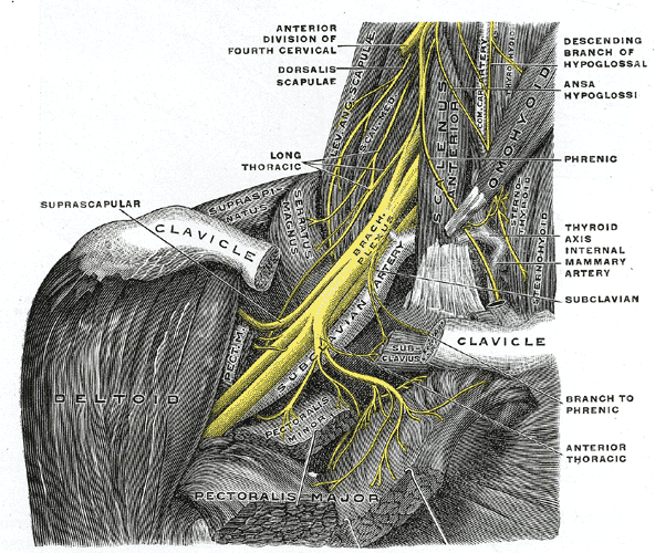
The brachial plexus is the network of nerves formed by the ventral rami of spinal nerves C5 through T1 that provides motor, sensory, and sympathetic innervation to the upper limb, shoulder, and upper chest [7]C4[13]D5.
Also Called / Synonyms
- Brachial plexus, plexus brachialis, BP
Key Terminology
- Roots: The five ventral rami (C5-T1) that emerge from the intervertebral foramina and converge to form trunks.
- Trunks: Superior (C5-C6), middle (C7), and inferior (C8-T1) trunks.
- Divisions: Each trunk splits into anterior and posterior divisions behind the clavicle.
- Cords: Lateral (anterior divisions of superior and middle trunks), medial (anterior division of inferior trunk), and posterior (all posterior divisions).
- Terminal branches: Musculocutaneous, axillary, radial, median, and ulnar nerves.
- Prefixed plexus: Receives a contribution from C4 (prevalence 11%, 95% CI 6-17%) [7]C4[14]D5.
- Postfixed plexus: Receives a contribution from T2 (prevalence 1%, 95% CI 0-1%) [7]C4[14]D5.
Classification of Plexus Types by Root Contribution
| Type | Root Contribution | Prevalence | Clinical Implication |
|---|---|---|---|
| Normal | C5-T1 | 84% (95% CI 79-89%) [7]C4 | Standard dermatomal and myotomal maps apply. |
| Prefixed | C4-T1 | 11% (95% CI 6-17%) [7]C4 | Higher risk of upper trunk injury during delivery; may alter nerve block levels [5]D5[14]D5. |
| Postfixed | C5-T2 | 1% (95% CI 0-1%) [7]C4 | Lower trunk more vulnerable; may mimic C8-T1 pathology. |
Clinical Significance
Brachial plexus injuries are a leading cause of upper limb disability, with traumatic avulsions and birth-related palsies (Erb's palsy) affecting thousands annually; anatomical variants such as a prefixed plexus lower the injury threshold during delivery [2]C4[5]D5[36]D5.
Pearl: The brachial plexus is most commonly formed by C5-T1, but prefixed (C4 contribution) occurs in 11% and postfixed (T2 contribution) in 1% of individuals, altering dermatomal maps and surgical approaches [7]C4[14]D5.
Gross Structure, Morphology & Function
- ▸The regular root-to-trunk arrangement occurs in 84% of cases; the regular division-to-cord arrangement is even more stable at 96%.
- ▸Supraclavicular nerve origins (long thoracic, dorsal scapular, suprascapular, subclavian) vary in 10-22% of individuals.
- ▸Infraclavicular branches have highly predictable usual origins, with radial, ulnar, and musculocutaneous nerves each >97% typical.
From the classification of the brachial plexus as a neural network formed by the ventral rami of C5-T1, the next level of analysis is its segmental organization into roots, trunks, divisions, cords, and terminal branches, a nested hierarchy that determines the functional mapping of every muscle and dermatome of the upper limb.
Roots and Trunks
The five roots (C5-T1) converge between the scalenus anterior and medius muscles to form three trunks. The regular arrangement, C5 and C6 joining to form the superior trunk, C7 continuing as the middle trunk, and C8 with T1 forming the inferior trunk, occurs in 84% of specimens (95% CI 79-89%) [7]C4. A prefixed plexus (receiving a branch from C4) is seen in 11% (95% CI 6-17%), and a postfixed plexus (receiving T2) in 1% (95% CI 0-1%) [7]C4. In fewer than 0.1% of cases, both C4 and T2 contribute [7]C4. Importantly, the human brachial plexus roots consistently comprise the 5th through 9th cervical spinal nerves, and vertebral formula anomalies do not produce a craniocaudal shift of this core composition [71]D5. Fetal studies show that trunk anomalies are rare, occurring in only 5.45% of plexuses [67]D5.
Divisions and Cords
Each trunk splits into an anterior and posterior division; the six divisions then reorganize into three cords. The regular pattern, anterior divisions of the superior and middle trunks form the lateral cord, anterior division of the inferior trunk becomes the medial cord, and all three posterior divisions unite as the posterior cord, is found in 96% of cases (95% CI 93-98%) [7]C4. The relationship of the cords to the axillary artery is regular (lateral, medial, posterior positions) in 96% (95% CI 89-100%), and the course through the interscalene triangle is regular in 86% (95% CI 66-98%) [7]C4. Additional communicating branches between plexus components appear in 5% of cases (95% CI 3-7%) [7]C4. The axillary artery’s segmental position, normally arising from intersegmental artery i7, is a major determinant of plexus morphology; deviation to i6 or i8 creates predictable variant configurations [24]D5.
Supraclavicular Branches
These nerves arise from the roots or trunks before the divisions and supply the scapular and thoracic wall muscles. Their origins show considerable variation [8]C4.
| Nerve | Usual origin | Prevalence (95% CI) | Function |
|---|---|---|---|
| Long thoracic | C5, C6, C7 | 78.1% (69.4-86.7%) | Serratus anterior (scapular protraction) |
| Dorsal scapular | C5 root | 85.2% (75.7-94.6%) | Rhomboids, levator scapulae |
| Suprascapular | Superior trunk | 89.8% (85.1-94.4%) | Supraspinatus, infraspinatus |
| Subclavian | Superior trunk | 98.3% (96.3-100%) | Subclavius |
An accessory long thoracic nerve occurs in 0.3% of cases, and an accessory suprascapular nerve in 0.2% [8]C4. The suprascapular nerve origin has a mean diameter of 1.33 mm and lies at a mean depth of 5.12 mm from the skin [48]C4.
Infraclavicular Branches
The terminal nerves of the upper limb arise from the cords. A meta-analysis of 4772 limbs provides the following prevalence data for usual origins [15]C4:
- Lateral cord: lateral pectoral nerve, 76.8% (95% CI 50-96%); musculocutaneous nerve, 98.8% (95% CI 98-100%).
- Medial cord: medial pectoral, 90.9% (68-100%); medial brachial cutaneous, 90.7% (73-100%); medial antebrachial cutaneous, 87.9% (67-99%); ulnar nerve, 97.7% (94-100%).
- Posterior cord: superior subscapular, 90.7% (80-98%); inferior subscapular, 76.1% (61-89%); thoracodorsal, 90.1% (84-95%); axillary, 79.8% (68-90%); radial nerve, 99.0% (96-100%).
- Median nerve: usual origin from lateral and medial cord roots, 89.7% (95% CI 84-95%) [15]C4.
The musculocutaneous nerve typically pierces the coracobrachialis muscle between its superficial and deep heads; this relationship is present in 100% of two- or three-headed coracobrachialis variants [51]C4. The lateral pectoral nerve supplies the clavicular of the deltoid in 86.2% of cases, a finding that may explain residual anteversion after axillary nerve injury [49]C4.
Functional Integration
The brachial plexus distributes motor innervation to all intrinsic and extrinsic muscles of the upper limb and provides sensory coverage from the shoulder to the fingertips. Its hierarchical structure allows for predictable patterns of deficit (e.g., Erb-Duchenne palsy affecting C5-C6; Klumpke palsy affecting C8-T1). The morphometric consistency of the plexus components with aging, demonstrated by proportional relationships between root, trunk, and cord dimensions, supports a stable scaffold for clinical localization [7]C4.
Pearl: When assessing a brachial plexus injury, the regular arrangement of divisions into cords is the most stable element (96% prevalence), while root/trunk variations (16% atypical) are common enough that a single anomalous trunk or communicating branch should not derail the diagnosis, trace the cords and their relationship to the axillary artery to confirm the level.
| Nerve | Usual origin | Prevalence (95% CI) | Function |
|---|---|---|---|
| Long thoracic | C5, C6, C7 | 78.1% (69.4-86.7%) | Serratus anterior |
| Dorsal scapular | C5 root | 85.2% (75.7-94.6%) | Rhomboids, levator scapulae |
| Suprascapular | Superior trunk | 89.8% (85.1-94.4%) | Supraspinatus, infraspinatus |
| Subclavian | Superior trunk | 98.3% (96.3-100%) | Subclavius |
| Cord | Nerve | Prevalence (95% CI) |
|---|---|---|
| Lateral cord | Lateral pectoral | 76.8% (50-96%) |
| Lateral cord | Musculocutaneous | 98.8% (98-100%) |
| Medial cord | Medial pectoral | 90.9% (68-100%) |
| Medial cord | Medial brachial cutaneous | 90.7% (73-100%) |
| Medial cord | Medial antebrachial cutaneous | 87.9% (67-99%) |
| Medial cord | Ulnar | 97.7% (94-100%) |
| Posterior cord | Superior subscapular | 90.7% (80-98%) |
| Posterior cord | Inferior subscapular | 76.1% (61-89%) |
| Posterior cord | Thoracodorsal | 90.1% (84-95%) |
| Posterior cord | Axillary | 79.8% (68-90%) |
| Posterior cord | Radial | 99.0% (96-100%) |
| Lateral + Medial | Median | 89.7% (84-95%) |
Relations, Borders & Spaces
- ▸The three compartments of the thoracic outlet (interscalene triangle, costoclavicular space, retropectoralis minor space) are the primary sites of brachial plexus compression in TOS.
- ▸Anatomical variations in the interscalene triangle occur in 47.7% of individuals, often involving the brachial plexus piercing the anterior scalene muscle, which may render traditional TOS positional tests falsely negative.
- ▸A fascial septum in the costoclavicular space separates the lateral cord from the medial and posterior cords in 94.16% of cases, mandating two separate injections for complete costoclavicular block.
From its emergence between the scalene muscles to its terminal branches in the axilla, the brachial plexus traverses a series of anatomically confined spaces that define its vulnerability to compression and guide surgical access. These compartments, the interscalene triangle, costoclavicular space, and retropectoralis minor space, correspond to the three sites of potential entrapment in (TOS) [46]D5. Understanding their borders and contents is essential for interpreting imaging, planning regional anesthesia, and diagnosing neurovascular compression.
Interscalene Triangle
This triangular space is bounded anteriorly by the , posteriorly by the , and inferiorly by the . The brachial plexus roots (C5-T1) and the pass through it; the subclavian vein lies anterior to the anterior scalene. Anatomical variations in this relationship are remarkably common: a cadaveric study of 65 specimens found that 47.7% of specimens deviated from the classic arrangement [85]C4. In 3.1% of cases, the entire superior trunk coursed completely anterior to the anterior scalene, placing it in a position of increased vulnerability. More frequently, a portion of or the entire superior trunk (and occasionally the middle trunk) pierced the anterior scalene muscle belly itself [85]C4. These variations are clinically important because they may not produce vascular compromise during traditional positional testing for TOS, potentially leading to false-negative results. Ultrasound evaluation of the interscalene triangle can identify such variants in symptomatic patients with negative provocative tests [85]C4.
Costoclavicular Space
The costoclavicular space is bounded superiorly by the clavicle, inferiorly by the first rib, and medially by the costoclavicular ligament. The three cords of the brachial plexus and the occupy this tight interval. A critical anatomical feature here is a fascial septum that separates the lateral cord from the medial and posterior cords. In an ultrasound-guided proximal infraclavicular costoclavicular block study, this septum was visualized in 46.2% of patients, but importantly, displacement of the septum preventing spread of local anesthetic between compartments occurred in 94.16% of cases [76]C4. Microanatomical cross-sections confirmed that the septum bundles the medial and posterior cords into one compartment and separates them from the lateral cord. This finding has direct clinical implications for regional anesthesia: two separate injections (or two catheter placements) into the superficial and deep compartments are recommended to ensure local anesthetic spreads around all three cords [76]C4. The costoclavicular block has gained interest as a diaphragm-sparing alternative to interscalene block for shoulder surgery, reliably blocking the suprascapular nerve while sparing the phrenic nerve [78]D5.
Retropectoralis Minor Space (Subcoracoid Space)
This space lies posterior to the , extending from the coracoid process to the chest wall. The brachial plexus cords and the pass through it. Compression here can produce the pectoralis minor syndrome, a variant of TOS. An anomalous muscle, the subclavius posticus muscle, occurs with a prevalence of 4.9% and can cross the superior thoracic aperture, compressing the brachial plexus and contributing to both neurogenic and vascular TOS [6]C4. The axillary arch, a musculofascial band extending from to the pectoralis major or coracoid process, can also cause compression in this region [79]C4.
Axilla and Quadrangular Space
Distal to the retropectoralis minor space, the brachial plexus cords divide into terminal branches within the axillary sheath, surrounding the . The axillary nerve and exit the axilla through the quadrangular space (bounded by teres minor, teres major, long of triceps, and humeral neck). This space is the target for posterior axillary nerve block, though an ultrasound-guided anterior approach targeting the first segment of the axillary nerve on the subscapularis muscle may provide more complete blockade of articular branches [75]D5. The radial nerve within the axillary fossa runs in close relation to the ; accessing it above the artery and over the tendon yields motor responses at significantly lower current intensity (0.44 ± 0.15 mA vs 0.57 ± 0.17 mA, P = 0.015) [77]C4.
Pearl: When performing a costoclavicular brachial plexus block, always plan for two separate injections because the fascial septum separating the lateral cord from the medial and posterior cords prevents adequate spread of a single bolus in over 94% of patients [76]C4.
| Compartment | Boundaries | Neurovascular Contents | Key Clinical Relevance |
|---|---|---|---|
| Interscalene triangle | Anterior scalene, middle scalene, first rib | Roots C5-T1, subclavian artery | Neurogenic TOS; 47.7% have variant anatomy [85]C4 |
| Costoclavicular space | Clavicle, first rib, costoclavicular ligament | Cords, subclavian vein | Site of costoclavicular block; fascial septum requires dual injection [76]C4 |
| Retropectoralis minor space | Pectoralis minor, coracoid, chest wall | Cords, axillary artery | Pectoralis minor syndrome; subclavius posticus muscle (4.9%) may compress here [6]C4 |
Blood Supply, Innervation & Lymphatic Drainage
- ▸The brachial plexus's vascular supply includes the subclavian and axillary arteries, with the dorsal scapular artery exhibiting a variable course through the plexus trunks in up to 63% of cases.
- ▸Venous variations are common: the axillary vein is duplicated in 17.5% of limbs, and brachial veins perforate the median nerve root in 15%, posing risks during axillary block and surgery.
- ▸Lymphatic drainage to the axillary nodes puts the long thoracic and thoracodorsal nerves at risk during dissection; the sentinel node blood supply is derived from the lateral thoracic and thoracodorsal arteries.
The interscalene triangle and axilla house the brachial plexus alongside a neurovascular supply that is both its life support and its point of vulnerability. The subclavian artery and vein, the axillary artery and vein, the dorsal scapular artery, and the axillary lymphatic chain each bear a precise relationship to the plexus roots, trunks, and cords. From the scalene compartment to the axillary sheath, the vessels and lymphatics that accompany the plexus determine its susceptibility to compression, iatrogenic injury, and patterns of metastatic spread.
Arterial Supply
The brachial plexus receives its nutrient blood supply (vasa nervorum) from branches of the subclavian and axillary arteries. The subclavian artery passes posterior to the anterior scalene muscle, with the plexus roots positioned above (C5), behind (C6-C7), and below (C8-T1) the artery. The dorsal scapular artery (DSA) is a critical vessel that supplies the levator scapulae and rhomboid muscles and exhibits a variable origin that directly affects the plexus. In a study of 252 sides, the DSA most commonly arose from the transverse cervical artery (69%), always coursing posterior or above the plexus. When it originated from the second (2.8%) or third (28.2%) part of the subclavian artery, it ran between the branches of the plexus: most frequently between the upper and middle trunks (63.2%), less often between the anterior division of the upper trunk and the middle trunk (1.3%) or between the C8 and T1 roots (2.6%) [112]C4. This inter-trunk course places the DSA at risk during scalenotomy or plexus exploration.
The axillary artery, the direct continuation of the subclavian, is classically described as passing through the brachial plexus with the cords named by their relation to it. However, the artery’s course pattern varies. In a cadaveric study of 49 axillae, the axillary artery penetrated the brachial plexus in only 51% of specimens (standard type). In 42.9%, a superficial subscapular artery (SSbsA) type was present, where the artery entered the deep layer without penetrating the plexus; in 2%, a superficial brachial artery type ran superficial to the plexus [111]C4. These variations alter the relationship between the cords and the artery, influencing the safety of infraclavicular block and axillary surgery.
Additional arterial variations with clinical relevance include the superficial brachioradial artery (radial artery with high origin from the axillary artery), reported in approximately 3% of specimens, which courses superficially in the arm and elevates the risk of bleeding complications during venous access or trauma [110]C4. The coexistence of arterial and neural variations is common: in 167 cadaver arms, 36% showed plexus variations, and these were frequently associated with aberrant arterial patterns (e.g., a common trunk of the axillary artery with a hidden median nerve loop) [105]C4.
Table 1. Origin of the Dorsal Scapular Artery and Relation to the Brachial Plexus
| Origin | Frequency | Course relative to plexus |
|---|---|---|
| Transverse cervical artery | 69% | Always posterior or above the plexus |
| Second part of subclavian artery | 2.8% | Between branches of plexus in 63.2% (upper and middle trunks) |
| Third part of subclavian artery | 28.2% | Between branches of plexus (see above) |
Data from Chaijaroonkhanarak et al. [112]C4
Venous Drainage
The venous drainage of the brachial plexus region converges on the axillary vein, which lies medial to the axillary artery and is separated from the cords by the axillary sheath. The axillary vein is formed by the union of the basilic and brachial veins. The brachial veins demonstrate two main termination patterns: they end separately into the basilic or axillary vein (Type A, 72.5%) or form a common brachial vein first (Type B, 27.5%) [91]C4. The basilic vein is absent in 5% of limbs, and duplication of the axillary vein occurs in 17.5% [91]C4.
A lateral venous channel running along the lateral wall of the axilla is present in 40% of specimens and may be mistaken for the axillary vein during central line placement [91]C4. The posterior circumflex humeral vein crosses posterior to the brachial plexus in 45% of cases to join the axillary vein, or in 42.5% to join the subscapular vein [91]C4. Critically, the brachial veins or their tributaries may perforate the lateral root of the median nerve, observed in 15% of dissections [91]C4. This venous loop through the median nerve roots can cause nerve compression when the vein is distended (e.g., during thrombosis or high-flow states) and may complicate axillary block or nerve transfer surgery.
Lymphatic Drainage
The lymphatic drainage of the upper limb and breast passes through the axillary lymph nodes, which are intimately related to the brachial plexus. The axillary nodes are divided into three levels by their relation to the pectoralis minor muscle: level I (lateral to pectoralis minor), level II (deep to pectoralis minor, including the interpectoral nodes), and level III (medial to pectoralis minor, infraclavicular). The brachial plexus cords lie posterior to the level I and II nodes, separated by the axillary sheath. During for breast cancer, the long thoracic nerve (C5-C7) is at risk along the chest wall, and the thoracodorsal nerve (C6-C8) is at risk on the lateral scapular wall. The arterial supply to the sentinel lymph nodes (level Ib) is derived from the lateral thoracic artery, thoracodorsal artery, inferior pectoral artery, and superficial thoracic artery, with the dominant supply shifting based on the axillary artery course type [111]C4.
Neural Relations and Sympathetic Innervation
The brachial plexus receives its autonomic innervation from the cervical sympathetic trunk. Postganglionic sympathetic fibers from the middle cervical ganglion and the stellate ganglion (cervicothoracic ganglion) enter the plexus via gray rami communicantes at the level of the spinal nerve roots (C5-T1). These fibers travel with the plexus to supply vasomotor, pilomotor, and sudomotor functions to the upper limb. Injury to the sympathetic chain (e.g., from a Pancoast tumor or iatrogenic dissection) produces Horner syndrome (ptosis, miosis, anhidrosis, enophthalmos) due to interruption of the oculosympathetic pathway.
The phrenic nerve (C3-C5) runs on the anterior surface of the anterior scalene muscle, crossing from lateral to medial as it descends. It is vulnerable during interscalene brachial plexus block, with reported rates of phrenic nerve palsy of up to 24% [94]A1b. The ultrasound-guided pericapsular nerve group (PENG) block avoids this complication, demonstrating 0% phrenic nerve palsy in a randomized trial [94]A1b. Conversely, ultrasound-guided phrenic nerve block (UPNB) can be performed intentionally to treat intractable hiccups or to improve oxygenation in patients with diaphragmatic dysfunction, without inducing dyspnea [98]B2a.
Several variant muscles in the axilla receive specific innervation that can be confused with the plexus. The axillary arch of Langer (a muscular slip from latissimus dorsi) is innervated by the thoracodorsal nerve in 85% of subjects [59]D5. The dorsoepitrochlearis (a slip from latissimus dorsi passing over the medial brachial plexus) is also supplied by the thoracodorsal nerve [90]C4. The subclavius posticus, a variant muscle with a prevalence of 4.9%, crosses the superior thoracic aperture and can compress the brachial plexus; its innervation is most commonly from the subclavian nerve (57.6%) or the suprascapular nerve (25.8%) [6]C4.
Pearl: The dorsal scapular artery passes between the upper and middle trunks in 63.2% of cases when it arises from the subclavian artery, a key landmark during supraclavicular exploration that, if inadvertently injured, can cause significant bleeding that obscures the plexus.
Microscopic & Histological Notes
- ▸The epineurium of the musculocutaneous nerve has 4-5 concentrically arranged laminae separated by adipose tissue on medial/lateral sides, creating an oval profile that permits length changes during muscle contraction.
- ▸Intraplexus fascial septae separate the lateral cord from the medial and posterior cords in the costoclavicular region, confirmed microanatomically.
- ▸Myeloid sarcoma of the brachial plexus shows a characteristic immunohistochemical profile (CD43+, CD45+, CD33+, CD117+, CD68+, focal MPO+, CD34-) with a high Ki‑67 index (80-90%).
The connective tissue architecture of the brachial plexus, its epineurium, perineurium, and endoneurium, defines both mechanical resilience and vulnerability to compression. Microanatomical studies reveal structural specializations that accommodate nerve gliding during limb movement and that explain variable injury patterns.
Connective Tissue Layers and Intraneural Architecture
Each nerve root and cord is enveloped by an epineurium composed of 4-5 concentrically arranged laminae of connective tissue [114]C4. Along the intramuscular course of the musculocutaneous nerve (MCN), this epineurium takes on an oval profile (mean circular factor 0.8) because laminae on the medial and lateral sides are separated by thin layers of adipose tissue, while those on the ventral and dorsal aspects are tightly packed [114]C4. This fibroadipose sheath functions as a “telescope” sliding system, allowing length changes during coracobrachialis contraction without nerve strain; loss of this compliance may predispose to compression syndrome [114]C4.
In the costoclavicular region, intraplexus fascial septae separate the lateral cord compartment from the medial and posterior cord compartments. These hyperechoic linear structures were displaced in 94.16% of patients during ultrasound-guided blocks, preventing anesthetic spread unless the septum is pierced [76]C4. Microanatomical cross‑sections confirm a consistent septum that bundles the medial and posterior cords together [76]C4.
Intrinsic Blood Supply
The intrinsic microvessels of the brachial plexus are best visualized by diaphanization and histology after gelatin‑lead oxide injection, which simultaneously displays extrinsic and intrinsic supply [119]C4. The anterior and posterior nerve roots receive blood from segmental branches of the vertebral artery, deep cervical artery, and ascending cervical artery, with a mean outer diameter of 0.61 mm [118]C4. This delicate intraneural network is vulnerable to stretch injury, contributing to ischemic damage in root avulsions.
Histomorphometry
In the rabbit brachial plexus, an accepted model for human nerve repair studies, histomorphometric analysis showed that the g‑ratio (axon diameter / fiber diameter) remains close to values in peripheral nerves, and that axon diameter, fiber diameter, and myelin sheath thickness peak at the trunk level then decrease progressively as nerves travel peripherally [39]C4. The number of axons, nerve area, and myelin fiber density also vary significantly across levels from C5 to T1 [39]C4. These data provide a baseline for evaluating regeneration after nerve grafting.
Histopathological Correlates of Brachial Plexus Lesions
Most primary brachial plexus tumors are benign nerve sheath tumors (schwannoma, neurofibroma). Schwannomas are indolent, encapsulated lesions arising from Schwann cells; immunohistochemistry is used for confirmation [43]C4. Malignant peripheral nerve sheath tumors (MPNSTs) show fascicular growth, nuclear atypia, and high mitotic activity; they are associated with neurofibromatosis and worse recurrence‑free survival [122]C4.
Brachial plexus lipomas are rare; four of 27 reported cases had a nonconventional lipoma type on histology [116]C4. Primary embryonal carcinoma of the brachial plexus has been documented histologically, highlighting that some primary tumors are malignant and require prompt treatment [121]C4. Myeloid sarcoma involving the brachial plexus was reported in a patient with acute myeloid leukemia; histology showed immature myeloid cells with a characteristic immunohistochemical profile:
| Marker | Expression |
|---|---|
| CD43 | + |
| CD45 | + |
| CD33 | + |
| CD117 | + |
| CD68 | + |
| MPO (focal) | + |
| CD34 | - |
| Ki‑67 | 80-90% (high proliferative index) |
[120]C4
Pearl: The MCN epineurium’s “telescope” sliding system, with its oval cross‑section and adipose‑separated laminae, is a histologic correlate of nerve compliance; disruption of this architecture may explain idiopathic compression syndromes and should be considered during surgical dissection of the coracobrachialis.
| Marker | Expression |
|---|---|
| CD43 | + |
| CD45 | + |
| CD33 | + |
| CD117 | + |
| CD68 | + |
| MPO (focal) | + |
| CD34 | - |
| Ki‑67 | 80-90% |
Development (Brief Embryology)
- ▸The brachial plexus main roots are fixed to the 5th-9th spinal nerves regardless of vertebral formula, unlike the lumbosacral plexus which shifts with vertebral count [71].
- ▸The scalene blastema appears at week 6 and is divided into anterior and medial primordia by the brachial plexus and subclavian artery, forming the scalene triangle [115].
- ▸Arterial variations (e.g., axillary artery arising from the ninth segmental artery) can produce abnormal root-trunk-cord relationships and single-corded plexuses [128].
From the microscopic organization of fibers and connective tissue, the developmental events that shape the brachial plexus become evident: the complex adult anatomy is laid down during the sixth week of gestation, when the spinal nerves and adjacent mesenchyme interact. The brachial plexus primordium arises from the ventral rami of the fifth through ninth spinal nerves (C5-T1 or, in some individuals, C4-C8), with contributions from the fourth and tenth nerves being small and variable [71]D5. Unlike the lumbosacral plexus, which exhibits a craniocaudal shift when the vertebral formula changes, the brachial plexus main roots remain fixed to the 5th-9th spinal nerves regardless of thoracolumbar variation, a stability that may reflect distinct regulation [71]D5.
Timing and Key Events
By the sixth week of development, the scalene blastema, a condensation of myoblasts, mesenchymal cells, and collagen fibers derived from both hypaxial myotomes and sclerotomes of the cervical somites, appears in the lateral neck [115]C4. The ventral rami of the spinal nerves that will form the brachial plexus, together with the subclavian artery, divide this blastema into two components: a medial portion ( primordium) and a lateral portion ( primordium). The pierces the medial portion, while the dorsal scapular nerve and the branches forming the long thoracic nerve pierce the lateral portion [115]C4. The space between the two primordia, the future scalene triangle, is thus created by the passage of the neurovascular bundle. The scalenus minimus muscle is identified in 10.6% of sides (7 of 66 sides) and appears as an accessory slip from the blastema [115]C4. The insertion of the scalenus anterior into the parietal pleura via the suprapleural membrane primordium is established early, linking the scalene muscles to respiratory mechanics [115]C4.
Vascular Relationships and Anomalies
The developing brachial plexus is intimately associated with the axillary artery. In rare cases, the axillary artery may develop from the ninth segmental artery instead of the usual seventh cervical intersegmental artery, causing the artery to lie inferomedial to the plexus rather than passing between the medial and lateral cords [128]C4. Such a shift can lead to a single-corded plexus or other root-trunk-cord variations [33]C4. The also shows developmental variation: when it arises from the proximal subclavian artery, it tends to pass below the brachial plexus, but atypical origins (e.g., from the internal thoracic artery) can cause the artery to course between the C5 and C6 roots, even suspending the subclavian artery in a high-arch configuration [34]C4.
Clinical Implications
These embryological events explain why congenital anomalies such as cervical ribs, anomalous scalene insertions, and fibrous bands are common in the thoracic outlet region. The fixed root composition of the brachial plexus in the face of vertebral variation may also account for the relative uniformity of plexus injury patterns across individuals. Understanding the developmental relationship between the scalene blastema and the neurovascular bundle is essential for interpreting variants and for planning surgical decompression [115]C4.
Pearl: Arterial variations (e.g., axillary artery arising from the ninth segmental artery) can produce abnormal root-trunk-cord relationships and single-corded plexuses [128]C4.
Variations & Anomalies
- ▸Prefixed plexus occurs in 11% and postfixed in 1%; trunk formation is regular in only 84% of cases [7].
- ▸Supraclavicular nerve origins vary in 10-22% of cases; variant origins alter landmarks for nerve blocks and transfers [8].
- ▸Muscular variants (subclavius posticus, axillary arch, accessory scalenes) are common causes of neurogenic thoracic outlet syndrome and must be recognised on imaging [6][145].
Embryological deviations in segmental contribution and fascial plane development give rise to a spectrum of brachial plexus variants. Meta-analysis of 3055 upper limbs provides pooled prevalence data essential for surgical planning and avoiding iatrogenic injury [7]C4.
Prefixation, Postfixation, and Trunk Formation
The regular C5-T1 root pattern forming three trunks occurs in 84% (95% CI 79-89%) of cases [7]C4. A prefixed plexus (C4 contribution) appears in 11% (95% CI 6-17%), and a postfixed plexus (T2 contribution) in 1% (95% CI 0-1%); both C4 and T2 contributions coexist in <0.1% [7]C4. Variant trunk formation is the most common structural anomaly and may alter interscalene block landmarks.
Supraclavicular Nerve Origins
| Nerve | Usual origin | Prevalence | Variation |
|---|---|---|---|
| Subclavian nerve | Superior trunk | 98.3% (95% CI 96.3-100%) | C5 root origin in 1.7% [8]C4 |
| Accessory long thoracic nerve occurs in 0.3% [8]C4. These variations matter when planning nerve transfers (e.g., spinal accessory to suprascapular nerve), where donor-recipient axon counts must be matched [132]D5[136]A1b[140]B2a. |
Muscular Variants with Neurovascular Impact
- Subclavius posticus muscle: prevalence 4.9%; inserts variably on scapular superior border (71.4%), coracoid process (25.4%), or clavicle (0.9%); can compress brachial plexus at the superior thoracic aperture and cause neurogenic [6]C4.
- Axillary arch (Langer's arch): muscular or fascial slip from latissimus dorsi to pectoralis major or coracoid; may compress the axillary vein or cords; innervated by the thoracodorsal nerve in 85% of cases [59]D5[79]C4[109]C4.
- Accessory middle scalene muscle: a triangular band compressing the lower trunk and subclavian artery, mimicking thoracic outlet syndrome [145]C4.
- Accessory subscapularis muscle: can entrap the axillary, lower subscapular, and thoracodorsal nerves [52]C4.
- Coracobrachialis multheadedness: two-headed in 63%, three-headed in 22%; associated with musculocutaneous nerve course variations [51]C4.
Vascular-Plexus Relationship Variants
In 96% (95% CI 89-100%), the axillary artery passes between the medial and lateral cords in the expected manner [7]C4. When the artery arises from a ninth intersegmental artery (1.8% of cases), it runs caudal to the plexus, missing the median nerve loop; a rare sixth intersegmental artery may pierce the musculocutaneous nerve [131]C4. Superficial brachioradial artery (high origin radial artery, ~3%) is another variant with risk of accidental cannulation [110]C4.
Terminal Nerve Interconnections
Communicating branches between components occur in 5% (95% CI 3-7%) [7]C4. Notable examples: musculocutaneous-median nerve anastomoses (types II-V in the accepted classification) [17]C4[26]C4[144]C4; radial-ulnar nerve communication in 3.7% [51]C4; trifid lateral root of median nerve [146]C4; and brachial artery piercing a fenestrated median nerve (rare) [135]C4.
Pearl: The single most common variation is a prefixed plexus (11% of cases); failing to recognise it can lead to missed root avulsion on MRI or unintended phrenic nerve palsy during supraclavicular block, as the C4 contribution lifts the plexus higher than expected [7]C4[9]D5.
| Nerve | Usual origin | Prevalence | Variation prevalence |
|---|---|---|---|
| Subclavian nerve | Superior trunk | 98.3% (95% CI 96.3-100%) | 1.7% [8]C4 |
Surface Anatomy & Imaging Correlation
- ▸Ultrasound-guided brachial plexus blockade relies on specific landmarks (interscalene groove, costoclavicular space, axillary artery) with real-time visualization of fascial septae to tailor injection strategy.
- ▸CT myelography remains the preferred initial study for differentiating pre- from postganglionic traumatic brachial plexus injury.
- ▸The subclavius posticus muscle, present in up to 8.3% of individuals on MRI, directly contacts the brachial plexus in 60% of cases and is an underrecognized cause of neurogenic thoracic outlet syndrome.
The anatomic variations just discussed underscore why imaging-guided confirmation of brachial plexus structure is essential for safe intervention. This section translates the three-dimensional anatomy into surface landmarks and cross-sectional imaging appearances used at the bedside and on MRI, CT, and ultrasound.
Surface Landmarks and Palpation
Palpable reference points guide every block approach. The interscalene groove is located between the anterior and middle scalene muscles at the level of the cricoid cartilage (C6). Here the roots of the plexus emerge as hypoechoic nodules on ultrasound. In the supraclavicular fossa, the plexus lies superior and posterior to the subclavian artery. The infraclavicular region positions the three cords around the axillary artery, while in the axilla the terminal branches surround the brachial artery. These four stations correspond to the four common brachial plexus block approaches used clinically [4]D5.
Ultrasound-Guided Identification
Real-time ultrasound has become the standard for guiding brachial plexus blockade. At the interscalene level, the origins of the suprascapular nerve (orSSN) can be identified as a hypoechoic structure with a mean diameter of 1.33 mm and depth of 5.12 mm from the skin, typically arising from the upper trunk or its posterior division [48]C4. A proximal SSN block targeting this origin is effective and reliable [150]D5. In the costoclavicular region, a fascial septum separates the lateral cord compartment from the medial and posterior cords. This septum is visible in 46.2% of patients on initial scanning but becomes displaced during injection in 94.16% of cases, preventing anesthetic spread unless two separate injections are made into the superficial and deep compartments [76]C4. In the axilla, radial nerve response differs by approach: accessing the nerve above the brachial artery and over the latissimus dorsi tendon elicits a triceps motor response at a significantly lower current intensity (0.44 ± 0.15 mA vs 0.57 ± 0.17 mA below the artery, P = 0.015) [77]C4. Artificial intelligence software can now identify brachial plexus anatomy on ultrasound with expert-level accuracy, providing real-time labeling for trainees [161]C4.
MRI and CT Appearance
MR neurography (3D STIR SPACE sequences) demonstrates the plexus with high fascicular detail. Diffusion tensor imaging (DTI) using the RESOLVE sequence at 3T yields a mean fractional anisotropy (FA) of 0.30 in healthy roots; angular measurements increase from proximal to distal roots with an intraclass correlation coefficient of 0.6 [156]C4. Three-dimensional reconstruction from non-contrast MRI shows high correspondence with cadaveric dissection and can be reproduced in vivo [155]C4. CT myelography remains the preferred initial study for traumatic brachial plexus injury. It differentiates preganglionic avulsion (pseudomeningoceles and absent root shadows) from postganglionic injury (neuroma, scar) with superior sensitivity [148]D5. For superior sulcus (Pancoast) tumors, MRI is superior to CT for evaluating tumor extension into the intervertebral foramina and the brachial plexus itself, while CT is optimal for depicting bone erosion, and PET/CT detects unsuspected nodal and distant metastases [149]D5.
Imaging of Common Variants and Pathology
The subclavius posticus muscle (SPM), present in 4.9% of cadavers and 8.3% of patients on coronal MRI, directly contacts the brachial plexus in 60% of cases [6]C4[158]C4. Its mean thickness is 6.10 ± 1.60 mm; when identified, SPM should be considered a potential contributor to neurogenic (TOS) [158]C4. For TOS evaluation, cervical radiography first screens for bone abnormalities, followed by CT angiography or MRI with postural maneuvers; ultrasound complements assessment of dynamic vessel compression [46]D5. Post-radiation plexopathy appears as diffuse T2 hyperintensity and thickening of the plexus on MRI. The brachial plexus is now routinely contoured as an avoidance structure in planning, with a recommended maximum dose limit of 60 Gy to reduce long-term toxicity [19]D5. In obstetric brachial plexus palsy, 3D CT reconstruction reveals scapular hypoplasia (average 14% reduction), acromial elongation (19%), and humeral subluxation (14%), all of which correlate with the scapular area visible over the clavicle [157]C4.
Impact on Regional Anesthesia
The imaging appearance directly affects block strategy. For shoulder arthroscopy, the pericapsular nerve group (PENG) block provides postoperative comparable to interscalene block but with a significantly lower incidence of phrenic nerve palsy (0% vs 24%, P = 0.022) and arm paresthesia (12% vs 36%, P = 0.047) [94]A1b. Reducing the anterior suprascapular nerve block volume from 10 mL to 5 mL of 0.5% bupivacaine lowers complete hemidiaphragmatic paralysis from 16.67% to 0% (P = 0.025) while preserving analgesia [153]A1b. For supraclavicular blocks, local anesthetic volumes of 25-30 mL accelerate sensory and motor block onset compared with 15 mL, but also produce volume-dependent bilateral increases in optic nerve sheath diameter, suggesting an intracranial pressure effect (P < 0.05) [159]A1b. For infraclavicular blocks, lidocaine 1% with epinephrine offers a shorter block duration than ropivacaine 0.5% without compromising patient satisfaction or reduction quality for [160]A1b.
Pearl: When performing an ultrasound-guided brachial plexus block in the costoclavicular region, always identify the fascial septum between the lateral cord and the medial/posterior cords; piercing it deliberately or planning two separate injections is necessary to achieve complete cord blockade [76]C4.
| Approach | Probe Position | Key Landmark | Coverage | Reference |
|---|---|---|---|---|
| Interscalene | Axial at cricoid level | Root nodules between scalene muscles | Shoulder & proximal upper limb | [4]D5 |
| Supraclavicular | Coronal oblique over supraclavicular fossa | Divisions lateral to subclavian artery | Mid-humerus & below | [4]D5 |
| Infraclavicular | Sagittal parasagittal below clavicle | Cords around axillary artery | Continuous anesthesia | [4]D5 |
| Axillary | Axial in axillary fossa | Terminal branches around brachial artery | Distal to elbow | [4]D5 |
Clinical Correlations
- ▸Brachial plexus injury patterns localize to specific roots or cords: upper trunk (C5‑C6) produces waiter's tip deformity; lower trunk (C8‑T1) claw hand with possible Horner's signaling preganglionic avulsion.
- ▸Anatomical variations, especially brachial plexus piercing the anterior scalene (47.7% of cadavers) and anomalous muscles (subclavius posticus 4.9%), are common and may cause false‑negative vascular tests in thoracic outlet syndrome.
- ▸Iatrogenic injury is preventable: avoid screws >1.4 cm in medial clavicle, limit interscalene block in patients with pulmonary reserve concerns, and contour brachial plexus to ≤60 Gy in radiotherapy planning.
Correlating the surface landmarks and imaging anatomy with patient presentation allows the clinician to localize brachial plexus pathology to specific roots, trunks, or cords, a skill essential for diagnosis, surgical planning, and avoiding iatrogenic injury.
Presenting Symptoms and Injury Patterns
Traumatic brachial plexus injury (BPI) most often results from high-energy traction, typically in young males involved in motorcycle collisions. The pattern of deficits follows the applied force: downward traction on the abducted arm stresses the upper trunk (C5‑C6), while upward traction on the abducted arm or forceful arm elevation injures the lower trunk (C8‑T1). Complete avulsions produce a flail, anesthetic limb and, when T1 is involved, ipsilateral Horner's syndrome (ptosis, miosis, anhidrosis). The presence of Horner's indicates a preganglionic root avulsion that cannot recover spontaneously and mandates urgent surgical consultation.
Brachial plexus birth injury (BPBI) occurs during difficult deliveries, particularly with . Meta-analysis of macrosomia trials shows that induction of labor does not significantly reduce the risk of brachial plexus palsy (RR 0.21, 95% CI 0.01‑4.28) but may improve Apgar scores in randomized controlled trials [167]A1a.
Position-related plexopathy emerged prominently during the COVID‑19 pandemic. In a systematic review of 30 patients who developed after prone positioning, the typical patient was male, over 50 years old, with , overweight, and diabetes. The most common symptoms were weakness, pain, and motion deficits; symptom onset occurred after prolonged or repeated prone sessions [2]C4.
(TOS) results from compression of the subclavian vessels or brachial plexus within the interscalene triangle, costoclavicular space, or retropectoralis minor space. Neurogenic TOS (nTOS) accounts for the majority of cases and most often involves the lower trunk. Vascular TOS (vTOS) may present with arm claudication, Raynaud's phenomenon, or thrombosis [44]D5.
Neoplastic involvement, most notably superior sulcus (Pancoast) tumor, can infiltrate the lower trunk and sympathetic chain. MRI is superior for evaluating tumor extension to the intervertebral foramen, spinal cord, and brachial plexus [149]D5.
Neurological Examination Findings
The examination follows a root-by-root and cord-by-cord sequence.
Motor:
- C5: Shoulder abduction (deltoid, MRC 0-5), external rotation (infraspinatus).
- C6: Elbow flexion (biceps brachii), forearm supination.
- C7: Elbow extension (triceps), wrist extension, finger extension.
- C8: Finger flexion (flexor digitorum profundus to digits 4‑5), thumb flexion.
- T1: Finger abduction/adduction (interossei), thumb opposition.
Sensory:
- C5: Lateral shoulder (axillary nerve) and lateral arm (superior lateral cutaneous nerve of arm).
- C6: Lateral forearm (lateral cutaneous nerve of forearm) and thumb/index.
- C7: Middle finger.
- C8: Medial forearm (medial cutaneous nerve of forearm) and ring/little fingers.
- T1: Medial arm (medial cutaneous nerve of arm).
Reflexes: Biceps jerk (C5‑C6), brachioradialis jerk (C6), triceps jerk (C7).
Autonomic: Horner's (ptosis, miosis, anhidrosis, enophthalmos) signals T1 preganglionic injury. Phrenic nerve involvement (C3‑C5) causes diaphragmatic paralysis; postoperative respiratory symptoms occurred in 4.9% of patients after phrenic nerve transfer, with 2.4% reporting persistent mild dyspnea [60]B2a.
Provocative maneuvers for TOS: Adson's test ( rotation and deep inspiration obliterates radial pulse), Roos test (elevated arm exercise reproduces symptoms), Wright test (hyperabduction compresses pectoralis minor). However, patients with brachial plexus piercing the anterior scalene muscle, a variant found in 47.7% of cadavers, may have false‑negative vascular tests because arterial compression is absent [85]C4.
Examination of scapular stabilizers: Winging suggests long thoracic nerve (C5‑C7) or dorsal scapular nerve (C5) entrapment. Anatomical entrapment of these nerves within scalene musculature occurs in 60.8% and 44.6% of specimens, respectively, and is linked to interscapular pain and scapulohumeral dysfunction [172]C4.
Phenotypic Variants
| Variant | Key Clinical Features | Common Cause |
|---|---|---|
| Erb‑Duchenne palsy (C5‑C6) | Waiter's tip arm: adducted, internally rotated shoulder; pronated forearm; flexed wrist; loss of shoulder abduction, external rotation, elbow flexion | Birth trauma, fall on shoulder |
| Extended Erb palsy (C5‑C7) | Above plus loss of elbow, wrist, and finger extension | Traction with greater force |
| Klumpke palsy (C8‑T1) | Claw hand (intrinsic minus); sensory loss on medial forearm and hand; Horner's if T1 avulsed | Upward traction on arm (e.g., grasping to break a fall) |
| Whole plexus injury | Flail, anesthetic limb; Horner's often present; severe functional loss | High‑energy traction (e.g., motorcycle) |
Red Flags
- Horner's syndrome, root avulsion; urgent surgical referral for possible nerve transfer or grafting.
- Expanding hematoma or pseudoaneurysm after clavicle fracture or penetrating trauma, immediate vascular assessment. The neurovascular bundle lies between the medial fourth and three‑fifths of the clavicle; screws longer than 1.4 cm risk iatrogenic injury [166]C4.
- Acute respiratory distress with diaphragmatic elevation, suspect bilateral phrenic nerve injury (rare but catastrophic; consider noninvasive ventilation).
- Rapidly progressive deficit over days, consider malignant infiltration (Pancoast, lymphoma) or inflammatory plexopathy (e.g., neuralgic amyotrophy).
Atypical Presentations
- Isolated nerve entrapments: Suprascapular notch (weakness of supraspinatus/infraspinatus), quadrilateral space (axillary nerve with deltoid atrophy), and median nerve in pronator syndrome can mimic brachial plexopathy. Imaging with MRI neurography helps differentiate [20]D5.
- Subclavius posticus muscle (prevalence 4.9%) crosses the superior thoracic aperture and can compress the brachial plexus, causing nTOS [6]C4.
- Accessory middle scalene muscle may compress the middle and lower trunks directly [145]C4.
- Variations in scalene anatomy: Upper trunk may pierce the anterior scalene (reported in some cadaver studies); the T1 nerve root may pierce the anterior scalene insertion tendon [173]C4. Such variations can alter the response to interscalene block and contribute to atypical TOS presentations.
- Langer's axillary arch, a musculotendinous slip from latissimus dorsi to coracoid, may compress the neurovascular bundle; in a recent report it crossed the basilic vein and lay over the lateral cord [109]C4.
Regional Anesthesia Considerations
Four principal blocks target different segments of the brachial plexus [4]D5:
| Block | Best Indication | Key Anatomic Risk |
|---|---|---|
| Interscalene | Shoulder and proximal humerus | Phrenic nerve palsy in 24%; hemidiaphragm elevation; avoid if poor pulmonary reserve [94]A1b |
| Supraclavicular | Mid‑humerus and below | Proximity to pleura; pneumothorax |
| Infraclavicular | Continuous catheter procedures | Risk to axillary vessels |
| Axillary | Distal elbow and hand | Incomplete block if variant anatomy |
A randomized trial comparing pericapsular nerve group (PENG) block to interscalene for shoulder arthroscopy found equivalent but significantly lower phrenic nerve palsy (0% vs 24%, p = 0.022) and paresthesia (12% vs 36%, p = 0.047) [94]A1b.
Surgical and Interventional Pearls
Nerve transfer surgery: For C5‑C6 injuries, the Oberlin transfer (fascicles of ulnar nerve to biceps branch) restores elbow flexion. A triple transfer adding brachioradialis reinnervation via a lateral cutaneous nerve of forearm interposition graft is anatomically feasible, with a calculated reinnervation distance of 94.1 mm (≈3 months) for brachioradialis [89]C4. Preoperative depression (PHQ‑9 ≥9) in patients undergoing partial ulnar nerve transfer was significantly associated with poorer recovery [138]B2b.
Reverse shoulder arthroplasty: Lowering the humerus below the glenoid equator can stretch the axillary nerve; lateralization is safer. Strains up to 15.3% and 19.3% occur in the lateral and medial roots of the median nerve, respectively [65]D5[70]D5.
Percutaneous tumor ablation: For tumors near the brachial plexus, nerve displacement with hydrodissection, balloon interposition, or electromyographic monitoring reduces thermal injury risk [162]D5.
Radiation therapy: The brachial plexus should be contoured as an avoidance structure in planning; maximum dose limited to 60 Gy to minimize radiation‑induced plexopathy [19]D5.
Prognosis and Recovery
Overall functional recovery (elbow flexion MRC ≥3) after brachial plexus reconstruction is approximately 42% at 24 months [138]B2b. End‑to‑end neurorrhaphy yields faster, stronger recovery than end‑to‑side repair in animal models, though functional improvement occurs with both techniques [58]D5. Neuromuscular electrical stimulation may be a useful adjunct in BPBI, but evidence is low certainty [55]B2a.
Controversies and Guideline Disagreement
| Question | Position A | Position B | Evidence Strength | Implication |
|---|---|---|---|---|
| Best block for shoulder arthroscopy | Interscalene (gold standard, complete analgesia) | PENG block (equivalent analgesia, fewer side effects) | RCT: moderate; small sample | Consider PENG in patients with respiratory comorbidity [94]A1b |
| Role of surgery for nTOS | Surgery after failed conservative therapy (scalenotomy, first rib resection) | Prolonged physiotherapy with neural mobilization and pain neuroscience education | Systematic review: moderate | Multidisciplinary approach; surgery reserved for refractory cases [170]B2a |
| Induction for macrosomia to prevent BPBI | Does not reduce Cesarean or shoulder dystocia | May improve neonatal Apgar (RCT subgroup) | Meta‑analysis: moderate; RR 0.21 (0.01‑4.28) | Do not routinely induce solely to prevent plexus injury [167]A1a |
These clinical patterns, eponymous signs, and procedural nuances lead directly to the nomenclature that labels brachial plexus disorders, the topic of the following section.
Pearl: Iatrogenic injury is preventable: avoid screws >1.4 cm in medial clavicle, limit interscalene block in patients with pulmonary reserve concerns, and contour brachial plexus to ≤60 Gy in radiotherapy planning.
Eponyms & Nomenclature
- ▸Thoracic outlet syndrome (TOS) is the most common eponym associated with brachial plexus compression, with neurogenic and vascular subtypes [44].
- ▸The anconeus medialis muscle has multiple synonyms (m. epitrochleoanconeus, caput mediale accessorium) and is distinct from triceps brachii [25].
- ▸Variant arterial nomenclature (superficial vs. main brachial artery) correlates with neural anomalies and has surgical implications [92].
Building on the clinical patterns described in the preceding section, a consistent nomenclature is essential for clear communication across surgical, radiological, and anatomical contexts. The following glossary reconciles eponyms and Terminologia Anatomica synonyms encountered in the brachial plexus literature.
Reconciled Glossary of Eponyms and Synonyms
| Eponym/Synonym | Structure/Context | Source |
|---|---|---|
| (TOS) | Compression of subclavian vessels and/or brachial plexus at thoracic outlet | [44]D5 |
| Neurogenic TOS (nTOS) | Subtype involving brachial plexus compression, primarily lower trunk | [44]D5 |
| Vascular TOS (vTOS) | Subtype involving subclavian artery or vein compression | [44]D5 |
| muscle / m. epitrochleoanconeus / caput mediale accessorium | Independent muscle innervated by ulnar nerve, not part of | [25]D5 |
| Coracobrachialis longus muscle | Atavistic variant of | [25]D5 |
| Caput breve of | Variant of biceps brachii | [25]D5 |
| / main brachial artery | Variant arterial pattern associated with anomalies | [92]C4 |
These terms illustrate the persistent variability in brachial plexus nomenclature. The anconeus medialis, for example, is described under at least three names in the literature, reflecting historical disagreement over its classification as part of the triceps brachii [25]D5. Similarly, the superficial brachial artery designation distinguishes a variant course that may alter surgical approach [92]C4.
Clinical Implications of Nomenclature Variability
Misuse of eponyms can lead to diagnostic confusion. For instance, TOS is often used imprecisely to describe any brachial plexus compression, but the term specifically denotes pathology at the thoracic outlet [44]D5. Recognizing the precise anatomical basis of each eponym improves interdisciplinary communication.
Pearl: Always specify the anatomical level (e.g., lower trunk) when using the eponym "thoracic outlet syndrome" to avoid ambiguity, as TOS can refer to neurogenic or vascular subtypes [44]D5.
References
- [1]
Sirico F, Castaldo C, Baioccato V et al.. “Prevalence of musculocutaneous nerve variations: Systematic review and meta-analysis.” Clinical anatomy (New York, N.Y.) (2018). PMID: 30113088 ↗
L4SR_OBSCited in: Definition & Classification - [2]
Toruńska E, Owczarek-Konopka M, Konopka M et al.. “Brachial plexopathy as a result of anatomical prone position in COVID-19 patients-Systematic review.” Clinical anatomy (New York, N.Y.) (2023). PMID: 37646090 ↗
L4SR_OBSCited in: Definition & Classification, Gross Structure, Morphology & Function, Blood Supply, Innervation & Lymphatic Drainage, Clinical Correlations - [3]
Sanchez-Sotelo J. “Reverse total shoulder arthroplasty.” Clinical anatomy (New York, N.Y.) (2009). PMID: 19090001 ↗
L5REVIEW_NARRATIVECited in: Definition & Classification - [4]
Mian A, Chaudhry I, Huang R et al.. “Brachial plexus anesthesia: A review of the relevant anatomy, complications, and anatomical variations.” Clinical anatomy (New York, N.Y.) (2013). PMID: 23959836 ↗
L5REVIEW_NARRATIVECited in: Definition & Classification, Surface Anatomy & Imaging Correlation, Clinical Correlations - [5]
Dunbar DC, Vilensky JA, Suárez-Quian CA et al.. “Risk factors for neonatal brachial plexus palsy attributed to anatomy, physiology, and evolution.” Clinical anatomy (New York, N.Y.) (2021). PMID: 33904192 ↗
L5REVIEW_NARRATIVECited in: Definition & Classification - [6]
Al-Redouan A, Benes M, Abbaspour E et al.. “Prevalence and anatomy of the anomalous subclavius posticus muscle and its clinical implications with emphasis in neurogenic thoracic outlet syndrome: Scoping review and meta-analysis.” Annals of anatomy = Anatomischer Anzeiger : official organ of the Anatomische Gesellschaft (2023). PMID: 36690046 ↗
L4SR_OBSCited in: Definition & Classification, Gross Structure, Morphology & Function, Relations, Borders & Spaces, Blood Supply, Innervation & Lymphatic Drainage, Variations & Anomalies, Surface Anatomy & Imaging Correlation, Clinical Correlations - [7]
Benes M, Kachlik D, Belbl M et al.. “A meta-analysis on the anatomical variability of the brachial plexus: Part I - Roots, trunks, divisions and cords.” Annals of anatomy = Anatomischer Anzeiger : official organ of the Anatomische Gesellschaft (2021). PMID: 33940116 ↗
L4SR_OBSCited in: Definition & Classification, Gross Structure, Morphology & Function, Variations & Anomalies - [8]
Benes M, Kachlik D, Belbl M et al.. “A meta-analysis on the anatomical variability of the brachial plexus: Part II - Branching of the supraclavicular part.” Annals of anatomy = Anatomischer Anzeiger : official organ of the Anatomische Gesellschaft (2021). PMID: 34186202 ↗
L4SR_OBSCited in: Definition & Classification, Gross Structure, Morphology & Function, Variations & Anomalies, Clinical Correlations - [9]
Gilcrease-Garcia BM, Deshmukh SD, Parsons MS. “Anatomy, Imaging, and Pathologic Conditions of the Brachial Plexus.” Radiographics : a review publication of the Radiological Society of North America, Inc (2020). PMID: 33001787 ↗
L5REVIEW_NARRATIVECited in: Definition & Classification, Gross Structure, Morphology & Function, Blood Supply, Innervation & Lymphatic Drainage, Variations & Anomalies, Surface Anatomy & Imaging Correlation, Eponyms & Nomenclature - [10]
Mohammed H, Skalski MR, Patel DB et al.. “Coracoid Process: The Lighthouse of the Shoulder.” Radiographics : a review publication of the Radiological Society of North America, Inc (2016). PMID: 27471875 ↗
L5REVIEW_NARRATIVECited in: Definition & Classification, Gross Structure, Morphology & Function, Clinical Correlations - [11]
Cummings KW, Sridhar S, Parsons MS et al.. “Cross-sectional Imaging Anatomy and Pathologic Conditions Affecting Thoracic Nerves.” Radiographics : a review publication of the Radiological Society of North America, Inc (2016). PMID: 27911674 ↗
L5REVIEW_NARRATIVECited in: Definition & Classification, Gross Structure, Morphology & Function, Surface Anatomy & Imaging Correlation, Clinical Correlations - [12]
Tubbs RS, Jones VL, Loukas M et al.. “Anatomy and landmarks for branches of the brachial plexus: a vade mecum.” Surgical and radiologic anatomy : SRA (2010). PMID: 20084513 ↗
L5REVIEW_NARRATIVECited in: Definition & Classification, Clinical Correlations - [13]
Johnson EO, Vekris M, Demesticha T et al.. “Neuroanatomy of the brachial plexus: normal and variant anatomy of its formation.” Surgical and radiologic anatomy : SRA (2010). PMID: 20237781 ↗
L5REVIEW_NARRATIVECited in: Definition & Classification, Clinical Correlations - [14]
Pellerin M, Kimball Z, Tubbs RS et al.. “The prefixed and postfixed brachial plexus: a review with surgical implications.” Surgical and radiologic anatomy : SRA (2010). PMID: 20087592 ↗
L5REVIEW_NARRATIVECited in: Definition & Classification, Blood Supply, Innervation & Lymphatic Drainage, Clinical Correlations - [15]
Benes M, Kachlik D, Belbl M et al.. “A meta-analysis on the anatomical variability of the brachial plexus: Part III - Branching of the infraclavicular part.” Annals of anatomy = Anatomischer Anzeiger : official organ of the Anatomische Gesellschaft (2022). PMID: 35787441 ↗
L4SR_OBSCited in: Definition & Classification, Gross Structure, Morphology & Function - [16]
Silveri OC, Dunemann G, Woo C et al.. “Rare case of divided phrenic nerve variation: a cadaveric case report.” Surgical and radiologic anatomy : SRA (2024). PMID: 38597949 ↗
L4CASE_REPORTCited in: Definition & Classification, Variations & Anomalies - [17]
Benes M, Kachlik D. “Atypical branching of the musculocutaneous and median nerves with associated unusual innervation of muscles in the anterior compartment of the arm: case report and plea for extension of the current classification system.” Surgical and radiologic anatomy : SRA (2021). PMID: 33689004 ↗
L4CASE_REPORTCited in: Definition & Classification, Relations, Borders & Spaces, Blood Supply, Innervation & Lymphatic Drainage, Variations & Anomalies - [18]
Wang Q, Feng L. “Evaluation of dexmedetomidine as an adjuvant to low-concentration lidocaine/ropivacaine mixtures in ultrasound-guided axillary brachial plexus block.” BMC anesthesiology (2025). PMID: 40676521 ↗
L1RCTCited in: Definition & Classification - [19]
Truong MT, Nadgir RN, Hirsch AE et al.. “Brachial plexus contouring with CT and MR imaging in radiation therapy planning for head and neck cancer.” Radiographics : a review publication of the Radiological Society of North America, Inc (2010). PMID: 20631370 ↗
L5OTHERCited in: Definition & Classification, Gross Structure, Morphology & Function, Surface Anatomy & Imaging Correlation, Clinical Correlations - [20]
Linda DD, Harish S, Stewart BG et al.. “Multimodality imaging of peripheral neuropathies of the upper limb and brachial plexus.” Radiographics : a review publication of the Radiological Society of North America, Inc (2010). PMID: 20833856 ↗
L5OTHERCited in: Definition & Classification, Clinical Correlations - [21]
Luo T, Zheng S, Zhao Y et al.. “Liposomal bupivacaine versus standard ropivacaine for pain control in adults receiving continuous supraclavicular brachial plexus blocks for open elbow arthrolysis: study protocol for a randomised controlled non-inferiority clinical trial.” BMJ open (2026). PMID: 41856584 ↗
L5TRIAL_NONRANDOMCited in: Definition & Classification - [22]
Hanna AS, Hellenbrand DJ, Schomberg DT et al.. “Brachial plexus anatomy in the miniature swine as compared to human.” Journal of anatomy (2021). PMID: 34355792 ↗
L4OTHERCited in: Definition & Classification, Gross Structure, Morphology & Function, Clinical Correlations - [23]
Wade RG, Bligh ER, Nar K et al.. “The Geometry of the roots of the Brachial Plexus.” Journal of anatomy (2020). PMID: 32628794 ↗
L4OTHERCited in: Definition & Classification - [24]
Leijnse JN, de Bakker BS, D'Herde K. “The brachial plexus - explaining its morphology and variability by a generic developmental model.” Journal of anatomy (2019). PMID: 31814126 ↗
L5OTHERCited in: Definition & Classification, Gross Structure, Morphology & Function, Blood Supply, Innervation & Lymphatic Drainage - [25]
Vélez García JF, Vélez Cabrera I, Miglino MA. “Updating the forelimb anatomy of the domestic cat (Felis catus, Felidae) based on evolutionary inferences of its muscles and nerves I: Shoulder and brachium.” Journal of anatomy (2026). PMID: 42003180 ↗
L5OTHERCited in: Definition & Classification, Blood Supply, Innervation & Lymphatic Drainage, Clinical Correlations, Eponyms & Nomenclature - [26]
Clarke E, Tubbs RS, Radek M et al.. “Unusual formation of the musculocutaneous and median nerves: a case report refined by intraneural dissection and literature review.” Folia morphologica (2020). PMID: 33084011 ↗
L4CASE_REPORTCited in: Definition & Classification, Blood Supply, Innervation & Lymphatic Drainage, Variations & Anomalies, Clinical Correlations - [27]
Stewart S, Choudhury B. “Mobile technology: Creation and use of an iBook to teach the anatomy of the brachial plexus.” Anatomical sciences education (2014). PMID: 25351850 ↗
L4OTHERCited in: Definition & Classification, Gross Structure, Morphology & Function - [28]
Ricci A, Minearo I, Hielscher A. “Focused anatomy workshops for clerkships and the USMLE Step 1 examination.” Anatomical sciences education (2024). PMID: 39578709 ↗
L4OTHERCited in: Definition & Classification, Clinical Correlations - [29]
Duraes M, Akkari M, Jeandel C et al.. “Dynamic three-dimensional virtual environment to improve learning of anatomical structures.” Anatomical sciences education (2022). PMID: 34850577 ↗
L4OTHERCited in: Definition & Classification, Gross Structure, Morphology & Function - [30]
Hanners T, Maraweera SSM, Mehrhoff F et al.. “Handheld three-dimensional scanning in medical education: Feasibility of student-generated donor-based digital 3D models.” Anatomical sciences education (2026). PMID: 42410747 ↗
L4OTHERCited in: Definition & Classification, Gross Structure, Morphology & Function - [31]
Buldu MT, Ezquerro Cortes MR, Panagiotidou A et al.. “Transforming peripheral nerve surgery with artificial intelligence: a review of surgical advances.” Panminerva medica (2025). PMID: 41186170 ↗
L4SR_OBSCited in: Definition & Classification - [32]
Sharma A, Singh U, Jain P et al.. “Unprecedented brachial plexus: a novel variant.” Anatomical science international (2025). PMID: 40381182 ↗
L4CASE_REPORTCited in: Definition & Classification, Variations & Anomalies, Clinical Correlations - [33]
Carroll MA, Blandino J, Flynn A et al.. “Neurovascular axillary variations: superficial brachial artery and single-corded brachial plexus.” Anatomical science international (2020). PMID: 32785843 ↗
L4CASE_REPORTCited in: Definition & Classification, Development (Brief Embryology), Clinical Correlations - [34]
Anetai H, Toriumi T, Suzuki R et al.. “Anatomic variations in origin and coursing of the transverse cervical artery: morphogenesis and relation to the high arch-shaped subclavian artery.” Anatomical science international (2025). PMID: 39871073 ↗
L4CASE_REPORTCited in: Definition & Classification, Gross Structure, Morphology & Function, Development (Brief Embryology) - [35]
Jeon CH, Kee KH, Kim JY et al.. “Bilateral variations of musculocutaneous nerves with rare pattern in a Korean female cadaver.” Anatomical science international (2013). PMID: 23338595 ↗
L4CASE_REPORTCited in: Definition & Classification - [36]
Beaumont HO, Wiberg A, Brown H et al.. “Anatomical predisposition and neurological vulnerability to brachial plexus birth injury: a contemporary narrative review.” The Journal of hand surgery, European volume (2026). PMID: 42080466 ↗
L5REVIEW_NARRATIVECited in: Definition & Classification - [37]
Petrov PP, Dimitrov D, Barbutska D et al.. “Anatomical Mechanisms Underlying Clinically Reported Complications of the Infraclavicular Brachial Plexus Block: A Narrative Review.” Journal of clinical medicine (2026). PMID: 41827348 ↗
L5REVIEW_NARRATIVECited in: Definition & Classification - [38]
Bakıcı C, Yunus HA, Batur B. “Origin and distribution of the brachial plexus in red-necked wallaby (Notamacropus rufogriseus, Marsupialia: Macropodidae).” Anatomical record (Hoboken, N.J. : 2007) (2024). PMID: 38742425 ↗
L4OTHERCited in: Definition & Classification, Blood Supply, Innervation & Lymphatic Drainage, Development (Brief Embryology), Clinical Correlations - [39]
Reichert P, Kiełbowicz Z, DziĘgiel P et al.. “The rabbit brachial plexus as a model for nerve repair surgery--histomorphometric analysis.” Anatomical record (Hoboken, N.J. : 2007) (2014). PMID: 25284580 ↗
L4OTHERCited in: Definition & Classification, Gross Structure, Morphology & Function, Microscopic & Histological Notes, Clinical Correlations - [40]
Santos AP, Suaid CA, Fazan VP et al.. “Microscopic anatomy of brachial plexus branches in Wistar rats.” Anatomical record (Hoboken, N.J. : 2007) (2007). PMID: 17436315 ↗
L4OTHERCited in: Definition & Classification, Gross Structure, Morphology & Function - [41]
da Cruz de Carvalho N, Estruc TM, Palmeira IB et al.. “Segmental composition and nerve distribution of the brachial plexus in Galictis cuja.” Anatomical record (Hoboken, N.J. : 2007) (2026). PMID: 42087352 ↗
L4OTHERCited in: Definition & Classification, Gross Structure, Morphology & Function, Blood Supply, Innervation & Lymphatic Drainage, Clinical Correlations - [42]
Emura K, Arakawa T, Terashima T. “Anatomical Study of the Brachial Plexus in the Common Marmoset (Callithrix Jacchus).” Anatomical record (Hoboken, N.J. : 2007) (2017). PMID: 28187501 ↗
L4OTHERCited in: Definition & Classification - [43]
Banik S, Sahoo S, Gaikwad MR et al.. “Schwannoma in an accessory branch of the posterior cord of the brachial plexus: a rare case report.” Folia morphologica (2021). PMID: 34060641 ↗
L4CASE_REPORTCited in: Definition & Classification, Microscopic & Histological Notes, Variations & Anomalies - [44]
Klaassen Z, Sorenson E, Tubbs RS et al.. “Thoracic outlet syndrome: a neurological and vascular disorder.” Clinical anatomy (New York, N.Y.) (2013). PMID: 23716186 ↗
L5REVIEW_NARRATIVECited in: Gross Structure, Morphology & Function, Blood Supply, Innervation & Lymphatic Drainage, Development (Brief Embryology), Variations & Anomalies, Clinical Correlations, Eponyms & Nomenclature - [45]
Hébert-Blouin MN, Chowdhry SA, Abrahams PH et al.. “An unusual anatomical explanation for contralateral upper extremity weakness after frontal craniotomy.” Clinical anatomy (New York, N.Y.) (2009). PMID: 19718769 ↗
L4CASE_REPORTCited in: Gross Structure, Morphology & Function, Relations, Borders & Spaces - [46]
Demondion X, Herbinet P, Van Sint Jan S et al.. “Imaging assessment of thoracic outlet syndrome.” Radiographics : a review publication of the Radiological Society of North America, Inc (2006). PMID: 17102047 ↗
L5REVIEW_NARRATIVECited in: Gross Structure, Morphology & Function, Relations, Borders & Spaces, Blood Supply, Innervation & Lymphatic Drainage, Surface Anatomy & Imaging Correlation, Clinical Correlations - [47]
Aboughalia H, Pathak P, Basavalingu D et al.. “Imaging Review of Obstetric Sequelae of Maternal Diabetes Mellitus.” Radiographics : a review publication of the Radiological Society of North America, Inc (2021). PMID: 34855544 ↗
L5REVIEW_NARRATIVECited in: Gross Structure, Morphology & Function - [48]
Laumonerie P, LapÈgue F, Chantalat E et al.. “Description and ultrasound targeting of the origin of the suprascapular nerve.” Clinical anatomy (New York, N.Y.) (2017). PMID: 28612344 ↗
L4OTHERCited in: Gross Structure, Morphology & Function, Surface Anatomy & Imaging Correlation - [49]
Larionov A, Yotovski P, Link K et al.. “Innervation of the clavicular part of the deltoid muscle by the lateral pectoral nerve.” Clinical anatomy (New York, N.Y.) (2020). PMID: 31894613 ↗
L4OTHERCited in: Gross Structure, Morphology & Function, Relations, Borders & Spaces, Blood Supply, Innervation & Lymphatic Drainage, Variations & Anomalies - [50]
Loukas M, Hullett J, Louis RG et al.. “The gross anatomy of the extrathoracic course of the intercostobrachial nerve.” Clinical anatomy (New York, N.Y.) (2006). PMID: 16470542 ↗
L4OTHERCited in: Gross Structure, Morphology & Function, Variations & Anomalies - [51]
Piagkou M, Tsakotos G, Triantafyllou G et al.. “Coracobrachialis muscle morphology and coexisted neural variants: a cadaveric case series.” Surgical and radiologic anatomy : SRA (2023). PMID: 37464221 ↗
L4CASE_REPORTCited in: Gross Structure, Morphology & Function, Relations, Borders & Spaces, Variations & Anomalies, Clinical Correlations - [52]
Zielinska N, Olewnik Ł, Karauda P et al.. “A very rare case of an accessory subscapularis muscle and its potential clinical significance.” Surgical and radiologic anatomy : SRA (2020). PMID: 32656573 ↗
L4CASE_REPORTCited in: Gross Structure, Morphology & Function, Relations, Borders & Spaces, Variations & Anomalies - [53]
Lombardo DJ, Buzas D, Siegel G et al.. “Aberrant radial-ulnar nerve communication in the upper arm presenting as an unusual radial nerve palsy: a case report.” Surgical and radiologic anatomy : SRA (2014). PMID: 25481257 ↗
L4CASE_REPORTCited in: Gross Structure, Morphology & Function - [54]
Rodrigues da Silva AC, Santos Ferreira da Silva R, de Oliveira Rodrigues H et al.. “Constraint-Induced Movement Therapy in Obstetrical Brachial Plexus Palsy: A Single-Blind Pilot Randomized Controlled Trial.” Physical & occupational therapy in pediatrics (2026). PMID: 41846483 ↗
L1RCTCited in: Gross Structure, Morphology & Function - [55]
Celbek B, Hoşbay Z, Urhun Keleş E et al.. “Neuromuscular Electrical Stimulation in Brachial Plexus Birth Injury Rehabilitation: A Systematic Review.” Medicina (Kaunas, Lithuania) (2026). PMID: 42356155 ↗
L2SR_OBSCited in: Gross Structure, Morphology & Function, Clinical Correlations - [56]
Hui L, Lin Z, Liang X et al.. “Effects of repetitive peripheral magnetic stimulation on upper extremity motor function recovery after stroke: a meta-analysis and dose-response study.” Frontiers in neurology (2026). PMID: 42147836 ↗
L2SR_OBSCited in: Gross Structure, Morphology & Function - [57]
Hirasawa T, Kuratani S. “A new scenario of the evolutionary derivation of the mammalian diaphragm from shoulder muscles.” Journal of anatomy (2013). PMID: 23448284 ↗
L5OTHERCited in: Gross Structure, Morphology & Function - [58]
Liao WC, Chen JR, Wang YJ et al.. “The efficacy of end-to-end and end-to-side nerve repair (neurorrhaphy) in the rat brachial plexus.” Journal of anatomy (2009). PMID: 19682138 ↗
L5OTHERCited in: Gross Structure, Morphology & Function, Clinical Correlations - [59]
Snoeck T, Balestra C, Calberson F et al.. “The innervation of the axillary arch determined by surface stimulodetection electromyography.” Journal of anatomy (2012). PMID: 22747747 ↗
L5OTHERCited in: Gross Structure, Morphology & Function, Blood Supply, Innervation & Lymphatic Drainage, Variations & Anomalies - [60]
Emamhadi M, Shoubash L, Haghani Dogahe M et al.. “Impact on pulmonary function from phrenic nerve transfer: a systematic review and meta-analysis.” Journal of neurosurgery (2026). PMID: 42214098 ↗
L2SR_OBSCited in: Gross Structure, Morphology & Function, Clinical Correlations - [61]
Carroll C, Desai A, Burks S et al.. “Surgical Treatment of Radiation-Induced Brachial Plexus Neuropathy in Breast Cancer Patients after Adjuvant Radiotherapy: A Systematic Review.” Indian journal of orthopaedics (2025). PMID: 41908998 ↗
L2SR_OBSCited in: Gross Structure, Morphology & Function - [62]
Li CG, Yuan W, Wei J et al.. “Comparison of anesthesia efficacy between subclavius muscle plane block and interscalene brachial plexus block for open reduction and internal fixation of clavicle fractures: study protocol for a randomized non-inferiority clinical trial.” Trials (2026). PMID: 42050621 ↗
L2TRIAL_NONRANDOMCited in: Gross Structure, Morphology & Function - [63]
Sakuma E, Omi K, Takeda N et al.. “Bilaterally existing sternoclavicularis anticus muscles.” Anatomical science international (2007). PMID: 18062154 ↗
L4CASE_REPORTCited in: Gross Structure, Morphology & Function - [64]
Toledo KS, Peracchi AL, Nogueira MR. “Morphological variation of the brachial plexus in four phyllostomid bat species (Chiroptera, Phyllostomidae).” Anatomical record (Hoboken, N.J. : 2007) (2022). PMID: 35112505 ↗
L5OTHERCited in: Gross Structure, Morphology & Function, Blood Supply, Innervation & Lymphatic Drainage - [65]
Van Hoof T, Gomes GT, Audenaert E et al.. “3D computerized model for measuring strain and displacement of the brachial plexus following placement of reverse shoulder prosthesis.” Anatomical record (Hoboken, N.J. : 2007) (2008). PMID: 18536051 ↗
L5OTHERCited in: Gross Structure, Morphology & Function, Surface Anatomy & Imaging Correlation, Clinical Correlations - [66]
Zielinska N, Szewczyk B, Olewnik Ł. “A previously undescribed case of the axillary arch muscle.” Folia morphologica (2023). PMID: 38078738 ↗
L4CASE_REPORTCited in: Gross Structure, Morphology & Function - [67]
Woźniak J, Kędzia A, Dudek K. “Brachial plexus variations during the fetal period.” Anatomical science international (2012). PMID: 22945314 ↗
L5OTHERCited in: Gross Structure, Morphology & Function - [68]
Elliott G, Lauber RK, Pinhal-Enfield G. “Dissection guide for en bloc brachial and lumbosacral plexuses and central nervous system.” Anatomical science international (2026). PMID: 42105079 ↗
L5OTHERCited in: Gross Structure, Morphology & Function, Clinical Correlations - [69]
Kimura S, Amatani H, Nakai H et al.. “A novel case of multiple variations in the brachial plexus with the middle trunk originating from the C7 and C8.” Anatomical science international (2020). PMID: 32333262 ↗
L5OTHERCited in: Gross Structure, Morphology & Function, Clinical Correlations - [70]
Marion B, Leclère FM, Casoli V et al.. “Potential axillary nerve stretching during RSA implantation: an anatomical study.” Anatomical science international (2014). PMID: 24497198 ↗
L5OTHERCited in: Gross Structure, Morphology & Function, Clinical Correlations - [71]
Kawashima T, Ishiguro K, Sato F. “Does the phenotypic morphology of the human brachial plexus reflect the theoretical development of concomitant regulation in thoracolumbar spines and nerves?” Folia morphologica (2021). PMID: 34545555 ↗
L5OTHERCited in: Gross Structure, Morphology & Function, Development (Brief Embryology) - [72]
Grzeczka AR, Zdun M. “The anatomical structure of the brachial plexus of the guinea pig (Cavia porcellus).” Folia morphologica (2024). PMID: 39674900 ↗
L5OTHERCited in: Gross Structure, Morphology & Function - [73]
Bertone VH, Ottone NE, Lo Tartaro M et al.. “The morphology and clinical importance of the axillary arch.” Folia morphologica (2008). PMID: 19085866 ↗
L5OTHERCited in: Gross Structure, Morphology & Function, Development (Brief Embryology) - [74]
Natsis K, Giannakopoulou A, Piagkou M et al.. “Connections between radial and ulnar nerve at high humeral level in cadavers: incidence, topography, and literature review.” Surgical and radiologic anatomy : SRA (2017). PMID: 29124342 ↗
L5REVIEW_NARRATIVECited in: Relations, Borders & Spaces, Clinical Correlations - [75]
González-Arnay E, Jiménez-Sánchez L, García-Simón D et al.. “Ultrasonography-guided anterior approach for axillary nerve blockade: An anatomical study.” Clinical anatomy (New York, N.Y.) (2019). PMID: 31050830 ↗
L5OTHERCited in: Relations, Borders & Spaces, Blood Supply, Innervation & Lymphatic Drainage, Surface Anatomy & Imaging Correlation - [76]
Monzó E, Boezaart AP, Tubbs RS et al.. “A reliable septum exists between the lateral cord and medial and posterior cords in the costoclavicular region: Clinical and microanatomical considerations in brachial plexus anesthetic blockade.” Clinical anatomy (New York, N.Y.) (2020). PMID: 32737896 ↗
L4OTHERCited in: Relations, Borders & Spaces, Microscopic & Histological Notes, Surface Anatomy & Imaging Correlation - [77]
Ruiz C, Varela V, Sala-Blanch X et al.. “"Triceps Brachii Muscle Response to Neurostimulation of the Radial Nerve during Axillary Plexus Blockade: Clinical, Anatomical and Histological Correlation".” Clinical anatomy (New York, N.Y.) (2019). PMID: 31408219 ↗
L4OTHERCited in: Relations, Borders & Spaces, Microscopic & Histological Notes, Surface Anatomy & Imaging Correlation - [78]
Pirotesak S, Fallah N, Alruqaie R et al.. “Costoclavicular brachial plexus block for shoulder surgery: a narrative review.” Korean journal of anesthesiology (2025). PMID: 40755089 ↗
L5SR_OBSCited in: Relations, Borders & Spaces - [79]
Kim PW. “Variations of the musculofascial axillary arch with the adjacent lymph nodes.” Surgical and radiologic anatomy : SRA (2020). PMID: 32804254 ↗
L4CASE_REPORTCited in: Relations, Borders & Spaces, Variations & Anomalies - [80]
Mohamed MG, Fahmy AM, Medhat MM. “Comparison of ultrasound-guided superior trunk block versus clavipectoral fascial plane block for clavicular surgery: a double-blind, randomized controlled trial.” The Korean journal of pain (2025). PMID: 40107773 ↗
L1RCTCited in: Relations, Borders & Spaces - [81]
Park KB, Cho HO, Kim MS et al.. “Rebound Pain After Arthroscopic Cuff Repair With Interscalene Brachial Plexus Block Anesthesia Is Reduced by Surgeon-Administered Multimodal Shoulder Injections: A Prospective Randomized Controlled Trial.” Arthroscopy : the journal of arthroscopic & related surgery : official publication of the Arthroscopy Association of North America and the International Arthroscopy Association (2024). PMID: 39128682 ↗
L1RCTCited in: Relations, Borders & Spaces - [82]
Mueed A, Inam K, Rehman SU et al.. “Efficacy and safety of clavipectoral fascial plane block for analgesia in clavicle fracture operations: a meta-analysis of randomized controlled trials.” Trauma surgery & acute care open (2026). PMID: 41878139 ↗
L1SR_MA_RCTCited in: Relations, Borders & Spaces - [83]
Zhang Z, Xu J, Huang Z et al.. “Ultrasound-Guided Modified Clavipectoral Fascial Plane Block With Superficial Cervical Plexus Block for Midshaft Clavicular Surgery: A Prospective Randomized Controlled Trial.” Medical science monitor : international medical journal of experimental and clinical research (2026). PMID: 41945503 ↗
L1RCTCited in: Relations, Borders & Spaces, Surface Anatomy & Imaging Correlation - [84]
Parchand MP, Patil ST. “Absence of musculocutaneous nerve with variations in course and distribution of the median nerve.” Anatomical science international (2012). PMID: 22237923 ↗
L4CASE_REPORTCited in: Relations, Borders & Spaces - [85]
Leonhard V, Smith R, Caldwell G et al.. “Anatomical variations in the brachial plexus roots: implications for diagnosis of neurogenic thoracic outlet syndrome.” Annals of anatomy = Anatomischer Anzeiger : official organ of the Anatomische Gesellschaft (2016). PMID: 27133185 ↗
L4OTHERCited in: Relations, Borders & Spaces, Surface Anatomy & Imaging Correlation, Clinical Correlations - [86]
Hager S, Backus TC, Futterman B et al.. “Posterior subscapular dissection: An improved approach to the brachial plexus for human anatomy students.” Annals of anatomy = Anatomischer Anzeiger : official organ of the Anatomische Gesellschaft (2014). PMID: 24698357 ↗
L5OTHERCited in: Relations, Borders & Spaces, Clinical Correlations - [87]
Koizumi M. “Comparative anatomy of the subscapularis, teres major and latissimus dorsi muscles from salamanders to mammals with special reference to their innervations from the brachial plexus.” Anatomical science international (2021). PMID: 34677758 ↗
L5OTHERCited in: Relations, Borders & Spaces - [88]
Bailey DN, Ishwarkumar S, De Gama BZ et al.. “The ulnar nerve in the cubital tunnel: a foetal study.” Folia morphologica (2021). PMID: 33438188 ↗
L4OTHERCited in: Relations, Borders & Spaces - [89]
Weekes MA, Al-Ani S, Burahee AS et al.. “Body Donor Feasibility Study of Triple Nerve Transfer for Elbow Flexion in Upper Brachial Plexus Injury.” Clinical anatomy (New York, N.Y.) (2026). PMID: 41981978 ↗
L4REVIEW_NARRATIVECited in: Blood Supply, Innervation & Lymphatic Drainage, Clinical Correlations - [90]
Haninec P, Tomás R, Kaiser R et al.. “Development and clinical significance of the musculus dorsoepitrochlearis in men.” Clinical anatomy (New York, N.Y.) (2009). PMID: 19373904 ↗
L4CASE_REPORTCited in: Blood Supply, Innervation & Lymphatic Drainage, Development (Brief Embryology) - [91]
Yang HJ, Gil YC, Jin JD et al.. “Novel findings of the anatomy and variations of the axillary vein and its tributaries.” Clinical anatomy (New York, N.Y.) (2012). PMID: 22623347 ↗
L4OTHERCited in: Blood Supply, Innervation & Lymphatic Drainage - [92]
Piagkou M, Totlis T, Panagiotopoulos NA et al.. “An arterial island pattern of the axillary and brachial arteries: a case report with clinical implications.” Surgical and radiologic anatomy : SRA (2016). PMID: 26831326 ↗
L4CASE_REPORTCited in: Blood Supply, Innervation & Lymphatic Drainage, Eponyms & Nomenclature - [93]
Haładaj R, Wysiadecki G, Polguj M et al.. “Hypoplastic superficial brachioradial artery coexisting with atypical formation of the median and musculocutaneous nerves: a rare combination of unusual topographical relationships.” Surgical and radiologic anatomy : SRA (2019). PMID: 30652211 ↗
L4CASE_REPORTCited in: Blood Supply, Innervation & Lymphatic Drainage - [94]
Hassan RMM, Alansary AM, Mahmoud HO et al.. “Comparison between ultrasound guided pericapsular nerve group (PENG) block and interscalene brachial plexus block for postoperative analgesia following shoulder arthroscopy: a randomized controlled trial.” BMC anesthesiology (2026). PMID: 42366342 ↗
L1RCTCited in: Blood Supply, Innervation & Lymphatic Drainage, Surface Anatomy & Imaging Correlation, Clinical Correlations - [95]
Xiang D, Zheng X, Liu S. “Liposomal bupivacaine as an adjunct to bupivacaine-based brachial plexus block in patients undergoing rotator cuff repair: a prospective double-blind randomized controlled clinical trial.” BMC anesthesiology (2025). PMID: 41353527 ↗
L1RCTCited in: Blood Supply, Innervation & Lymphatic Drainage - [96]
Vergari A, Frassanito L, Piersanti A et al.. “Continuous versus intermittent noninvasive blood pressure monitoring during beach chair position for shoulder surgery: A randomised controlled trial.” European journal of anaesthesiology (2025). PMID: 40855940 ↗
L1RCTCited in: Blood Supply, Innervation & Lymphatic Drainage - [97]
Wang P, Gu J, Wang J et al.. “Effect of controlled hypotension on postoperative awakening quality in shoulder arthroscopy with lateral decubitus rotator cuff repair.” BMC anesthesiology (2025). PMID: 40804619 ↗
L4RCTCited in: Blood Supply, Innervation & Lymphatic Drainage - [98]
Rafla C, Zitek T, Shalaby M. “A systematic review of the respiratory effects of ultrasound-guided phrenic nerve block.” Anesthesia and pain medicine (2025). PMID: 41035293 ↗
L2SR_OBSCited in: Blood Supply, Innervation & Lymphatic Drainage - [99]
Wang KC, Salunkhe AR, Morrison JJ et al.. “Ontology-based image navigation: exploring 3.0-T MR neurography of the brachial plexus using AIM and RadLex.” Radiographics : a review publication of the Radiological Society of North America, Inc (2015). PMID: 25590394 ↗
L5OTHERCited in: Blood Supply, Innervation & Lymphatic Drainage, Eponyms & Nomenclature - [100]
Limthongthang R, Wongtrakul S, Laohaprasitiporn P et al.. “Free Functional Muscle Transfer in Brachial Plexus Injury Patients With Subclavian Artery Injury Using Arteriovenous Loop Grafts.” Microsurgery (2025). PMID: 39775958 ↗
L4TRIAL_NONRANDOMCited in: Blood Supply, Innervation & Lymphatic Drainage - [101]
Salopek D, Dujmovic A, Hadjina J et al.. “Bilateral arterial and nervous variations in the human upper limb: a case report.” Annals of anatomy = Anatomischer Anzeiger : official organ of the Anatomische Gesellschaft (2007). PMID: 17534038 ↗
L4CASE_REPORTCited in: Blood Supply, Innervation & Lymphatic Drainage - [102]
de Souza Junior P, da Cruz de Carvalho N, de Mattos K et al.. “Brachial Plexus in the Pampas Fox (Lycalopex gymnocercus): a Descriptive and Comparative Analysis.” Anatomical record (Hoboken, N.J. : 2007) (2016). PMID: 27788289 ↗
L4OTHERCited in: Blood Supply, Innervation & Lymphatic Drainage, Development (Brief Embryology) - [103]
Saberi M, Pu Q, Valasek P et al.. “The hypaxial origin of the epaxially located rhomboid muscles.” Annals of anatomy = Anatomischer Anzeiger : official organ of the Anatomische Gesellschaft (2017). PMID: 28655569 ↗
L5OTHERCited in: Blood Supply, Innervation & Lymphatic Drainage - [104]
Hayashi M, Shionoya K, Hayashi S et al.. “A novel classification of musculocutaneous nerve variations: The relationship between the communicating branch and transposed innervation of the brachial flexors to the median nerve.” Annals of anatomy = Anatomischer Anzeiger : official organ of the Anatomische Gesellschaft (2016). PMID: 27765675 ↗
L4OTHERCited in: Blood Supply, Innervation & Lymphatic Drainage, Clinical Correlations - [105]
Claassen H, Schmitt O, Wree A et al.. “Variations in brachial plexus with respect to concomitant accompanying aberrant arm arteries.” Annals of anatomy = Anatomischer Anzeiger : official organ of the Anatomische Gesellschaft (2016). PMID: 27507152 ↗
L4OTHERCited in: Blood Supply, Innervation & Lymphatic Drainage - [106]
Kamali Y. “Revisiting the anatomy of the pectoral nerves and nerve loops of the brachial plexus in the goat (Capra hircus).” Annals of anatomy = Anatomischer Anzeiger : official organ of the Anatomische Gesellschaft (2024). PMID: 39332650 ↗
L4OTHERCited in: Blood Supply, Innervation & Lymphatic Drainage - [107]
Sakamoto Y. “Spatial relationships between the morphologies and innervations of the scalene and anterior vertebral muscles.” Annals of anatomy = Anatomischer Anzeiger : official organ of the Anatomische Gesellschaft (2011). PMID: 22209543 ↗
L4OTHERCited in: Blood Supply, Innervation & Lymphatic Drainage, Clinical Correlations - [108]
Natsis K, Piagkou M, Totlis T et al.. “A prefix brachial plexus with two trunks and one anterior cord.” Folia morphologica (2019). PMID: 31322725 ↗
L4CASE_REPORTCited in: Blood Supply, Innervation & Lymphatic Drainage - [109]
Seu Y, Hur MS. “Layered relationship of Langer's axillary arch with the axillary neurovascular bundle: a cadaveric case report.” Folia morphologica (2026). PMID: 41685979 ↗
L4CASE_REPORTCited in: Blood Supply, Innervation & Lymphatic Drainage, Variations & Anomalies, Clinical Correlations - [110]
Konarik M, Knize J, Baca V et al.. “Superficial brachioradial artery (radial artery originating from the axillary artery): a case-report and its embryological background.” Folia morphologica (2009). PMID: 19722162 ↗
L4CASE_REPORTCited in: Blood Supply, Innervation & Lymphatic Drainage, Variations & Anomalies, Clinical Correlations - [111]
Abe M, Miura M, Hamada F et al.. “Arterial blood supply patterning of the mammary sentinel lymph nodes with special reference to the relation of the formation mechanism of the superficial subscapular artery.” Anatomical science international (2021). PMID: 34227059 ↗
L4OTHERCited in: Blood Supply, Innervation & Lymphatic Drainage - [112]
Chaijaroonkhanarak W, Kunatippapong N, Ratanasuwan S et al.. “Origin of the dorsal scapular artery and its relation to the brachial plexus in Thais.” Anatomical science international (2013). PMID: 23990382 ↗
L4OTHERCited in: Blood Supply, Innervation & Lymphatic Drainage - [113]
Koizumi M. “A new classification of the shoulder girdle muscles in domestic fowl based on their innervation from the brachial plexus.” Anatomical science international (2023). PMID: 37982992 ↗
L4OTHERCited in: Blood Supply, Innervation & Lymphatic Drainage - [114]
Macchi V, Tiengo C, Porzionato A et al.. “Musculocutaneous nerve: histotopographic study and clinical implications.” Clinical anatomy (New York, N.Y.) (2007). PMID: 17022027 ↗
L4OTHERCited in: Microscopic & Histological Notes - [115]
Martínez-Sanz E, Barrio-Asensio MC, Maldonado E et al.. “Morphogenesis of Human Scalene Muscles Between Weeks 6 and 13 of Development: Anatomical Aspects and Clinical-Functional Relevance.” Clinical anatomy (New York, N.Y.) (2025). PMID: 41208303 ↗
L4OTHERCited in: Microscopic & Histological Notes, Development (Brief Embryology) - [116]
Dusseau NB, Khalafallah AM, Xu KY et al.. “Brachial Plexus Lipoma Causing Neurological Impairment: A Case Series and Systematic Review.” Neurosurgery (2025). PMID: 40202307 ↗
L4SR_OBSCited in: Microscopic & Histological Notes - [117]
Szerb JJ, Greenberg JL, Kwofie MK et al.. “Histological confirmation of needle tip position during ultrasound-guided interscalene block: a randomized comparison between the intraplexus and the periplexus approach.” Canadian journal of anaesthesia = Journal canadien d'anesthesie (2015). PMID: 26335906 ↗
L1RCTCited in: Microscopic & Histological Notes - [118]
Zhong LY, Wang AP, Hong L et al.. “Microanatomy of the brachial plexus roots and its clinical significance.” Surgical and radiologic anatomy : SRA (2016). PMID: 27866248 ↗
L4OTHERCited in: Microscopic & Histological Notes - [119]
Peng TH, Ding HM, Chen SH et al.. “Demonstration of three injection methods for the analysis of extrinsic and intrinsic blood supply of the peripheral nerve.” Surgical and radiologic anatomy : SRA (2009). PMID: 19255704 ↗
L4OTHERCited in: Microscopic & Histological Notes - [120]
Aktoklu M, Bıyık MO, Topsakal AS et al.. “Myeloid sarcoma of the skull base: a brief review of the literature and a rare case confused radiologically with end plaque meningioma.” Neurological sciences : official journal of the Italian Neurological Society and of the Italian Society of Clinical Neurophysiology (2025). PMID: 41454030 ↗
L4CASE_REPORTCited in: Microscopic & Histological Notes, Eponyms & Nomenclature - [121]
Chen H, Yin G, Cui N et al.. “Rare Primary Embryonal Carcinoma in the Brachial Plexus: A Case Report and Literature Review.” Neurosurgery (2020). PMID: 31555806 ↗
L4CASE_REPORTCited in: Microscopic & Histological Notes - [122]
Montano N, D'Alessandris QG, D'Ercole M et al.. “Tumors of the peripheral nervous system: analysis of prognostic factors in a series with long-term follow-up and review of the literature.” Journal of neurosurgery (2015). PMID: 26636382 ↗
L4REVIEW_NARRATIVECited in: Microscopic & Histological Notes - [123]
Shing CH, Wang F, Lam PM et al.. “Addition of liposomal bupivacaine in brachial plexus blocks: a systematic review and meta-analysis of randomized controlled trials.” Regional anesthesia and pain medicine (2026). PMID: 40701802 ↗
L1SR_MA_RCTCited in: Development (Brief Embryology) - [124]
Menéndez-Cámara J, Arribas-Romano A, González-Zamorano Y et al.. “Effect of non-invasive brain stimulation (NIBS) techniques on neuropathic pain in patients with peripheral neuropathies. A systematic review with meta-analysis.” Neuromodulation : journal of the International Neuromodulation Society (2026). PMID: 42207058 ↗
L1SR_OBSCited in: Development (Brief Embryology) - [125]
Arrigoni C, Facchi R, Catena N. “The Role of Glenoid Osteotomy in the Treatment of Shoulder Dysplasia in Brachial Plexus Birth Palsy: A Systematic Review of the Literature.” Journal of clinical medicine (2025). PMID: 40869436 ↗
L2SR_OBSCited in: Development (Brief Embryology) - [126]
Cid-Rodríguez FX, Armas-Salazar A, González-Morales HF et al.. “Clinical assessment in brachial plexus injury surgery: systematic review and proposal for integrated evaluation among different medical departments.” European journal of orthopaedic surgery & traumatology : orthopedie traumatologie (2025). PMID: 40272565 ↗
L5SR_OBSCited in: Development (Brief Embryology) - [127]
Miller C, Cross J, Jeffrey J et al.. “Outcome measurement instruments for adult brachial plexus injury: a systematic review of development and measurement properties.” Disability and rehabilitation (2025). PMID: 39895357 ↗
L2SR_OBSCited in: Development (Brief Embryology) - [128]
Aggarwal A, Puri N, Aggarwal AK et al.. “Anatomical variation in formation of brachial plexus and its branching.” Surgical and radiologic anatomy : SRA (2010). PMID: 20521147 ↗
L4CASE_REPORTCited in: Development (Brief Embryology), Variations & Anomalies, Clinical Correlations - [129]
Carroll MA, Lebron EM, Jensen TE et al.. “Chondroepitrochlearis and a supernumerary head of the biceps brachii.” Anatomical science international (2019). PMID: 30863930 ↗
L4CASE_REPORTCited in: Development (Brief Embryology), Variations & Anomalies - [130]
Artico M, Santarelli MT, Stevanato G et al.. “The role of congenital malformations of the thoracic outlet in the development of the syndrome.” Folia morphologica (2021). PMID: 33438186 ↗
L5OTHERCited in: Development (Brief Embryology) - [131]
Yang HJ, Gil YC, Lee HY. “Intersegmental origin of the axillary artery and accompanying variation in the brachial plexus.” Clinical anatomy (New York, N.Y.) (2009). PMID: 19484799 ↗
L4OTHERCited in: Variations & Anomalies - [132]
Pruksakorn D, Sananpanich K, Khunamornpong S et al.. “Posterior approach technique for accessory-suprascapular nerve transfer: a cadaveric study of the anatomical landmarks and number of myelinated axons.” Clinical anatomy (New York, N.Y.) (2007). PMID: 16944529 ↗
L5OTHERCited in: Variations & Anomalies - [133]
Olinger A, Benninger B. “Branching patterns of the lateral thoracic, subscapular, and posterior circumflex humeral arteries and their relationship to the posterior cord of the brachial plexus.” Clinical anatomy (New York, N.Y.) (2010). PMID: 20235185 ↗
L4OTHERCited in: Variations & Anomalies - [134]
Wyles CC, Maldonado AA, Wagner ER et al.. “Proposed surgical technique to facilitate targeted reinnervation of the infraspinatus: A cadaveric feasibility study.” Clinical anatomy (New York, N.Y.) (2019). PMID: 30362668 ↗
L5OTHERCited in: Variations & Anomalies - [135]
Rutkowski WE, Howe CA. “Variation of the brachial artery emerging through an opening in the median nerve: a case report.” Surgical and radiologic anatomy : SRA (2023). PMID: 37552268 ↗
L4CASE_REPORTCited in: Variations & Anomalies, Clinical Correlations - [136]
Kashyap VH, Singh VK, Haq A et al.. “Outcomes in restoration of shoulder abduction in upper trunk brachial plexus injuries: a randomized study comparing ipsilateral C7 fascicle and spinal accessory nerve as donor nerves.” Journal of clinical neuroscience : official journal of the Neurosurgical Society of Australasia (2025). PMID: 40850018 ↗
L1RCTCited in: Variations & Anomalies - [137]
Genedy MKA, Abdelwahab MI, Salama EY et al.. “Anterior vs. posterior approach for spinal accessory nerve transfer to suprascapular nerve in brachial plexus injury: a systematic review and meta-analysis of comparative studies.” Neurosurgical review (2025). PMID: 40415160 ↗
L2SR_OBSCited in: Variations & Anomalies - [138]
Jianmongkol S, Wattanakamolchai S, Wainipitapong S et al.. “Impact of Preoperative Depression on Motor Recovery Following Brachial Plexus Reconstruction: A 24-Month Prospective Cohort Study.” Hand (New York, N.Y.) (2026). PMID: 42244077 ↗
L2COHORTCited in: Variations & Anomalies, Clinical Correlations - [139]
Guerra PLN, Silva ICSD, Souza FHM et al.. “Evaluation of nerve transfer techniques for shoulder function recovery in traumatic brachial plexus injuries: A Systematic review and individual patient data meta-analysis.” Journal of clinical neuroscience : official journal of the Neurosurgical Society of Australasia (2025). PMID: 41218420 ↗
L2SR_OBSCited in: Variations & Anomalies - [140]
Vincent J, Mathew A, Chintapalli R. “Efficacy of spinal accessory nerve to suprascapular nerve transfer to restore shoulder function in brachial plexus injury: A systematic review and meta-analysis.” Clinical neurology and neurosurgery (2025). PMID: 40024164 ↗
L2SR_OBSCited in: Variations & Anomalies - [141]
Mendiratta D, Singh R, Abdelmalek G et al.. “Spinal accessory nerve transfer for shoulder abduction has no benefit over supraclavicular exploration and nerve grafting in brachial plexus birth injury: a systematic review.” Frontiers in pediatrics (2024). PMID: 39748811 ↗
L5SR_OBSCited in: Variations & Anomalies - [142]
Honma S, Kawai K, Koizumi M et al.. “The deep axillary artery.” Anatomical science international (2006). PMID: 16526593 ↗
L4CASE_REPORTCited in: Variations & Anomalies - [143]
Namking M, Mo-thong W, Chaijaroonkhanarak W et al.. “A rare variation of posterior cord brachial plexus branching coexisting with the intercalated ectopic muscle.” Anatomical science international (2012). PMID: 23086721 ↗
L4CASE_REPORTCited in: Variations & Anomalies, Clinical Correlations - [144]
Radunovic M, Vukasanovic-Bozaric A, Radojevic N et al.. “A new anatomical variation of the musculocutaneous and the median nerve anastomosis.” Folia morphologica (2013). PMID: 23740508 ↗
L4CASE_REPORTCited in: Variations & Anomalies, Clinical Correlations - [145]
Paraskevas G, Ioannidis O, Papaziogas B et al.. “An accessory middle scalene muscle causing thoracic outlet syndrome.” Folia morphologica (2007). PMID: 17985318 ↗
L4CASE_REPORTCited in: Variations & Anomalies, Clinical Correlations - [146]
Chrysikos D, Papapostolou S, Samolis A et al.. “Anatomical variation of a trifid (trifurcation) lateral root origin of the median nerve.” Folia morphologica (2020). PMID: 32037498 ↗
L4CASE_REPORTCited in: Variations & Anomalies, Clinical Correlations - [147]
McDermott M, Frederick SW, Griebie E et al.. “Neurogenic thoracic outlet syndrome secondary to an unilateral cervical rib in a poodle mix.” Journal of veterinary internal medicine (2026). PMID: 42023676 ↗
L4CASE_REPORTCited in: Variations & Anomalies - [148]
Yoshikawa T, Hayashi N, Yamamoto S et al.. “Brachial plexus injury: clinical manifestations, conventional imaging findings, and the latest imaging techniques.” Radiographics : a review publication of the Radiological Society of North America, Inc (2006). PMID: 17050511 ↗
L5REVIEW_NARRATIVECited in: Surface Anatomy & Imaging Correlation, Clinical Correlations - [149]
Bruzzi JF, Komaki R, Walsh GL et al.. “Imaging of non-small cell lung cancer of the superior sulcus: part 2: initial staging and assessment of resectability and therapeutic response.” Radiographics : a review publication of the Radiological Society of North America, Inc (2008). PMID: 18349458 ↗
L5REVIEW_NARRATIVECited in: Surface Anatomy & Imaging Correlation, Clinical Correlations - [150]
Laumonerie P, Ferré F, Cances J et al.. “Ultrasound-guided proximal suprascapular nerve block: A cadaveric study.” Clinical anatomy (New York, N.Y.) (2018). PMID: 29732608 ↗
L5OTHERCited in: Surface Anatomy & Imaging Correlation - [151]
Santos Pereira G, Amorim Mazuy NÁ, Araújo Júnior IL et al.. “Percutaneous peripheral nerve stimulation for shoulder pain: A systematic review of randomized controlled trials.” Journal of back and musculoskeletal rehabilitation (2026). PMID: 42223136 ↗
L1SR_MA_RCTCited in: Surface Anatomy & Imaging Correlation, Clinical Correlations - [152]
Stimpson J, Kamath P. “A spiralling origin of the median nerve around an accessory coracobrachialis brevis muscle.” Surgical and radiologic anatomy : SRA (2015). PMID: 26202484 ↗
L4CASE_REPORTCited in: Surface Anatomy & Imaging Correlation - [153]
Tunprasit C, Areeruk P, Chawachart C et al.. “Incidence of Hemidiaphragmatic Paresis After Low Volume of Anterior Suprascapular Nerve Block for Arthroscopic Shoulder Surgery: A Randomized, Double-Blind Controlled Trial.” Journal of pain research (2026). PMID: 41868301 ↗
L1TRIAL_NONRANDOMCited in: Surface Anatomy & Imaging Correlation - [154]
Benkhadra M, Savoldelli G, Fournier R et al.. “A new anatomical technique to investigate nerves by imagery.” Surgical and radiologic anatomy : SRA (2008). PMID: 18827954 ↗
L4OTHERCited in: Surface Anatomy & Imaging Correlation - [155]
Van de Velde J, Bogaert S, Vandemaele P et al.. “Brachial plexus 3D reconstruction from MRI with dissection validation: a baseline study for clinical applications.” Surgical and radiologic anatomy : SRA (2015). PMID: 26298831 ↗
L4OTHERCited in: Surface Anatomy & Imaging Correlation - [156]
Payen M, Didier M, Vialle R et al.. “MRI of brachial plexus using diffusion tensor imaging: a pilot study for the use of resolve sequence surgical and radiologic anatomy.” Surgical and radiologic anatomy : SRA (2023). PMID: 37884742 ↗
L4OTHERCited in: Surface Anatomy & Imaging Correlation - [157]
Nath RK, Paizi M. “Scapular deformity in obstetric brachial plexus palsy: a new finding.” Surgical and radiologic anatomy : SRA (2007). PMID: 17262175 ↗
L4OTHERCited in: Surface Anatomy & Imaging Correlation - [158]
Ulusoy OL, Alis D, Oz A et al.. “The prevalence and characteristics of the subclavius posticus muscle in the adult population on MRI.” Surgical and radiologic anatomy : SRA (2018). PMID: 29974181 ↗
L4OTHERCited in: Surface Anatomy & Imaging Correlation - [159]
Çopur İ, Erbay RH, İlhan S et al.. “Association Between Local Anesthetic Volume-Dose Combinations and Optic Nerve Sheath Diameter as an Indirect Marker of Intracranial Pressure During Ultrasound-Guided Supraclavicular Brachial Plexus Block: A Randomized Trial.” Medicina (Kaunas, Lithuania) (2026). PMID: 42356116 ↗
L1RCTCited in: Surface Anatomy & Imaging Correlation - [160]
Steensbæk MT, Yousef S, Bahuet AR et al.. “Effect of Ultrasound-Guided Lateral Infraclavicular Brachial Plexus Block With Lidocaine or Ropivacaine for Closed Reduction of Distal Radius Fractures: A Randomized Controlled Noninferiority Trial.” Acta anaesthesiologica Scandinavica (2026). PMID: 42104772 ↗
L1RCTCited in: Surface Anatomy & Imaging Correlation - [161]
Gungor I, Gunaydin B, Buyukgebiz Yeşil BM et al.. “Evaluation of the effectiveness of artificial intelligence for ultrasound guided peripheral nerve and plane blocks in recognizing anatomical structures.” Annals of anatomy = Anatomischer Anzeiger : official organ of the Anatomische Gesellschaft (2023). PMID: 37572764 ↗
L4OTHERCited in: Surface Anatomy & Imaging Correlation - [162]
Kurup AN, Morris JM, Schmit GD et al.. “Neuroanatomic considerations in percutaneous tumor ablation.” Radiographics : a review publication of the Radiological Society of North America, Inc (2013). PMID: 23842979 ↗
L5REVIEW_NARRATIVECited in: Clinical Correlations - [163]
Pather N, Partab P, Singh B et al.. “Cervico-thoracic ganglion: its clinical implications.” Clinical anatomy (New York, N.Y.) (2006). PMID: 16317739 ↗
L4OTHERCited in: Clinical Correlations - [164]
Tubbs RS, Loukas M, Shahid K et al.. “Anatomy and quantitation of the subscapular nerves.” Clinical anatomy (New York, N.Y.) (2007). PMID: 17352414 ↗
L4OTHERCited in: Clinical Correlations - [165]
Loukas M, Louis RG, Fitzsimmons J et al.. “The surgical anatomy of the ansa pectoralis.” Clinical anatomy (New York, N.Y.) (2006). PMID: 16583423 ↗
L4OTHERCited in: Clinical Correlations - [166]
Schmiedl A, Buchhorn A, Schönberger M. “The relationship between the subclavian vessels and brachial plexus and the overlying clavicle: Anatomical study with application to plate osteosynthesis.” Clinical anatomy (New York, N.Y.) (2022). PMID: 36104939 ↗
L4OTHERCited in: Clinical Correlations - [167]
Al Kadri HM, El-Metwally AA, Farook FF et al.. “Maternal and neonatal outcomes among women with suspected macrosomia undergoing induction of labor: a systematic review and meta-analysis.” Frontiers in global women's health (2026). PMID: 42396224 ↗
L1SR_OBSCited in: Clinical Correlations - [168]
Yap EN, Wei J, Zeltser DW et al.. “Regional anesthesia for distal radius fracture repair: a multicenter retrospective cohort study of perioperative outcomes and postoperative healthcare utilization.” Regional anesthesia and pain medicine (2026). PMID: 42303317 ↗
L2COHORTCited in: Clinical Correlations - [169]
Sujka K, Zielinska N, Tubbs RS et al.. “Summary of known anastomoses between the long branches of the brachial plexus and their clinical significance.” Folia morphologica (2024). PMID: 38567934 ↗
L5REVIEW_NARRATIVECited in: Clinical Correlations - [170]
Joshi AV, Shinde S, Chaudhary NS et al.. “Evidence-Based Diagnostic and Therapeutic Methods for Thoracic Outlet Syndrome: A Systematic Review.” Cureus (2026). PMID: 42245866 ↗
L2SR_OBSCited in: Clinical Correlations - [171]
Radunovic M, Vukcevic B, Abramovic M et al.. “Bilateral anatomic variation in the relation of the upper trunk of the brachial plexus to the anterior scalene muscle.” Folia morphologica (2018). PMID: 30009368 ↗
L4CASE_REPORTCited in: Clinical Correlations - [172]
Williams AA, Smith HF. “Anatomical entrapment of the dorsal scapular and long thoracic nerves, secondary to brachial plexus piercing variation.” Anatomical science international (2019). PMID: 31338726 ↗
L4OTHERCited in: Clinical Correlations - [173]
Kanazawa J, Aizawa Y, Kimura E. “A rare anomaly of the cervicobrachial plexus: penetration of the anterior scalene muscle tendon by the T1 nerve root.” Anatomical science international (2026). PMID: 41849066 ↗
L4OTHERCited in: Clinical Correlations - [174]
Garabedian C, Sénat MV, Sananès N et al.. “[Preexisting diabetes: Expert consensus from the College of French Gynecologists and Obstetricians and from the French Society of Diabetology].” Gynecologie, obstetrique, fertilite & senologie (2025). PMID: 41365423 ↗
L1GUIDELINECited in: Eponyms & Nomenclature - [175]
Gad M, Pommier G, Choi R et al.. “Neurolymphomatosis: A Comprehensive Review of Clinical and Imaging Features.” Radiology. Imaging cancer (2026). PMID: 42101313 ↗
L5REVIEW_NARRATIVECited in: Eponyms & Nomenclature - [176]
Sung CW, Chang TN, Chuang DC et al.. “Free Vascularized Ulnar Nerve Flap in Total Brachial Plexus Injury-The Chang Gung Experience.” Seminars in plastic surgery (2025). PMID: 42037622 ↗
L4REVIEW_NARRATIVECited in: Eponyms & Nomenclature