On this page
Quick Reference
Overview and Recommendations
Background
- •Cellulitis is an acute bacterial infection of the deep dermis and subcutaneous tissue, characterized by spreading erythema, warmth, edema, and tenderness — it is the most common acute bacterial skin and skin structure infection (ABSSSI), accounting for 14.5 million ambulatory visits and 650,000 hospitalizations annually in the United States, with costs exceeding $3.74 billion.
- •The predominant pathogens are β-hemolytic streptococci (groups A, C, G) and Staphylococcus aureus; community-acquired MRSA (USA300 clone) is a common cause of purulent cellulitis and abscesses, while immunocompromised hosts may harbor unusual organisms such as Cryptococcus neoformans, Helicobacter cinaedi, or mycobacteria.
- •Pathogenesis begins with a breach in the epidermal barrier (often trivial), followed by bacterial adherence via surface adhesins, immune evasion through M protein and capsular polysaccharide, and rapid tissue spread driven by hyaluronidase and streptokinase — the resulting inflammatory response produces the cardinal signs of erythema, warmth, edema, and pain.
- •Chronic leg edema ( or ) is the strongest modifiable risk factor, increasing recurrence risk by 1.6-fold; obesity (BMI ≥30) confers a 2.4-fold increased risk, and injection drug use carries a pooled cellulitis prevalence of 8.2%.
- •Recurrence occurs in 20–30% of patients within 3 years, driven by progressive lymphatic damage from each episode — this creates a vicious cycle that can be interrupted by prophylactic or compression therapy.
Evaluation
- •Suspect cellulitis in any patient with acute onset of localized erythema, warmth, edema, and tenderness that progresses over hours to days, most commonly on the lower extremities.
- •Ask about the inciting event: trauma, insect bite, ulcer, , or injection drug use; also inquire about systemic symptoms (fever, chills, malaise) and prior episodes of cellulitis.
- •Examine the affected area for poorly defined, spreading margins (distinguishing it from which has a raised, well-demarcated border); note the presence of bullae, petechiae, or hemorrhage.
- •Always examine the interdigital spaces for maceration, fissures, or tinea pedis — these are common portals of entry, especially for gram-negative toe web infections.
- •Palpate for fluctuance (suggests abscess) and crepitus (suggests gas-forming infection); assess for lymphangitic streaking and regional lymphadenopathy.
- •Evaluate for red flags that suggest : pain out of proportion to examination, rapid progression of erythema, skin necrosis, bullae, crepitus, or systemic toxicity (hypotension, altered mental status) — obtain urgent surgical consultation if present.
- •Assess severity using the : mild (no systemic signs, no uncontrolled comorbidities), moderate (fever, tachycardia, or comorbidities), severe (marked systemic toxicity, rapid progression, or immunocompromise).
- •Order blood cultures only if the patient has systemic toxicity (temperature >38.5°C, hypotension), immunocompromise, extremes of age, or failure to respond to initial therapy — yield is only 5–10%.
- •Calculate the (Laboratory Risk Indicator for Necrotizing Fasciitis) if necrotizing infection is suspected: a score ≥6 has ~90% sensitivity and ~80% specificity for necrotizing fasciitis.
- •Consider imaging: ultrasound to differentiate cellulitis from abscess or deep vein thrombosis; CT or MRI if necrotizing fasciitis is in the differential.
- •Consider alternative diagnoses if presentation is atypical: stasis dermatitis (bilateral, chronic, no fever), contact dermatitis (pruritus, well-demarcated), deep vein thrombosis (unilateral swelling without fever), gout (acute monoarticular arthritis), or pyogenic flexor tenosynovitis (Kanavel signs).
- •Reassess at 48–72 hours: clinical response (reduction in erythema, pain, and fever) confirms the diagnosis and guides therapy duration; failure to improve should prompt a search for abscess, resistant organisms, or an alternative diagnosis.
Management
- •For mild cellulitis (Dundee class I), initiate oral beta-lactam therapy: 500 mg four times daily or 500 mg three times daily for 5–7 days.
- •For penicillin-allergic patients with mild cellulitis, use 300–450 mg three times daily or 100 mg twice daily as alternatives.
- •For moderate cellulitis (Dundee class II), start intravenous therapy: 2 g every 8 hours or 1–2 g daily; consider outpatient parenteral antimicrobial therapy (OPAT) for suitable patients.
- •Switch from IV to oral therapy when the patient is afebrile for ≥24 hours, the cellulitis margin is no longer advancing, pain is improving, and oral intake is tolerated — typically within 24–48 hours.
- •For severe cellulitis (Dundee class III–IV), admit to hospital and initiate empiric broad-spectrum therapy covering both streptococci and MRSA: 15–20 mg/kg IV every 8–12 hours (target trough 15–20 μg/mL) plus 4.5 g IV every 6 hours or 2 g IV every 8 hours.
- •Alternative MRSA-active agents for severe disease include 600 mg IV/oral every 12 hours, 6 mg/kg IV daily, or 1200 mg IV as a single dose for patients requiring prolonged coverage who can be discharged early.
- •Total duration of therapy is 5–7 days for most uncomplicated cellulitis; longer courses (10–14 days) are reserved for slow responders, immunocompromised patients, or those with significant comorbidities.
- •If no clinical improvement after 48–72 hours of appropriate therapy, reassess with imaging (ultrasound, CT) to exclude abscess or necrotizing fasciitis, obtain blood cultures if not already done, and consider broadening coverage (e.g., adding MRSA coverage if not present).
- •For any purulent collection, perform incision and drainage — alone are insufficient for abscesses.
- •Provide supportive care: elevate the affected limb to reduce edema, use analgesics (acetaminophen 650–1000 mg every 6 hours or ibuprofen 400–600 mg every 6–8 hours) for pain and fever.
- •Avoid routine addition of MRSA coverage for nonpurulent cellulitis without risk factors (prior MRSA, purulence, injection drug use, severe disease) — beta-lactam monotherapy is first-line.
- •Do not use monotherapy for suspected streptococcal cellulitis due to inferior anti-streptococcal activity.
- •Do not prolong IV therapy beyond 48–72 hours if clinical improvement occurs; early oral switch reduces length of stay and costs without compromising outcomes.
- •For patients with two or more episodes of leg cellulitis, prescribe prophylactic V 250 mg twice daily for 12 months to reduce recurrence (HR 0.55, NNT = 8).
- •Address predisposing factors: treat interdigital , manage chronic edema with compression therapy (graduated compression stockings 20–30 mmHg), and optimize glycemic control in diabetes.
Board Review — High Yield
- •Nonpurulent cellulitis — most commonly caused by β-hemolytic streptococci; beta-lactam monotherapy is first-line.
- •LRINEC score ≥6 — raises suspicion for necrotizing fasciitis; requires urgent surgical consultation.
- •Dundee classification — stratifies severity (mild, moderate, severe) and predicts mortality (0.3% vs 6.7%).
- •Early IV-to-oral switch — safe after 24–48 hours of clinical improvement; reduces length of stay.
- •Penicillin prophylaxis — 250 mg BID for 12 months reduces recurrence by 45% (NNT=8).
- •Compression therapy — halves recurrence risk in patients with chronic leg edema.
- •Do not routinely add MRSA coverage for nonpurulent cellulitis without risk factors.
- •Do not use TMP-SMX monotherapy for suspected streptococcal cellulitis.
- •Blood cultures — low yield (5–10%); obtain only if systemic toxicity or immunocompromise.
- •Obesity and heart failure — independent predictors of treatment failure.
Deep Dive — Evidence Details
Definition, Classification and Causative Organisms
- ▸Cellulitis is an acute bacterial infection of the deep dermis and subcutaneous tissue, most commonly caused by β-hemolytic streptococci and Staphylococcus aureus.
- ▸Classification by depth (erysipelas vs cellulitis), site, severity, and setting guides empiric antibiotic selection.
- ▸Unusual pathogens (e.g., Cryptococcus, Helicobacter, Mycoplasma) should be considered in immunocompromised patients or with atypical exposures.
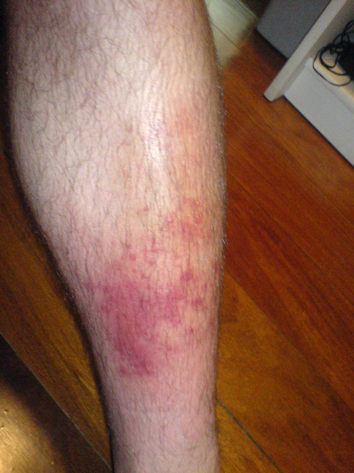
Cellulitis is an acute bacterial infection of the deep dermis and subcutaneous tissue, characterized by spreading erythema, warmth, edema, and tenderness [22]D5. It is a subset of acute bacterial skin and skin structure infections (ABSSSI) [1]A1b[8]A1b. Synonyms include nonnecrotizing cellulitis (to distinguish it from [6]D5) and the superficial variant , which involves the upper dermis with a well-demarcated, raised border.
Classification
Cellulitis is classified along several axes that guide empiric therapy (Table 1). Depth distinguishes erysipelas from deeper cellulitis. Anatomic site matters: lower limb cellulitis is most common, but facial, periorbital, and perianal infections have distinct microbiologic profiles. Severity ranges from mild (uncomplicated, amenable to oral ) to severe (requiring hospitalization and intravenous therapy) [41]B2b[42]B2b. Setting—community-acquired versus hospital-acquired—influences the likelihood of MRSA and gram-negative pathogens [17]C4[24]C4.
Table 1: Classification Axes for Cellulitis
| Axis | Categories | Key Features |
|---|---|---|
| Depth | Erysipelas vs. cellulitis | Erysipelas: superficial, raised border; cellulitis: deeper, diffuse |
| Site | Lower limb, face, periorbital, perianal, etc. | Site-specific microbiology (e.g., anaerobes in perianal) |
| Severity | Uncomplicated vs. complicated | Uncomplicated: oral therapy; complicated: IV, possible surgery |
| Setting | Community-acquired vs. hospital-acquired | CA-MRSA common in community; nosocomial pathogens in hospital |
Causative Organisms
The majority of cellulitis cases are nonculturable, but when an organism is identified, β-hemolytic streptococci (groups A, C, G) and Staphylococcus aureus predominate [21]B2b[22]D5[16]B3b. Streptococcus pyogenes (group A) is the classic cause, but Streptococcus dysgalactiae subsp. equisimilis (group G) is increasingly recognized, especially in lower limb cellulitis in older adults [44]C4[45]B2b. MRSA, particularly the USA300 clone, is a common cause of purulent cellulitis and abscesses in community settings [24]C4[34]D5[37]B2b. In immunocompromised patients, unusual pathogens such as Cryptococcus neoformans [29]C4, Mycoplasma arginini [31]C4, Helicobacter cinaedi [46]C4, and Sphingobacterium hotanense [32]C4 have been reported. Bite wounds introduce oral flora including Pasteurella multocida and anaerobes [35]D5. Marine exposure can lead to Psychrobacter sanguinis [33]C4 or Vibrio species. Gram-negative toe web infections may progress to cellulitis [18]A1c.
Pearl: The most common causes of cellulitis are β-hemolytic streptococci and Staphylococcus aureus; however, in immunocompromised hosts or atypical exposures, clinicians must consider a broader range of pathogens, including fungi and fastidious bacteria [21]B2b[22]D5[29]C4[46]C4.
Microbiology and Pathogenesis
- ▸Streptococcus pyogenes and Staphylococcus aureus are the primary pathogens; GAS is more common in cellulitis without an open wound, while S. aureus predominates when a portal of entry is present.
- ▸Virulence factors such as M protein (GAS) and protein A (S. aureus) enable immune evasion, while hyaluronidase and streptolysins facilitate tissue spread and inflammation.
- ▸Impaired lymphatic drainage (from venous insufficiency, obesity, or prior episodes) is the strongest host risk factor, creating a cycle of recurrent infection.

Despite decades of investigation, the causative pathogen is identified in fewer than 30% of cellulitis cases using conventional culture methods [50]B3b. This diagnostic gap reflects both the low bacterial burden in diffuse dermal infection and the fastidious nature of the primary pathogens. Molecular profiling of the skin microbiota has revealed that the lesion site in cellulitis harbors a distinct bacterial community compared with the contralateral unaffected limb, but no single organism consistently dominates [15]B3b. The challenge of identifying the culprit has shaped our understanding of pathogenesis: cellulitis is not a monomicrobial infection but a syndrome driven by a limited set of organisms that share the ability to breach the epidermis, evade host defenses, and incite a vigorous inflammatory response.
Microbiology: The Usual Suspects
Streptococcus pyogenes ( , GAS) and Staphylococcus aureus account for the vast majority of community-acquired cellulitis. GAS is the classic pathogen in cellulitis without an obvious portal of entry, particularly when lymphangitis or rapid spread is present. S. aureus predominates when cellulitis arises from an open wound, ulcer, or pre-existing dermatitis. Other β-hemolytic streptococci (groups B, C, G) are increasingly recognized, especially in older adults and patients with diabetes or peripheral vascular disease. In injection drug users, gram-negative bacilli (e.g., , Klebsiella spp.) and anaerobes are common, reflecting contamination of injection equipment and skin flora [52]B2b. Mycobacterial species, such as Mycobacterium ulcerans (Buruli ulcer) and M. marinum, cause indolent cutaneous infections that mimic cellulitis but follow a distinct pathogenesis involving mycolactone toxin and granulomatous inflammation [14]D5.
Virulence Factors and Host–Pathogen Interaction
| Pathogen | Key Virulence Factors | Mechanism |
|---|---|---|
| Streptococcus pyogenes | M protein, hyaluronidase, streptolysins (SLO, SLS), superantigens (SpeA, SpeB) | M protein inhibits phagocytosis; hyaluronidase degrades connective tissue matrix, facilitating spread; streptolysins lyse host cells; superantigens trigger massive cytokine release, contributing to systemic toxicity [61]C4. |
| Staphylococcus aureus | Protein A, coagulase, Panton-Valentine leukocidin (PVL), exfoliative toxins (ETA, ETB) | Protein A binds IgG Fc, blocking opsonization; coagulase promotes fibrin deposition; PVL causes neutrophil lysis and necrosis; exfoliative toxins cleave desmoglein-1, leading to superficial blistering. |
The ability of GAS to spread rapidly through tissue planes is driven by hyaluronidase and the antiphagocytic M protein, which also binds fibrinogen to mask the bacterial surface. S. aureus, by contrast, tends to produce more localized, purulent infections due to its arsenal of toxins that destroy neutrophils and form abscesses. In recurrent GAS infections, the same emm type (e.g., M1UK) can cause sequential episodes, suggesting that virulence factors may overwhelm host immunity even after appropriate antibiotic therapy [61]C4.
Pathogenesis: From Barrier Breach to Clinical Syndrome
- Breach of the epidermal barrier – The initiating event is a disruption of the skin, often trivial (scratch, fissure, insect bite, or dermatophyte infection). In many cases, the portal is clinically inapparent.
- Adherence and invasion – Bacteria adhere to extracellular matrix proteins (fibronectin, collagen) via surface adhesins (e.g., GAS M protein, S. aureus fibronectin-binding proteins). They then invade the dermis and subcutaneous tissue.
- Immune evasion – M protein and capsular polysaccharide resist opsonophagocytosis. Superantigens bypass conventional antigen presentation, activating large numbers of T cells and causing a cytokine storm.
- Inflammation – Recruited neutrophils release reactive oxygen species and proteases, causing collateral tissue damage. The resulting vasodilation and increased capillary permeability produce the cardinal signs: erythema, warmth, edema, and pain. Lymphatic involvement leads to lymphangitis (red streaks) and regional lymphadenopathy.
- Tissue spread – Bacterial enzymes (hyaluronidase, streptokinase) degrade the extracellular matrix, allowing the infection to advance along fascial planes. This explains the characteristic rapid expansion of the erythematous border.
Host Susceptibility and Recurrence
Chronic and lymphedema are the strongest risk factors for cellulitis because impaired lymphatic drainage reduces clearance of bacteria and antigens [51]A1a. Obesity (BMI ≥30 kg/m²) increases risk by 2.5-fold, likely due to altered skin barrier function, low-grade inflammation, and impaired immune responses [51]A1a. Recurrent cellulitis occurs in 20–30% of patients within 3 years, often at the same site, and is driven by progressive lymphatic damage from each episode. This creates a vicious cycle: infection damages lymphatics, which impairs bacterial clearance, which predisposes to reinfection.
Controversies and Guideline Disagreement
| Question | Position A | Position B | Strength | Implication |
|---|---|---|---|---|
| Is S. aureus or Streptococcus the dominant pathogen in non-purulent cellulitis? | IDSA guidelines emphasize Streptococcus as the primary cause in classic cellulitis without drainage or abscess. | Some studies using PCR on skin biopsies find S. aureus DNA as frequently as Streptococcus, suggesting a larger role for S. aureus than previously thought [50]B3b. | Moderate | Empiric should cover both organisms; anti-MRSA coverage is reserved for purulent infection or specific risk factors. |
| Should skin biopsy with PCR be used to guide therapy? | Not recommended in routine practice due to low yield and lack of impact on outcomes [50]B3b. | Proponents argue that molecular methods could identify atypical pathogens in immunocompromised or refractory cases. | Weak | Biopsy is reserved for research or when mycobacterial/fungal infection is suspected. |
Pearl: The pathogenesis of cellulitis hinges on a breach in the skin barrier, followed by invasion of β-hemolytic streptococci or S. aureus that deploy virulence factors to evade phagocytosis and spread through tissue planes; the low yield of cultures underscores the importance of empiric therapy targeting the most likely pathogens based on clinical context [50]B3b[61]C4.
| Pathogen | Key Virulence Factors | Mechanism |
|---|---|---|
| Streptococcus pyogenes | M protein, hyaluronidase, streptolysins (SLO, SLS), superantigens (SpeA, SpeB) | M protein inhibits phagocytosis; hyaluronidase degrades connective tissue matrix; streptolysins lyse host cells; superantigens trigger massive cytokine release [61]C4. |
| Staphylococcus aureus | Protein A, coagulase, Panton-Valentine leukocidin (PVL), exfoliative toxins (ETA, ETB) | Protein A binds IgG Fc, blocking opsonization; coagulase promotes fibrin deposition; PVL causes neutrophil lysis; exfoliative toxins cleave desmoglein-1. |
Epidemiology, Transmission and Risk Factors
- ▸Cellulitis accounts for 14.5 million ambulatory visits and 650,000 hospitalizations annually in the US, with costs exceeding $3.7 billion.
- ▸Incidence peaks in summer (July 35% higher than February) and has nearly doubled over two decades.
- ▸Chronic leg edema (compression reduces recurrence by 50%), obesity (OR 2.4), injection drug use (8.2% prevalence), and household contact with scarlet fever (RR 12.2) are key risk factors.
Cellulitis accounts for 14.5 million ambulatory care visits annually in the United States, with hospitalization rates that have nearly doubled from 1998 to 2013, reaching an estimated 650,000 admissions per year and $3.74 billion in annual costs [22]D5[83]B2c. The incidence of cellulitis-related hospitalization is 162.2 per 10,000 persons (95% CI 159.3–165.1) in the most recent estimates, up from 152.5 per 10,000 in 1998 [67]B2c. Rates are highest among adults aged 60 years and older and in males, who account for 58% of invasive group A streptococcal (iGAS) cases [82]B2c. Indigenous populations in remote regions experience disproportionately high rates: in Northwestern Ontario, Canada, the incidence of iGAS disease (including cellulitis) is 12-fold higher than in non-Indigenous populations [87]B2c.
Temporal and Seasonal Trends
Hospitalizations for cellulitis demonstrate a striking seasonal pattern, peaking in July with an incidence 35% higher than the February trough [68]B2c[83]B2c. Warmer monthly average temperatures independently predict admission risk, likely reflecting increased skin exposure, minor trauma, and arthropod bites [68]B2c. During the pandemic, cellulitis diagnoses collapsed by 40–60% in Ontario, Canada, coinciding with physical distancing and reduced healthcare-seeking for minor infections [85]B2c.
Risk Factors
Chronic edema of the leg is the strongest modifiable risk factor for recurrent cellulitis; compression therapy reduces recurrence by 50% (HR 0.50, 95% CI 0.27–0.93; NNT = 6 over 3 years) [64]A1b. Obesity (BMI ≥30 kg/m²) increases SSTI risk approximately 2- to 3-fold (OR 2.4, 95% CI 1.8–3.2) in European populations [75]B2a. Among people who inject drugs (PWID), the pooled prevalence of cellulitis is 8.2% (95% CI 5.6–11.3), with any SSTI occurring in 26.5% [70]A1a. Additional risk factors include diabetes, heart failure, hepatitis C, alcohol abuse, homelessness, and pre-existing lymphedema [80]B3b[82]B2c[86]B2b. Household contacts of scarlet fever cases have a 12.2-fold increased risk of iGAS disease (including cellulitis) within 60 days, with infants and adults >75 years at highest risk [48]B2b.
| Risk Factor | Odds Ratio / Relative Risk | Evidence Level |
|---|---|---|
| Chronic leg edema | HR 0.50 (compression benefit) | 1b [64]A1b |
| Obesity (BMI ≥30) | OR 2.4 (95% CI 1.8–3.2) | 2a [75]B2a |
| Injection drug use | Prevalence 8.2% for cellulitis | 1a [70]A1a |
| Diabetes | OR 1.8 (95% CI 1.3–2.5) | 2b [86]B2b |
| Heart failure | OR 2.1 (95% CI 1.4–3.1) | 3b [80]B3b |
| Household contact of scarlet fever | RR 12.2 (95% CI 6.1–24.4) | 2b [48]B2b |
| Indigenous ethnicity (remote communities) | Incidence rate ratio 12.0 | 2c [87]B2c |
Pearl: Cellulitis is a common and costly infection with a strong seasonal summer peak; chronic leg edema and obesity are the most important modifiable risk factors, and compression therapy halves recurrence risk in patients with edema [64]A1b[75]B2a.
Clinical Presentation
- ▸Cellulitis presents acutely with poorly demarcated erythema, warmth, edema, and tenderness, most often on the lower extremities.
- ▸Pain out of proportion, rapid progression, crepitus, or systemic toxicity are red flags for necrotizing fasciitis.
- ▸Recurrent or atypical cellulitis in immunocompromised patients warrants investigation for unusual pathogens such as Helicobacter cinaedi, fungi, or viruses.
The patient with cellulitis typically presents with acute onset of localized erythema, warmth, edema, and tenderness that progresses over hours to days [2]B2b[22]D5. Most cases involve the lower extremities, followed by the upper limbs and face [22]D5. Systemic symptoms—fever, chills, malaise, and tachycardia—accompany roughly one-third of cases and are more common when the infection is extensive or the patient is immunocompromised [2]B2b[22]D5. The incubation period after a breach in the skin barrier (e.g., trauma, ulcer, ) is usually 24–72 hours [22]D5.
Physical Examination Findings
The hallmark of cellulitis is an area of erythema with poorly defined, spreading margins, distinguishing it from , which has a raised, well-demarcated border [22]D5. The skin is warm, tender, and edematous; in severe cases, bullae, petechiae, or frank hemorrhage may appear [2]B2b[22]D5. Always examine the interdigital spaces for maceration, fissures, or tinea pedis—these are common portals of entry, especially in gram-negative toe web infections [18]A1c. Regional lymphadenopathy and lymphangitic streaking (red lines tracking proximally) are present in up to 20% of cases and signal deeper lymphatic involvement [22]D5. Palpate for fluctuance or crepitus; the latter, along with pain out of proportion to examination, should raise immediate concern for [6]D5.
Phenotypic Variants
| Variant | Key Features | Frequency | Notes |
|---|---|---|---|
| Erysipelas | Well-demarcated, raised, indurated border; often on face or legs; more superficial | Common | Usually streptococcal [22]D5 |
| Cellulitis with abscess | Fluctuant nodule or purulent drainage | Common in MRSA | Requires incision and drainage [93]B3b |
| Bullous cellulitis | Tense or hemorrhagic blisters | Uncommon | May indicate deeper infection or specific pathogens (e.g., Haematospirillum jordaniae [95]C4, Vibrio spp.) |
| Recurrent cellulitis | Repeated episodes at same site | 10–30% | Often due to lymphedema, , or untreated tinea pedis [22]D5; consider Helicobacter cinaedi in immunocompromised [46]C4 |
| Dissecting cellulitis of scalp | Painful nodules, interconnected sinus tracts, scarring alopecia | Rare | Associated with follicular occlusion triad [54]D5 |
| Pseudocellulitis | Erythema and edema without infection | Variable | Mimics include -induced radiation recall [89]C4, red breast syndrome after acellular dermal matrix [25]C4, [53]D5, primary cutaneous cryptococcosis [99]D5, and mycobacterial infections [14]D5 |
Red Flags
Several features demand urgent action:
- Rapid progression of erythema beyond the initial margin within hours, severe pain out of proportion, crepitus, skin necrosis, or systemic toxicity (hypotension, altered mental status) → suspect necrotizing fasciitis; immediate surgical consultation is required [6]D5.
- Failure to improve after 48–72 hours of appropriate → reconsider the diagnosis (e.g., abscess, foreign body, atypical pathogen) or assess for host factors such as obesity or heart failure that predict treatment failure [2]B2b[80]B3b.
- Fever >38.5°C, hypotension, or tachycardia → obtain blood cultures before antibiotics; the yield is highest in patients with systemic inflammatory response syndrome [19]B2a[65]D5.
- Immunocompromised hosts (e.g., anti-CD20 therapy, STAT3 deficiency, transplant recipients) may present with minimal cutaneous inflammation yet harbor unusual pathogens: Borealpox virus [10]C4, Nannizziopsis obscura [30]C4, Mycoplasma arginini [31]C4, Sphingobacterium hotanense [32]C4, or Helicobacter cinaedi [46]C4.
Atypical Presentations
Cellulitis can be missed when it presents in unusual contexts. Recurrent cellulitis without an obvious portal should prompt evaluation for lymphedema, chronic venous insufficiency, or factitious injection [22]D5. Facial cellulitis may originate from a dental abscess, sinusitis, or—rarely—cowpox virus inoculation [4]C4. Cellulitis after marine or soil exposure raises the possibility of Psychrobacter sanguinis [33]C4, Vibrio vulnificus, or Mycobacterium marinum [14]D5. In patients receiving chemotherapy, gemcitabine can produce a striking erysipeloid reaction that mimics infectious cellulitis but resolves without antibiotics [89]C4. Periorbital erythema and edema may represent or, in endemic areas, acute (Romaña's sign) [79]C4.
Pearl: The diagnosis of cellulitis rests on the constellation of acute-onset, spreading erythema with poorly defined borders, warmth, tenderness, and edema; the absence of a well-demarcated edge, the presence of lymphangitic streaks, and a portal of entry help distinguish it from mimics. Failure to respond to antibiotics within 48–72 hours should prompt a search for abscess, necrotizing infection, or an alternative diagnosis [2]B2b[22]D5.
Diagnosis and Workup
- ▸Cellulitis is diagnosed clinically; no single laboratory test or imaging study confirms it.
- ▸Blood cultures are low-yield (5–10% positive) and should be reserved for patients with systemic toxicity, immunocompromise, or failure to respond.
- ▸The LRINEC score (≥6) helps distinguish cellulitis from necrotizing fasciitis, which requires urgent surgical intervention.
The diagnosis of cellulitis is made at the bedside; no laboratory test or imaging study can replace the clinical assessment. The hallmark is an area of expanding erythema with indistinct borders, accompanied by warmth, tenderness, and edema, typically evolving over hours to days [22]D5. Systemic signs such as fever and chills may be present but are not required. The diagnosis rests on the characteristic clinical presentation and the exclusion of mimics, particularly necrotizing soft-tissue infection (NSTI), which demands urgent surgical intervention [6]D5.
Gold-Standard Test
There is no single gold-standard diagnostic test. Clinical diagnosis by an experienced clinician remains the reference standard [22]D5. Microbiologic confirmation (blood cultures, tissue culture, or serology) is neither sensitive nor required for diagnosis but can guide therapy when positive [21]B2b[50]B3b.
Laboratory Studies
- Blood cultures: Positive in only 5–10% of cellulitis cases [65]D5. Routine blood cultures are not recommended for uncomplicated cellulitis. Obtain them when the patient has systemic toxicity (temperature >38.5°C, hypotension), immunocompromise, extremes of age, or failure to respond to initial therapy [65]D5[107]B3b. In elderly patients, the yield may be higher (up to 15%) [107]B3b.
- Inflammatory markers: White blood cell count, C-reactive protein, and procalcitonin are nonspecific and do not confirm the diagnosis but can support severity assessment and monitor response [2]B2b.
- LRINEC score (Laboratory Risk Indicator for ): A score ≥6 raises suspicion for NSTI (sensitivity ~90%, specificity ~80%) [6]D5. Components include CRP, WBC, hemoglobin, sodium, creatinine, and glucose.
- Dundee classification: A simple severity tool that stratifies patients into mild, moderate, or severe cellulitis based on clinical and laboratory parameters; higher grades correlate with increased mortality and length of stay [81]B3b.
- BRRISC score: Predicts hospital-attended recurrence using data available at presentation; validated externally [104]B2b.
- Novel biomarkers: Plasma cell-free DNA (cfDNA) levels are elevated in cellulitis and correlate with inflammatory markers and clinical response, but this is not yet standard [26]C4.
Imaging
Imaging is not routinely indicated. Consider:
- Ultrasound: To differentiate cellulitis from abscess (fluctuance, anechoic collection) or deep vein thrombosis (DVT) [22]D5.
- CT or MRI: Reserved for cases where NSTI is suspected (gas in tissues, fascial thickening, fluid collections) [6]D5.
Biopsy / Histology
Skin biopsy with culture and PCR is not recommended for routine diagnosis. In a prospective study of 50 patients, biopsy culture identified a pathogen in only 2% of cases, and PCR did not improve yield [50]B3b. Biopsy may be useful in refractory or atypical cases (e.g., suspected fungal, mycobacterial, or parasitic infection) [14]D5[29]C4[105]C4.
Diagnostic Algorithm
- Clinical assessment: History (onset, progression, systemic symptoms, risk factors) and physical exam (margins, tenderness, warmth, edema, bullae, crepitus).
- Severity assessment: Apply Dundee classification or LRINEC score. If LRINEC ≥6 or pain out of proportion, suspect NSTI and obtain urgent surgical consultation [6]D5.
- Blood cultures: Obtain only if criteria met (see above).
- Imaging: Perform if abscess, DVT, or NSTI is in the differential.
- Reassessment at 48–72 hours: Clinical response (reduction in erythema, pain, and fever) confirms the diagnosis and guides therapy duration [2]B2b.
Differential Diagnosis
| Condition | Key Differentiating Features |
|---|---|
| Necrotizing fasciitis | Pain out of proportion, bullae, crepitus, systemic toxicity, LRINEC ≥6 [6]D5 |
| Deep vein thrombosis | Unilateral swelling without fever; positive ultrasound [22]D5 |
| Stasis dermatitis | Bilateral, chronic, no fever, responds to elevation [22]D5 |
| Pruritus, well-demarcated, no systemic signs [22]D5 | |
| Gout | Acute monoarticular arthritis, urate crystals [22]D5 |
| Well-demarcated, raised border, more superficial [22]D5 | |
| Abscess | Fluctuance, purulent drainage on aspiration [22]D5 |
| Prepatellar or olecranon swelling, bursal aspiration positive [96]B3b | |
| Pyogenic flexor tenosynovitis | Kanavel signs (fusiform swelling, flexed posture, tenderness along sheath, pain on extension) [100]D5 |
| Dissecting cellulitis of scalp | Painful nodules, sinus tracts, cicatricial alopecia [54]D5[110]C4 |
| ESRD, painful violaceous plaques, rapid progression [111]C4 | |
| Bilateral, heat-induced burning, relieved by cooling [53]D5 | |
| Cat bite, lymphocutaneous spread, fails [105]C4 | |
| Cryptococcal cellulitis | Immunocompromised, high serum CrAg [29]C4 |
| Cutaneous mycobacterial infection | Chronic, nodular, ulcerative; biopsy with AFB stain [14]D5 |
| Helicobacter cinaedi | Recurrent cellulitis in immunocompromised, requires extended blood culture [46]C4 |
| Haematospirillum jordaniae | Bullous cellulitis, bacteremia [95]C4 |
| (Romaña's sign) | Periorbital swelling, fails antibiotics, exposure in endemic area [79]C4 |
Pearl: Cellulitis is a clinical diagnosis; the most critical step is to exclude necrotizing fasciitis using the LRINEC score and clinical judgment, as delay in surgical debridement increases mortality [6]D5[22]D5.
Severity Assessment and Risk Stratification
- ▸Dundee classification stratifies cellulitis into mild, moderate, and severe with stepwise increases in 30-day mortality (0.3% to 6.7%) and length of stay [81].
- ▸BRRISC score predicts recurrence risk (c-statistic 0.72) and is not improved by adding acute clinical response markers [104].
- ▸Pitt bacteremia score accurately predicts death in cellulitis-associated bacteremia (AUC 0.85) [45].
Several validated severity assessment tools guide site-of-care decisions and treatment intensity in cellulitis. The Dundee classification stratifies patients into three severity groups (mild, moderate, severe) based on clinical parameters including temperature, heart rate, respiratory rate, white blood cell count, and extent of erythema. In a large retrospective validation (n=1462), 30-day all-cause mortality was 2% overall, but rose stepwise with severity: 0.3% in mild, 1.5% in moderate, and 6.7% in severe disease (p<0.001) [81]B3b. Severe-class patients also had significantly longer median length of stay (6 vs 3 days) and higher readmission rates (OR 2.1, 95% CI 1.4–3.2) [81]B3b. The tool is simple to apply at the bedside and directly informs whether a patient requires intravenous therapy and hospital admission.
BRRISC Score
The Baseline Recurrence Risk in Cellulitis (BRRISC) score predicts hospital-attended recurrence using data available at initial presentation. In an external validation cohort, the score demonstrated good discrimination (c-statistic 0.72, 95% CI 0.67–0.77) [104]B2b. Adding markers of acute clinical response at 48–72 hours (e.g., reduction in erythema size, limb temperature) did not significantly improve performance, suggesting that baseline factors dominate recurrence risk [104]B2b. The BRRISC score helps identify patients who may benefit from extended antibiotic courses or prophylactic measures.
Pitt Bacteremia Score and Biomarkers
For patients with suspected bacteremia, the Pitt bacteremia score accurately predicts death (area under receiver operating characteristic curve 0.85) [45]B2b. In Streptococcus dysgalactiae subspecies equisimilis bacteremia, blood leukocyte counts on days 2 and 3 and C-reactive protein levels on days 3 and 4 after admission were independent predictors of severe disease [45]B2b. Emerging biomarkers such as cell-free DNA (cfDNA) show correlation with clinical severity: in a prospective study, cfDNA levels at admission were significantly higher in patients with cellulitis than controls (median 1.8 vs 0.4 ng/mL, p<0.001) and decreased by 72 hours after antibiotic initiation [26]C4. However, cfDNA is not yet validated for routine clinical use.
Clinical Decision Rules for Site of Care
No single tool is universally adopted, but the Dundee classification is the most extensively validated for initial triage. Patients classified as mild can be managed as outpatients with oral ; moderate severity often requires intravenous therapy with consideration for outpatient parenteral antibiotic therapy (OPAT); severe disease mandates inpatient admission. The IDSA guidelines emphasize that active antimicrobial therapy against MRSA improves outcomes in community-onset MRSA SSTIs, but do not provide a specific severity scoring system [93]B3b.
Pearl: The Dundee classification reliably stratifies mortality risk (0.3% mild vs 6.7% severe) and length of stay, making it a practical bedside tool for site-of-care decisions in cellulitis [81]B3b.
| Tool | Purpose | Key Parameters | Outcome Predicted | Validation |
|---|---|---|---|---|
| Dundee classification | Initial triage and site-of-care | Temperature, HR, RR, WBC, erythema extent | 30-day mortality, LOS, readmission | Large cohort (n=1462) [81]B3b |
| BRRISC score | Recurrence risk | Age, comorbidities, prior cellulitis, lymphedema | Hospital-attended recurrence within 1 year | External validation (c=0.72) [104]B2b |
| Pitt bacteremia score | Bacteremia severity | Temperature, hypotension, mechanical ventilation, cardiac arrest, mental status | In-hospital death | AUC 0.85 in SDSE bacteremia [45]B2b |
| cfDNA (emerging) | Disease activity | Plasma cell-free DNA level | Correlation with clinical severity | Small prospective study (n=36) [26]C4 |
Empiric and Acute Management
- ▸Empiric antibiotic selection is guided by Dundee severity class: oral beta-lactam for mild, IV beta-lactam for moderate, and broad-spectrum MRSA coverage for severe cellulitis.
- ▸Early switch from IV to oral therapy (within 24–48 hours) is safe and reduces length of stay; total duration of 5–7 days is sufficient for most uncomplicated cases.
- ▸Failure to improve by 72 hours warrants reassessment for abscess, necrotizing fasciitis, or alternative diagnoses; avoid unnecessary MRSA coverage in nonpurulent cellulitis without risk factors.
Once severity is classified using the Dundee system (Section 6), empiric antibiotic selection follows a structured pathway guided by the presence or absence of purulence, systemic signs, and host factors. The goal is to initiate therapy within 1 hour of presentation for moderate-to-severe cases, as delays are associated with prolonged hospitalization [2]B2b (2b).
Step 1: Severity-Based Disposition and Empiric Antibiotic Selection
Mild cellulitis (Dundee class I) — no systemic signs, no uncontrolled comorbidities — is managed with oral in the outpatient setting. First-line therapy targets β-hemolytic streptococci, the predominant pathogen in nonpurulent cellulitis [21]B2b (2b). Cephalexin 500 mg orally four times daily or 500 mg three times daily for 5–7 days achieves cure rates exceeding 90% [133]A1b (1b). For patients with , clindamycin 300–450 mg three times daily or 100 mg twice daily are effective alternatives; doxycycline showed noninferiority to cephalexin in a propensity-matched analysis (6% failure in each arm; OR 1.34, 95% CI 0.21–8.69) [143]B2b (2b).
Moderate cellulitis (Dundee class II) — systemic signs (fever, tachycardia) or comorbidities (diabetes, obesity, heart failure) — requires initial intravenous therapy. Cefazolin 2 g IV every 8 hours or 1–2 g IV daily are first-line choices. The CHOICE trial demonstrated that once-daily ceftriaxone at home was noninferior to inpatient flucloxacillin in children (cure rate 97% vs 96%; risk difference 1.0%, 95% CI –4.5% to 6.5%) [123]A1b (1b), supporting outpatient parenteral antimicrobial therapy (OPAT) pathways. Early switch to oral therapy (within 24–48 hours) is safe when clinical improvement is documented: the Switch Trial found that 24 hours of IV antibiotics followed by oral therapy was noninferior to ≥72 hours IV (cure rate 92% vs 88%; difference 4%, 95% CI –10% to 18%) [136]A1b (1b).
Severe cellulitis (Dundee class III–IV) — marked systemic toxicity, rapid progression, or immunocompromise — mandates hospital admission, often to an ICU. Empiric therapy must cover both streptococci and MRSA. 15–20 mg/kg IV every 8–12 hours (target trough 15–20 μg/mL) plus 4.5 g IV every 6 hours or 2 g IV every 8 hours provides broad coverage. Alternative MRSA-active agents include 600 mg IV/oral every 12 hours or 6 mg/kg IV daily. Single-dose oritavancin 1200 mg IV is an option for patients requiring prolonged MRSA coverage who can be discharged early; registry data show 94% clinical success in cellulitis [152]C4 (4).
Step 2: Escalation Triggers and Second-Line Therapy
Failure to improve within 48–72 hours — defined as persistent fever, expanding erythema, or rising inflammatory markers — warrants reassessment. Consider:
- Imaging (ultrasound, CT, MRI) to exclude abscess, , or deep vein thrombosis.
- Blood cultures if not already obtained; yield is low (<10%) but higher in elderly patients [107]B3b (3b).
- Broadening coverage: add MRSA coverage if not already present, or switch to an agent with enhanced Gram-negative activity (e.g., ceftaroline 600 mg IV every 12 hours) if water exposure or immunocompromise is present [8]A1b (1b).
Step 3: Monitoring and Titration
Clinical response is assessed daily: reduction in erythema margin, pain, swelling, and fever. The median time to clinical response is 48–72 hours [2]B2b (2b). Biomarkers such as C-reactive protein (CRP) and white blood cell count typically peak at day 2–3 and decline thereafter [138]B2b (2b). Switch from IV to oral therapy when:
- Afebrile for ≥24 hours
- Erythema and pain improving
- Hemodynamically stable
- Able to tolerate oral medications
An algorithm-based switch protocol reduced median IV duration from 4.0 to 2.0 days without increasing 30-day treatment failure (composite of death, readmission, or antibiotic change: 6.3% vs 7.8%; P = 0.74) [150]B2b (2b).
Step 4: Duration of Therapy
5–7 days is sufficient for most uncomplicated cellulitis. Longer courses (10–14 days) are reserved for slow responders, immunocompromised patients, or those with significant comorbidities. A retrospective study found no benefit of >8 days in obese or heart failure patients (failure rate 12% vs 14%; P = 0.68) [148]B2b (2b).
Step 5: Source Control and Supportive Care
- Elevation of the affected limb reduces edema and pain.
- Compression therapy is contraindicated in acute infection but may prevent recurrence once resolved [64]A1b (1b).
- Incision and drainage of any purulent collection is essential; antibiotics alone are insufficient for abscesses [128]A1b (1b).
- Analgesics (acetaminophen, NSAIDs) for pain and fever.
Algorithm
Figure 1: Empiric management pathway for cellulitis based on severity (adapted from [81]B3b[137]B2b).
Drug Comparison Table
| Option | Indication | Dose | Key Trial | Outcome | Evidence Level |
|---|---|---|---|---|---|
| Cephalexin | Mild nonpurulent | 500 mg PO QID | [133]A1b | Cure 85% vs 82% (plus TMP-SMX) | 1b |
| Amoxicillin | Mild nonpurulent | 500 mg PO TID | [38]B2c | Guideline-recommended | 2c |
| Clindamycin | Mild-moderate, penicillin allergy | 300-450 mg PO TID | [125]A1b | Cure 89% vs 88% (TMP-SMX) | 1b |
| Doxycycline | Mild-moderate, MRSA risk | 100 mg PO BID | [143]B2b | Failure 6% vs 6% (cephalexin) | 2b |
| Cefazolin | Moderate, IV | 2 g IV q8h | [135]A1b | Noninferior to oral cephalexin | 1b |
| Ceftriaxone | Moderate, IV/OPAT | 1-2 g IV daily | [123]A1b | Noninferior to flucloxacillin | 1b |
| Vancomycin | Severe, MRSA coverage | 15-20 mg/kg IV q8-12h | [130]B2b | Cure 91% in abscess | 2b |
| Linezolid | Severe, MRSA | 600 mg IV/PO q12h | [122]A1b | Noninferior to vancomycin | 1b |
| Oritavancin | Severe, single-dose | 1200 mg IV once | [66]A1b | Similar safety to vancomycin | 1b |
Dosing Table
| Drug | Starting dose | Target/max dose | Renal adjustment | Hepatic adjustment | Key monitoring |
|---|---|---|---|---|---|
| Cephalexin | 500 mg PO QID | 1 g QID | CrCl <30: 250-500 mg q12h | None | Allergy, diarrhea |
| Amoxicillin | 500 mg PO TID | 1 g TID | CrCl <30: 250-500 mg q12h | None | Rash, diarrhea |
| Clindamycin | 300-450 mg PO TID | 600 mg PO QID | None | Severe: reduce dose | C. difficile colitis |
| Doxycycline | 100 mg PO BID | 100 mg BID | None | None | Photosensitivity |
| Cefazolin | 2 g IV q8h | 2 g q6h | CrCl <30: 1 g q12h | None | Allergy, renal function |
| Ceftriaxone | 1-2 g IV daily | 2 g daily | None | None | Biliary sludging |
| Vancomycin | 15-20 mg/kg IV q8-12h | Trough 15-20 μg/mL | CrCl <30: extended interval | None | Trough levels, renal function |
| Linezolid | 600 mg IV/PO q12h | 600 mg q12h | None | None | Myelosuppression (weekly CBC) |
| Oritavancin | 1200 mg IV once | Single dose | None | None | Infusion reactions |
Treatment Failure Protocol
If no improvement by 72 hours:
- Obtain blood cultures and imaging (ultrasound/CT) to rule out abscess or necrotizing fasciitis.
- Review culture results; if MRSA isolated and not covered, add vancomycin, linezolid, or daptomycin.
- Consider alternative diagnoses: venous stasis dermatitis, deep vein thrombosis, , or lymphedema.
- If necrotizing fasciitis suspected (LRINEC score ≥6, bullae, crepitus), obtain urgent surgical consultation [6]D5 (5).
What NOT to Do
- Do not routinely add MRSA coverage for nonpurulent cellulitis without risk factors (prior MRSA, purulence, injection drug use, severe disease). The IDSA guideline recommends beta-lactam monotherapy for nonpurulent cellulitis [120]D5 (5).
- Do not prolong IV therapy beyond 48–72 hours if clinical improvement occurs; early oral switch reduces length of stay and costs without compromising outcomes [136]A1b[150]B2b (1b).
- Do not use TMP-SMX monotherapy for suspected streptococcal cellulitis; it has inferior activity against group A streptococci compared with beta-lactams [147]B2a (2a).
- Do not routinely obtain blood cultures in mild uncomplicated cellulitis; yield is <5% and rarely changes management [107]B3b (3b).
Controversies and Guideline Disagreement
| Question | Position A | Position B | Strength of disagreement | Implication for practice |
|---|---|---|---|---|
| Empiric MRSA coverage for nonpurulent cellulitis | IDSA 2014 — recommends beta-lactam alone unless risk factors present [120]D5 | Some experts — advocate adding TMP-SMX or clindamycin in areas with high MRSA prevalence | Moderate (different regional thresholds) [133]A1b[147]B2a | In low-prevalence settings, beta-lactam monotherapy is appropriate; in high-prevalence settings, consider dual therapy or MRSA-active agent |
| Optimal duration of therapy | 5–7 days supported by multiple RCTs [133]A1b[136]A1b | Common practice — 10–14 days, especially in hospitalized patients | Moderate (evidence vs practice) [148]B2b | Short-course therapy is safe for most; extend only for slow responders or immunocompromise |
| Role of TMP-SMX for streptococcal infections | IDSA — not recommended due to inferior anti-streptococcal activity [147]B2a | Some trials — show noninferiority when combined with beta-lactam [133]A1b | Mild (combination vs monotherapy) | TMP-SMX should not be used alone; combination with beta-lactam may be considered in select cases |
Pearl: For nonpurulent cellulitis, beta-lactam monotherapy remains first-line; adding MRSA coverage is reserved for purulent infections, known colonization, or failure to respond within 48–72 hours [120]D5[133]A1b.
| Severity | First-line | Alternative | Duration | Evidence |
|---|---|---|---|---|
| Mild (Class I) | Cephalexin 500 mg PO QID | Amoxicillin 500 mg PO TID, Clindamycin 300-450 mg PO TID | 5-7 days | [133]A1b (1b) |
| Moderate (Class II) | Cefazolin 2 g IV q8h or Ceftriaxone 1-2 g IV daily | Clindamycin 600 mg IV q8h | 5-7 days (IV then oral) | [123]A1b[135]A1b (1b) |
| Severe (Class III-IV) | Vancomycin 15-20 mg/kg IV q8-12h + Piperacillin-tazobactam 4.5 g IV q6h | Linezolid 600 mg IV/PO q12h, Daptomycin 6 mg/kg IV daily | 7-14 days | [130]B2b (2b) |
Definitive Therapy, Duration and De-escalation
- ▸Once culture results are available, narrow therapy to target the specific pathogen: penicillin/amoxicillin for streptococci, cefazolin/cephalexin for MSSA, and doxycycline/TMP-SMX/linezolid for MRSA.
- ▸Total duration of 5–7 days is sufficient for uncomplicated cellulitis; longer courses do not reduce failure rates and increase antibiotic exposure.
- ▸Switch from IV to oral antibiotics as soon as clinical improvement is evident (afebrile, stable/improving erythema, tolerating oral intake) to reduce length of stay and costs.
Step 1: Transition from Empiric to Pathogen-Directed Therapy
Once culture and susceptibility results return—typically within 48–72 hours—narrow therapy to target the identified organism. For β-hemolytic streptococci (the most common cause of nonpurulent cellulitis), penicillin G 2–4 million units IV every 4–6 hours or oral 500–875 mg twice daily is definitive [22]D5 (5). For methicillin-susceptible Staphylococcus aureus (MSSA), cefazolin 1–2 g IV every 8 hours or oral cephalexin 500 mg four times daily is appropriate [5]D5 (5). For MRSA, options include 100 mg twice daily, clindamycin 300–450 mg three times daily, (TMP-SMX) 1–2 double-strength tablets twice daily, or 600 mg twice daily [125]A1b (1b), [36]B2b (2b). For gram-negative or anaerobic coverage (e.g., diabetic foot, bite wounds), tailor to susceptibility; 875/125 mg twice daily or 400 mg daily may be used [35]D5 (5).
Step 2: Determining Total Duration of Therapy
Five to seven days is sufficient for most uncomplicated cellulitis. A retrospective cohort of 34 VA hospitals found that durations ≤8 days were not associated with higher failure rates compared with longer courses (OR 1.12, 95% CI 0.78–1.60) [41]B2b (2b). In patients with obesity or heart failure, short courses (≤8 days) also did not increase failure (adjusted OR 0.91, 95% CI 0.52–1.59) [148]B2b (2b). The Switch trial demonstrated that 24 hours of IV therapy followed by oral to complete 7–10 total days was non-inferior to ≥72 hours IV (clinical cure 92% vs 90%; difference 2%, 95% CI –12% to 16%) [136]A1b (1b). For cellulitis requiring hospitalization, the Auckland pathway reduced median length of stay from 1.8 to 0.7 days by enforcing early oral switch and short total duration [137]B2b (2b).
Step 3: Intravenous-to-Oral Switch (De-escalation)
Switch from IV to oral antibiotics when the patient is afebrile for ≥24 hours, the cellulitis margin is no longer advancing, pain is improving, and oral intake is tolerated [150]B2b (2b). An algorithm-based approach using these criteria achieved successful switch within 48 hours in 78% of patients, with 30-day treatment failure of only 6% [150]B2b (2b). Oral options for step-down include amoxicillin 500–875 mg twice daily (streptococci), cephalexin 500 mg four times daily (MSSA), or doxycycline 100 mg twice daily (MRSA). For patients who cannot take oral medications, outpatient parenteral antimicrobial therapy (OPAT) with once-daily 50 mg/kg (max 2 g) is safe and cost-effective [123]A1b (1b), [124]A1b (1b).
Step 4: Treatment Failure and Escalation
If no clinical improvement after 48–72 hours of appropriate therapy, reassess for undrained abscess, , or an alternative diagnosis (e.g., venous stasis dermatitis, deep vein thrombosis) [2]B2b (2b). Risk factors for failure include obesity (BMI >30), heart failure, and peripheral vascular disease [80]B3b (3b). Escalate by broadening coverage (e.g., adding or if MRSA suspected) and obtaining imaging (ultrasound, CT) to rule out suppurative complications. Surgical consultation is mandatory if necrotizing infection is suspected [63]B2b (2b).
Step 5: Prevention of Recurrence
For patients with two or more episodes of leg cellulitis, prophylactic penicillin V 250 mg twice daily for 12 months reduces recurrence (HR 0.55, 95% CI 0.35–0.86; NNT = 8 to prevent one recurrence) [126]A1b (1b). Address predisposing factors: treat interdigital , manage edema with compression, and optimize glycemic control in diabetes.
Controversies and Guideline Disagreement
| Question | Position A | Position B | Strength | Implication for practice |
|---|---|---|---|---|
| Optimal total duration for uncomplicated cellulitis | IDSA 2014 recommends 5–6 days for cellulitis with clinical response | UK guidelines (e.g., CREST) suggest 7–10 days, though evidence supports shorter courses | Moderate (different recommendations, but both allow early switch) [97]D5 (5), [136]A1b (1b) | Clinicians can safely prescribe 5–7 days; longer courses are rarely needed and increase resistance and cost. |
| Role of MRSA coverage in nonpurulent cellulitis | Moran et al. 2017 found no benefit of adding TMP-SMX to cephalexin (cure 83.5% vs 85.5%) | IDSA 2014 recommends empiric MRSA coverage only if purulence or risk factors present | Strong (RCT evidence vs guideline nuance) [133]A1b (1b), [22]D5 (5) | Do not routinely add anti-MRSA agents for nonpurulent cellulitis without systemic signs or purulent drainage. |
Pearl: For uncomplicated cellulitis, 5–7 days of pathogen-directed therapy with early IV-to-oral switch (often within 24–48 hours) is sufficient; longer courses do not improve outcomes and should be avoided [136]A1b[148]B2b.
| Pathogen | First-line agent | Dose | Alternative | Evidence level |
|---|---|---|---|---|
| β-hemolytic streptococci | Penicillin G IV or amoxicillin PO | 2–4 million U IV q4–6h; 500–875 mg PO BID | Cephalexin 500 mg PO QID | 5 [22]D5 |
| MSSA | Cefazolin IV or cephalexin PO | 1–2 g IV q8h; 500 mg PO QID | Clindamycin 300–450 mg PO TID | 5 [5]D5 |
| MRSA | Doxycycline PO or TMP-SMX PO | 100 mg PO BID; 1–2 DS tabs PO BID | Linezolid 600 mg PO BID | 1b [125]A1b, 2b [36]B2b |
| Gram-negative (e.g., Vibrio) | Ceftriaxone IV or ciprofloxacin PO | 1–2 g IV q24h; 500–750 mg PO BID | Doxycycline + ceftazidime | 4 [78]C4 |
| Anaerobes (bite wounds) | Amoxicillin-clavulanate PO | 875/125 mg PO BID | Moxifloxacin 400 mg PO daily | 5 [35]D5 |
| Drug | Starting dose | Target/max dose | Renal adjustment | Hepatic adjustment | Key monitoring |
|---|---|---|---|---|---|
| Penicillin G | 2 million U IV q4h | 4 million U IV q4h | CrCl <10: 2 million U q8h | None | K+, seizure risk at high doses |
| Amoxicillin | 500 mg PO BID | 875 mg PO BID | CrCl <30: 500 mg q24h | None | Rash, diarrhea |
| Cefazolin | 1 g IV q8h | 2 g IV q8h | CrCl <30: 1 g q12h | None | Renal function |
| Cephalexin | 500 mg PO QID | 500 mg PO QID | CrCl <30: 250–500 mg q12h | None | GI intolerance |
| Doxycycline | 100 mg PO BID | 100 mg PO BID | None | None | Photosensitivity |
| TMP-SMX | 1 DS tab PO BID | 2 DS tabs PO BID | CrCl 15–30: 1 DS tab daily; <15: avoid | None | K+, Cr, rash |
| Linezolid | 600 mg PO/IV BID | 600 mg PO/IV BID | None | None | CBC weekly (thrombocytopenia) |
Antimicrobial Resistance and Stewardship
- ▸Community-acquired MRSA is the most clinically relevant resistant pathogen in cellulitis, requiring empiric coverage in high-prevalence settings.
- ▸Antimicrobial stewardship interventions, including clinical pathways (e.g., Auckland Cellulitis Pathway), electronic decision support (WISCA), and diagnostic stewardship (dermatology consultation), reduce unnecessary antibiotic use and improve outcomes.
- ▸Duration of therapy should be minimized to 5–7 days for uncomplicated cellulitis to reduce resistance pressure; default order durations in EHRs effectively shorten treatment courses.
Antimicrobial resistance complicates the empiric of cellulitis, particularly with the rise of community-acquired MRSA and other multidrug-resistant organisms. The most clinically relevant resistant pathogen is community-acquired MRSA (CA-MRSA), which now accounts for a substantial proportion of skin and soft tissue infections in many regions [24]C4[86]B2b. CA-MRSA strains carry the mecA gene encoding PBP2a, conferring resistance to all β-lactams [24]C4. Tetracycline resistance, mediated by tet(K) and tet(M) genes, is less common but emerging; and minocycline remain active against >90% of MRSA isolates in most US centers [36]B2b[143]B2b. Other multidrug-resistant variants include Acinetobacter baumannii in war-related wounds [3]B3b, Pseudomonas otitidis producing the carbapenemase POM-1 [173]C4, Myroides injenensis with broad resistance [171]C4, and Vibrio spp. with intermediate resistance to penicillins and first-generation cephalosporins [78]C4. Group G streptococci (Streptococcus dysgalactiae subsp. equisimilis) show high resistance to quinupristin-dalfopristin [109]C4. These organisms, though less common, require targeted therapy based on culture and susceptibility results.
Principles
Stewardship in cellulitis focuses on appropriate antibiotic selection, minimizing duration, and avoiding unnecessary treatment of mimics. The Auckland Cellulitis Pathway, which stratifies patients by Dundee severity class, reduced median length of stay from 1.8 to 0.7 days and decreased total antibiotic days without increasing readmission or mortality [137]B2b. The WISCA (Weighted Incidence Syndromic Combination Antibiogram) tool, a real-time electronic decision support system, improved empiric antibiotic selection and reduced hospital length of stay for nonpurulent cellulitis [62]A1b. Diagnostic stewardship through dermatology consultation reduces misdiagnosis and inappropriate antibiotic use; a meta-analysis found that dermatologist evaluation led to treatment modification in 30–50% of suspected cellulitis cases [169]A1a.
Stewardship Interventions and Outcomes
Default antibiotic order durations in electronic health records significantly reduce long prescriptions. A cluster randomized trial in pediatric primary care found that default durations of 5 days for cellulitis decreased the proportion of long prescriptions from 62% to 38% [170]A1b. Similarly, a clinical practice guideline for pediatric reduced use of broad-spectrum (dual/triple therapy and MRSA-active agents) without increasing treatment failure [166]C4. A systematic review of antibiotic duration trials concluded that 85% of studies support shorter courses (5–7 days) for common bacterial infections, including cellulitis [168]A1a. Despite these data, guideline discordance persists; a review of 70 hospital guidelines found marked variability in recommended agents and durations, reflecting a lack of clear best practices [177]B2c.
Pearl: Antimicrobial resistance in cellulitis is driven by both community-acquired MRSA and less common multidrug-resistant organisms; stewardship programs that incorporate clinical pathways, diagnostic consultation, and default short durations can significantly reduce unnecessary antibiotic exposure without compromising outcomes [62]A1b[137]B2b[169]A1a[170]A1b.
Complications and Supportive Care
- ▸Complications of cellulitis include local progression (necrotizing fasciitis, lymphedema) and systemic spread (bacteremia, endocarditis, osteomyelitis), with highest risk in people who inject drugs.
- ▸Supportive care—respiratory monitoring, DVT prophylaxis, pain management, and rehabilitation—reduces hospital-acquired morbidity and prevents recurrence.
Complications of cellulitis range from local progression to life-threatening systemic infection, and supportive care is critical to prevent hospital-acquired morbidity. Local complications include lymphangitis, abscess formation, and , the latter occurring in <1% of cases but carrying mortality up to 30% [22]D5[181]B2c. Recurrent cellulitis damages lymphatic vessels, leading to chronic lymphedema, which itself is a major risk factor for further episodes (HR 1.63, 95% CI 1.58–1.68) [185]B2b. Systemic complications arise in 5–10% of hospitalized patients: bacteremia, sepsis, endocarditis, osteomyelitis, and septic arthritis are well-documented, especially among people who inject drugs [22]D5[52]B2b[180]B2c. Lemierre-like syndrome with has been reported following due to MRSA [28]C4. In pediatric , adjunctive corticosteroids reduce the need for surgical intervention (RR 0.68, 95% CI 0.49–0.94) and PICU admission (RR 0.51, 95% CI 0.30–0.86) [182]A1a.
Respiratory Monitoring
Patients with severe cellulitis and sepsis require close respiratory surveillance. Forced vital capacity (FVC) should be measured if there is concern for ascending infection or airway compromise (e.g., facial or neck involvement). Intubation criteria follow standard sepsis guidelines: inability to protect airway, refractory hypoxemia (PaO₂/FiO₂ <200), or progressive hypercapnia [22]D5.
Autonomic Complications
Sepsis-associated autonomic dysfunction manifests as tachyarrhythmias, blood pressure instability requiring vasopressors, ileus, and urinary retention. These complications are managed with goal-directed fluid resuscitation, vasopressor support (norepinephrine 0.05–0.5 µg/kg/min), and bowel/bladder protocols [22]D5.
DVT/PE Prophylaxis
Hospitalized patients with cellulitis and reduced mobility should receive pharmacologic thromboprophylaxis. 40 mg subcutaneously once daily (or 5000 U subcutaneously three times daily) is standard, reducing venous thromboembolism risk by approximately 60% [42]B2b.
Pain
Cellulitis causes significant pain from inflammation and tissue edema. First-line includes acetaminophen 650–1000 mg every 6 hours or ibuprofen 400–600 mg every 6–8 hours. For severe pain, short-course opioids (e.g., 5–10 mg every 4–6 hours as needed) may be used, with attention to avoiding overuse [22]D5.
Rehabilitation
After infection resolution, rehabilitation focuses on restoring mobility and managing lymphedema. Compression therapy (graduated compression stockings, 20–30 mmHg) reduces recurrent cellulitis risk by approximately 50% (HR 0.50, 95% CI 0.26–0.95) [64]A1b. Physical therapy for gait training and strength is initiated once acute inflammation subsides.
Hospital-Acquired Complications
Prevention of pressure injuries, catheter-associated urinary tract infection, and hospital-acquired pneumonia is paramount. Early mobilization, avoidance of indwelling urinary catheters, and oral care protocols reduce these events. Implementation of a cellulitis management guideline decreased unnecessary antibiotic use without increasing treatment failure [42]B2b.
| Complication | Frequency | Prevention | Management |
|---|---|---|---|
| Necrotizing fasciitis | <1% | Prompt recognition of red flags (pain out of proportion, bullae, gas) | Surgical debridement, broad-spectrum [22]D5 |
| Bacteremia/sepsis | 5–10% | Early appropriate antibiotics, source control | IV fluids, vasopressors, ICU care [22]D5 |
| Endocarditis | 1–3% in injection drug users | Aseptic injection technique, treat bacteremia promptly | Echocardiography, prolonged IV antibiotics [52]B2b[180]B2c |
| Osteomyelitis/septic arthritis | 2–5% in injection drug users | Avoid contiguous spread from deep abscess | MRI, surgical drainage, targeted antibiotics [52]B2b |
| Lymphedema | 10–20% after recurrent cellulitis | Compression therapy, skin care | Graduated compression, manual lymphatic drainage [64]A1b[185]B2b |
| Antibiotic-associated adverse events | 5–15% | Narrow-spectrum therapy, shortest effective duration | Discontinue offending agent, treat C. difficile if indicated [42]B2b |
Pearl: The most critical complication to identify early is necrotizing fasciitis, which requires immediate surgical intervention; all other complications are mitigated by prompt antibiotic therapy, meticulous supportive care, and prevention of hospital-acquired conditions [22]D5[42]B2b.
Prognosis and Natural History
- ▸Clinical improvement occurs within 48–72 hours in >80% of patients; failure to respond warrants reassessment [2, 138].
- ▸Obesity and heart failure are strong predictors of treatment failure; prolonged antibiotic courses do not mitigate this risk [80, 148].
- ▸Recurrence affects 30–40% of patients within 3 years; penicillin prophylaxis and compression therapy each reduce recurrence by approximately 50% [126, 64].
With appropriate antibiotic therapy, the majority of cellulitis episodes resolve without sequelae, but the trajectory of recovery and risk of recurrence vary substantially by patient and pathogen factors.
Treated Course
Clinical improvement is typically evident within 48–72 hours of effective antibiotic therapy. In a prospective study of 247 patients with mild-to-moderate unilateral lower limb cellulitis, pain, swelling, erythema, and warmth all showed measurable improvement by day 3, with complete resolution in most by day 7–14 [138]B2b. Early clinical response—defined as cessation of spreading erythema and reduction in local signs—occurs in >80% of hospitalized patients by day 3 [2]B2b. Failure to improve within this window should prompt reassessment for resistant organisms, undrained pus, or alternative diagnoses.
Predictors of Outcome
Obesity (BMI ≥30 kg/m²) and heart failure independently predict treatment failure in outpatient cellulitis, with failure rates of 24% overall and obesity present in 83% of failures [80]B3b. Prolonged antibiotic courses (>8 days) do not reduce failure risk in these populations [148]B2b. The Dundee classification stratifies in-hospital mortality (overall 2% at 30 days) and readmission risk: class III (severe) patients have significantly longer length of stay and higher readmission rates [81]B3b. The BRRISC score, incorporating age, comorbidities, and prior episodes, accurately predicts hospital-attended recurrence within 1 year [104]B2b. Bacteremic cellulitis, most often due to beta-hemolytic streptococci, carries a case-fatality rate of 6% and is associated with Pitt bacteremia score as a strong mortality predictor [45]B2b.
Recurrence and Long-Term Outcomes
Recurrence is common: 30–40% of patients experience a second episode within 3 years without preventive measures. Low-dose penicillin (250 mg twice daily) reduces recurrence by approximately 50% over 12 months [126]A1b. In patients with chronic leg edema, daily compression therapy similarly halves recurrence risk [64]A1b. For those with secondary lymphedema, microsurgical procedures—lymphovenous bypass or vascularized lymph node transfer—reduce cellulitis episodes by 70–80% in long-term follow-up [188]A1a[189]B2b.
Pearl: Most cellulitis improves within 72 hours of appropriate ; failure to respond should trigger reassessment. Obesity and heart failure predict worse outcomes, and recurrence is common—prophylactic penicillin or compression therapy can halve the risk [126]A1b[64]A1b.
| Factor | Impact on Prognosis | Evidence |
|---|---|---|
| Obesity (BMI ≥30) | 2–3× increased risk of treatment failure | [80]B3b[148]B2b |
| Heart failure | 2–3× increased risk of treatment failure | [80]B3b[148]B2b |
| Dundee class III (severe) | Higher mortality (2% overall), longer LOS, more readmissions | [81]B3b |
| Bacteremia (beta-hemolytic strep) | 6% case-fatality rate; Pitt score predicts death | [45]B2b |
| Chronic leg edema | 2× increased recurrence risk; compression reduces risk | [64]A1b |
| Prior cellulitis (≥2 episodes) | 30–40% recurrence within 3 years; BRRISC score stratifies | [104]B2b[126]A1b |
Prevention, Infection Control and Special Populations
- ▸Penicillin prophylaxis (250 mg twice daily for 12 months) reduces recurrent cellulitis by 45% (HR 0.55; NNT=9) [126].
- ▸Immunocompromised patients require a broader diagnostic workup including biopsy and culture for atypical pathogens such as Cryptococcus, nontuberculous mycobacteria, and Helicobacter-like organisms [29, 196, 106].
- ▸In pregnancy, penicillins and cephalosporins are safe; tetracyclines and fluoroquinolones are contraindicated [76].
Prevention of cellulitis centers on addressing modifiable risk factors and, in selected patients, antibiotic prophylaxis. For those with two or more episodes in the preceding year, penicillin 250 mg twice daily for 12 months reduces recurrence by 45% (HR 0.55, 95% CI 0.35–0.86; NNT = 9 to prevent one recurrence) [126]A1b. Secondary prevention also includes managing lymphedema, , and , as these interventions reduce the risk of recurrent limb cellulitis [195]B2a.
Infection Control Precautions
Standard precautions apply to all patients with draining wounds. Contact precautions are indicated when methicillin-resistant Staphylococcus aureus (MRSA) or other multidrug-resistant organisms are identified. Hand hygiene and patient education on wound care and skin barrier protection are essential to prevent spread and recurrence.
Special Populations
Pediatrics
Cellulitis in children most often involves the face or periorbital region. In unvaccinated children, type b (Hib) remains a cause of facial cellulitis, though routine vaccination has dramatically reduced its incidence [88]B2c. Treatment is weight-based: 45 mg/kg/day divided every 12 hours or cephalexin 25–50 mg/kg/day divided every 6–8 hours. Differential diagnoses include Wells syndrome (eosinophilic cellulitis) and dissecting cellulitis of the scalp, which require distinct [192]B2a[193]A1a. Prognosis is excellent with prompt antibiotic therapy.
Pregnancy
Penicillins and cephalosporins are safe throughout pregnancy and are first-line for cellulitis. For mild infection, cephalexin 500 mg orally four times daily or -clavulanate 875/125 mg twice daily for 7–10 days. For severe infection, IV penicillin G 2–4 million units every 4 hours or cefazolin 1–2 g every 8 hours. Tetracyclines and fluoroquinolones are contraindicated. In obese women undergoing cesarean delivery, adding postoperative oral cephalexin 500 mg every 6 hours plus 500 mg every 8 hours for 48 hours reduces surgical site infection (RR 0.52; NNT not calculable from reported data) [76]A1a. All penicillins and cephalosporins are compatible with .
Elderly
Older adults often present with atypical features: less fever, more confusion, and a higher risk of bacteremia [167]C4. Treatment should account for age-related renal impairment; for example, cefazolin requires dose adjustment when creatinine clearance is below 30 mL/min. Broader empiric coverage for gram-negative organisms may be warranted in the presence of comorbidities such as diabetes or chronic kidney disease. with PCV13 followed by PPSV23 is recommended and well tolerated in this age group [84]B2b.
Immunocompromised
In immunocompromised hosts—including solid organ transplant recipients, those on anti-CD20 therapy, and patients with hematologic malignancies—cellulitis may be caused by unusual pathogens that require a dedicated diagnostic approach. Reported organisms include Cryptococcus neoformans [29]C4, Mycobacterium haemophilum [196]C4, Sphingobacterium hotanense [32]C4, Myroides injenensis [171]C4, Helicobacter cinaedi [106]C4, Campylobacter fetus [167]C4, and Mycoplasma arginini [31]C4. Biopsy for histopathology and culture (including mycobacterial and fungal media) is mandatory when infection is nonresolving or atypical. Empiric therapy should cover both typical bacteria and, based on risk, antifungal or antimycobacterial agents. Prolonged IV and source control are often required. Vaccination with PCV13 (three doses 1 month apart starting 3–6 months post-HSCT) followed by PPSV23 1 month later is recommended for allogeneic hematopoietic stem cell transplant recipients [179]B2b.
Pearl: Penicillin prophylaxis (250 mg twice daily) reduces recurrent cellulitis by 45% (NNT=9) [126]A1b; in immunocompromised patients, a low threshold for biopsy and culture is essential to identify atypical pathogens that require targeted therapy [29]C4[196]C4.
References
- [1]
Abrahamian FM, Sakoulas G, Tzanis E et al.. “Omadacycline for Acute Bacterial Skin and Skin Structure Infections.” Clinical infectious diseases : an official publication of the Infectious Diseases Society of America (2019). PMID: 31367742 ↗
L1RCTCited in: Definition, Classification and Causative Organisms, Empiric and Acute Management, Definitive Therapy, Duration and De-escalation, Complications - [2]
Bruun T, Oppegaard O, Hufthammer KO et al.. “Early Response in Cellulitis: A Prospective Study of Dynamics and Predictors.” Clinical infectious diseases : an official publication of the Infectious Diseases Society of America (2016). PMID: 27402819 ↗
L2COHORTCited in: Definition, Classification and Causative Organisms, Clinical Presentation, Diagnosis and Workup, Empiric and Acute Management, Definitive Therapy, Duration and De-escalation, Prognosis and Natural History - [3]
Sebeny PJ, Riddle MS, Petersen K. “Acinetobacter baumannii skin and soft-tissue infection associated with war trauma.” Clinical infectious diseases : an official publication of the Infectious Diseases Society of America (2008). PMID: 18611157 ↗
L3CASE_REPORTCited in: Definition, Classification and Causative Organisms, Epidemiology, Transmission and Risk Factors, Diagnosis and Workup, Empiric and Acute Management, Definitive Therapy, Duration and De-escalation, Antimicrobial Resistance and Stewardship, Complications - [4]
Pahlitzsch R, Hammarin AL, Widell A. “A case of facial cellulitis and necrotizing lymphadenitis due to cowpox virus infection.” Clinical infectious diseases : an official publication of the Infectious Diseases Society of America (2006). PMID: 16912948 ↗
L4CASE_REPORTCited in: Definition, Classification and Causative Organisms, Epidemiology, Transmission and Risk Factors, Clinical Presentation, Diagnosis and Workup, Empiric and Acute Management - [5]
Cardona AF, Wilson SE. “Skin and soft-tissue infections: a critical review and the role of telavancin in their treatment.” Clinical infectious diseases : an official publication of the Infectious Diseases Society of America (2015). PMID: 26316560 ↗
L5REVIEW_NARRATIVECited in: Definition, Classification and Causative Organisms, Epidemiology, Transmission and Risk Factors, Diagnosis and Workup, Empiric and Acute Management, Definitive Therapy, Duration and De-escalation - [6]
Anaya DA, Dellinger EP. “Necrotizing soft-tissue infection: diagnosis and management.” Clinical infectious diseases : an official publication of the Infectious Diseases Society of America (2007). PMID: 17278065 ↗
L5REVIEW_NARRATIVECited in: Definition, Classification and Causative Organisms, Epidemiology, Transmission and Risk Factors, Clinical Presentation, Diagnosis and Workup, Empiric and Acute Management - [7]
O'Toole RV, O'Hara NN, Carlini AR et al.. “Intrawound Tobramycin Plus Vancomycin to Prevent Surgical Site Infection in Tibial Fractures: The TOBRA Randomized Clinical Trial.” JAMA (2026). PMID: 41984450 ↗
L1RCTCited in: Definition, Classification and Causative Organisms, Epidemiology, Transmission and Risk Factors, Diagnosis and Workup, Empiric and Acute Management, Definitive Therapy, Duration and De-escalation, Complications, Prognosis and Natural History - [8]
Friedland HD, O'Neal T, Biek D et al.. “CANVAS 1 and 2: analysis of clinical response at day 3 in two phase 3 trials of ceftaroline fosamil versus vancomycin plus aztreonam in treatment of acute bacterial skin and skin structure infections.” Antimicrobial agents and chemotherapy (2012). PMID: 22314524 ↗
L1RCTCited in: Definition, Classification and Causative Organisms, Empiric and Acute Management, Definitive Therapy, Duration and De-escalation - [9]
Prokocimer P, Bien P, Surber J et al.. “Phase 2, randomized, double-blind, dose-ranging study evaluating the safety, tolerability, population pharmacokinetics, and efficacy of oral torezolid phosphate in patients with complicated skin and skin structure infections.” Antimicrobial agents and chemotherapy (2010). PMID: 21115795 ↗
L1RCTCited in: Definition, Classification and Causative Organisms, Empiric and Acute Management, Definitive Therapy, Duration and De-escalation, Complications - [10]
Rogers JH, Westley B, Mego T et al.. “Fatal Borealpox in an Immunosuppressed Patient Treated With Antivirals and Vaccinia Immunoglobulin-Alaska, 2023.” Clinical infectious diseases : an official publication of the Infectious Diseases Society of America (2025). PMID: 39499668 ↗
L4CASE_REPORTCited in: Definition, Classification and Causative Organisms, Clinical Presentation, Diagnosis and Workup, Empiric and Acute Management, Definitive Therapy, Duration and De-escalation, Prevention, Infection Control and Special Populations - [11]
Gloviczki P, Gloviczki ML, Mohos B et al.. “Microsurgical treatment of lower extremity lymphedema: A PEER umbrella systematic review supporting the American Venous Forum and the American Vein and Lymphatic Society clinical practice guidelines on management of lymphedema.” Journal of vascular surgery. Venous and lymphatic disorders (2026). PMID: 42208734 ↗
L1GUIDELINECited in: Definition, Classification and Causative Organisms, Epidemiology, Transmission and Risk Factors, Empiric and Acute Management - [12]
Friedman ER, Juliano AF, Hagiwara M et al.. “ACR Appropriateness Criteria® Vision Loss.” Journal of the American College of Radiology : JACR (2025). PMID: 41193055 ↗
L1GUIDELINECited in: Definition, Classification and Causative Organisms - [13]
Hafkin B, Kaplan N, Murphy B. “Efficacy and Safety of AFN-1252, the First Staphylococcus-Specific Antibacterial Agent, in the Treatment of Acute Bacterial Skin and Skin Structure Infections, Including Those in Patients with Significant Comorbidities.” Antimicrobial agents and chemotherapy (2015). PMID: 26711777 ↗
L4TRIAL_NONRANDOMCited in: Definition, Classification and Causative Organisms, Empiric and Acute Management, Definitive Therapy, Duration and De-escalation, Complications - [14]
Franco-Paredes C, Marcos LA, Henao-Martínez AF et al.. “Cutaneous Mycobacterial Infections.” Clinical microbiology reviews (2018). PMID: 30429139 ↗
L5REVIEW_NARRATIVECited in: Definition, Classification and Causative Organisms, Microbiology and Pathogenesis, Clinical Presentation, Diagnosis and Workup, Empiric and Acute Management - [15]
Cranendonk DR, Hugenholtz F, Prins JM et al.. “The Skin Microbiota in Patients Hospitalized for Cellulitis and Association With Outcome.” Clinical infectious diseases : an official publication of the Infectious Diseases Society of America (2019). PMID: 30321312 ↗
L3OTHERCited in: Definition, Classification and Causative Organisms, Microbiology and Pathogenesis, Diagnosis and Workup, Prognosis and Natural History - [16]
Siljander T, Karppelin M, Vähäkuopus S et al.. “Acute bacterial, nonnecrotizing cellulitis in Finland: microbiological findings.” Clinical infectious diseases : an official publication of the Infectious Diseases Society of America (2008). PMID: 18260753 ↗
L3OTHERCited in: Definition, Classification and Causative Organisms, Diagnosis and Workup - [17]
Jenkins TC, Sabel AL, Sarcone EE et al.. “Skin and soft-tissue infections requiring hospitalization at an academic medical center: opportunities for antimicrobial stewardship.” Clinical infectious diseases : an official publication of the Infectious Diseases Society of America (2010). PMID: 20839951 ↗
L4OTHERCited in: Definition, Classification and Causative Organisms, Diagnosis and Workup, Empiric and Acute Management, Definitive Therapy, Duration and De-escalation - [18]
Zeyen C, Abeck D, Becker K et al.. “S1 Guideline on Infected Interdigital Intertrigo (also called Gram-Negative Toe Web Infection).” Journal der Deutschen Dermatologischen Gesellschaft = Journal of the German Society of Dermatology : JDDG (2026). PMID: 41919364 ↗
L1GUIDELINECited in: Definition, Classification and Causative Organisms, Clinical Presentation, Diagnosis and Workup, Empiric and Acute Management - [19]
Coburn B, Morris AM, Tomlinson G et al.. “Does this adult patient with suspected bacteremia require blood cultures?” JAMA (2012). PMID: 22851117 ↗
L2CASE_REPORTCited in: Definition, Classification and Causative Organisms, Clinical Presentation, Diagnosis and Workup, Complications, Prevention, Infection Control and Special Populations - [20]
Ciccarese G, Sbarra G, Liguori G et al.. “Bullous Wells' Syndrome: Case Report and Systematic Review.” Journal of clinical medicine (2025). PMID: 41375673 ↗
L4SR_OBSCited in: Definition, Classification and Causative Organisms, Definitive Therapy, Duration and De-escalation - [21]
Bruun T, Oppegaard O, Kittang BR et al.. “Etiology of Cellulitis and Clinical Prediction of Streptococcal Disease: A Prospective Study.” Open forum infectious diseases (2015). PMID: 26734653 ↗
L2COHORTCited in: Definition, Classification and Causative Organisms, Diagnosis and Workup, Empiric and Acute Management - [22]
Raff AB, Kroshinsky D. “Cellulitis: A Review.” JAMA (2016). PMID: 27434444 ↗
L5REVIEW_NARRATIVECited in: Definition, Classification and Causative Organisms, Epidemiology, Transmission and Risk Factors, Clinical Presentation, Diagnosis and Workup, Empiric and Acute Management, Definitive Therapy, Duration and De-escalation, Complications - [23]
Alkholaiwi F, Zamil Alzamil L, Mohammed Alotaibi R et al.. “Navigating the orbital complications of endoscopic sinus surgery: a systematic review of 204,286 patients.” Frontiers in surgery (2026). PMID: 41815776 ↗
L2SR_OBSCited in: Definition, Classification and Causative Organisms, Epidemiology, Transmission and Risk Factors - [24]
Moellering RC. “A 39-year-old man with a skin infection.” JAMA (2007). PMID: 18056894 ↗
L4CASE_REPORTCited in: Definition, Classification and Causative Organisms, Epidemiology, Transmission and Risk Factors, Clinical Presentation, Diagnosis and Workup, Empiric and Acute Management, Definitive Therapy, Duration and De-escalation, Antimicrobial Resistance and Stewardship - [25]
Zanetti E, Quaglia D, De Francesco F et al.. “An Overview of Red Breast Syndrome: A Qualitative Systematic Review of the Literature and a Single-center Case Series.” Aesthetic surgery journal (2026). PMID: 41784074 ↗
L4SR_OBSCited in: Definition, Classification and Causative Organisms, Microbiology and Pathogenesis, Clinical Presentation - [26]
Gordon CB, Ollech A, Test G et al.. “Cell-free DNA Levels in Cellulitis: A Cross-sectional Prospective Study.” Acta dermato-venereologica (2026). PMID: 42231633 ↗
L4COHORTCited in: Definition, Classification and Causative Organisms, Clinical Presentation, Diagnosis and Workup, Severity Assessment and Risk Stratification, Empiric and Acute Management, Definitive Therapy, Duration and De-escalation - [27]
Bansal AA, Sachdeva V, Kekunnaya R. “Infection post strabismus surgery: A review and case series.” Survey of ophthalmology (2026). PMID: 41747837 ↗
L4CASE_REPORTCited in: Definition, Classification and Causative Organisms, Diagnosis and Workup - [28]
Newman N, Bantikassegn A, West TG et al.. “An Unusual Etiology of Lemierre-Like Syndrome: Preseptal Cellulitis due to Methicillin-Resistant Staphylococcus aureus.” Open forum infectious diseases (2022). PMID: 35531375 ↗
L4CASE_REPORTCited in: Definition, Classification and Causative Organisms, Complications - [29]
Hamad HM, Specht CA, Carlson D et al.. “Primary Cryptococcal Cellulitis With High Antigenemia in an Immunocompromised Host: A Case Report and Laboratory Investigation of the Causative Cryptococcus neoformans Strain.” Open forum infectious diseases (2026). PMID: 42311648 ↗
L4CASE_REPORTCited in: Definition, Classification and Causative Organisms, Diagnosis and Workup, Prevention, Infection Control and Special Populations - [30]
Mascitti H, Sivadon-Tardy V, Bougnoux ME et al.. “Arthritis Caused by Nannizziopsis obscura, France.” Emerging infectious diseases (2022). PMID: 35997528 ↗
L4CASE_REPORTCited in: Definition, Classification and Causative Organisms, Clinical Presentation, Diagnosis and Workup, Empiric and Acute Management, Definitive Therapy, Duration and De-escalation, Prognosis and Natural History - [31]
Vivoda T, Rojko T, Ulčar BK et al.. “Mycoplasma arginini Cellulitis, Tenosynovitis, and Arthritis in Kidney Transplant Recipient, Slovenia, 2024.” Emerging infectious diseases (2025). PMID: 40439509 ↗
L4CASE_REPORTCited in: Definition, Classification and Causative Organisms, Epidemiology, Transmission and Risk Factors, Clinical Presentation, Diagnosis and Workup, Prevention, Infection Control and Special Populations - [32]
Abu-Zeinah K, Lueck BD, Fuentes SA et al.. “Sphingobacterium hotanense Infections in Immunocompromised Patients, United States.” Emerging infectious diseases (2026). PMID: 41612589 ↗
L4CASE_REPORTCited in: Definition, Classification and Causative Organisms, Epidemiology, Transmission and Risk Factors, Clinical Presentation, Diagnosis and Workup, Empiric and Acute Management, Definitive Therapy, Duration and De-escalation, Prevention, Infection Control and Special Populations - [33]
Bonwitt J, Tran M, Droz A et al.. “Psychrobacter sanguinis Wound Infection Associated with Marine Environment Exposure, Washington, USA.” Emerging infectious diseases (2018). PMID: 30226173 ↗
L4CASE_REPORTCited in: Definition, Classification and Causative Organisms, Clinical Presentation, Diagnosis and Workup, Empiric and Acute Management, Definitive Therapy, Duration and De-escalation, Antimicrobial Resistance and Stewardship - [34]
Guo Y, Ramos RI, Cho JS et al.. “In vivo bioluminescence imaging to evaluate systemic and topical antibiotics against community-acquired methicillin-resistant Staphylococcus aureus-infected skin wounds in mice.” Antimicrobial agents and chemotherapy (2012). PMID: 23208713 ↗
L5OTHERCited in: Definition, Classification and Causative Organisms, Diagnosis and Workup, Empiric and Acute Management, Definitive Therapy, Duration and De-escalation - [35]
Goldstein EJC, Citron DM, Tyrrell KL et al.. “Comparative In Vitro Activity of Omadacycline against Dog and Cat Bite Wound Isolates.” Antimicrobial agents and chemotherapy (2018). PMID: 29439969 ↗
L5OTHERCited in: Definition, Classification and Causative Organisms, Diagnosis and Workup, Empiric and Acute Management, Definitive Therapy, Duration and De-escalation, Antimicrobial Resistance and Stewardship - [36]
Ruhe JJ, Menon A. “Tetracyclines as an oral treatment option for patients with community onset skin and soft tissue infections caused by methicillin-resistant Staphylococcus aureus.” Antimicrobial agents and chemotherapy (2007). PMID: 17576834 ↗
L2OTHERCited in: Definition, Classification and Causative Organisms, Diagnosis and Workup, Empiric and Acute Management, Definitive Therapy, Duration and De-escalation, Antimicrobial Resistance and Stewardship - [37]
Bouchiat C, Curtis S, Spiliopoulou I et al.. “MRSA infections among patients in the emergency department: a European multicentre study.” The Journal of antimicrobial chemotherapy (2016). PMID: 27798212 ↗
L2OTHERCited in: Definition, Classification and Causative Organisms, Epidemiology, Transmission and Risk Factors, Diagnosis and Workup, Definitive Therapy, Duration and De-escalation - [38]
Bettuzzi T, Strady C, Diamantis S et al.. “Differences in prescription for impetigo and cellulitis across years in three European countries.” The Journal of antimicrobial chemotherapy (2026). PMID: 41182839 ↗
L2OTHERCited in: Definition, Classification and Causative Organisms, Empiric and Acute Management, Definitive Therapy, Duration and De-escalation - [39]
Lee R, Arnold DE, Parta M et al.. “Pre- and peri-hematopoietic cell transplant management of disseminated non-Helicobacter pylori Helicobacter infection in X-linked agammaglobulinemia: Case series and literature review.” Clinical immunology (Orlando, Fla.) (2026). PMID: 41713716 ↗
L4CASE_REPORTCited in: Definition, Classification and Causative Organisms - [40]
Taggart M, Langworthy K, Hui S et al.. “Serological Responses to Streptococcus pyogenes Vaccine Candidate Antigens Suggests That Streptococcus dysgalactiae Is the Predominant Cause of Lower Limb Cellulitis.” Open forum infectious diseases (2024). PMID: 38872850 ↗
L3OTHERCited in: Definition, Classification and Causative Organisms, Diagnosis and Workup - [41]
Sutton JD, Carico R, Burk M et al.. “Inpatient Management of Uncomplicated Skin and Soft Tissue Infections in 34 Veterans Affairs Medical Centers: A Medication Use Evaluation.” Open forum infectious diseases (2020). PMID: 32010738 ↗
L2OTHERCited in: Definition, Classification and Causative Organisms, Empiric and Acute Management, Definitive Therapy, Duration and De-escalation, Prognosis and Natural History - [42]
Housman E, Livings SE, Knee A et al.. “Improving Management of Hospitalized Adults With Uncomplicated Cellulitis or Cutaneous Abscess.” Open forum infectious diseases (2017). PMID: 29497628 ↗
L2OTHERCited in: Definition, Classification and Causative Organisms, Complications - [43]
Sirijatuphat R, Nookeu P, Thamlikitkul V. “Effectiveness of Implementing a Locally Developed Antibiotic Use Guideline for Community-Acquired Cellulitis at a Large Tertiary Care University Hospital in Thailand.” Open forum infectious diseases (2020). PMID: 33134410 ↗
L2OTHERCited in: Definition, Classification and Causative Organisms, Complications - [44]
Wajima T, Morozumi M, Hanada S et al.. “Molecular Characterization of Invasive Streptococcus dysgalactiae subsp. equisimilis, Japan.” Emerging infectious diseases (2016). PMID: 26760778 ↗
L4OTHERCited in: Definition, Classification and Causative Organisms, Epidemiology, Transmission and Risk Factors, Clinical Presentation, Diagnosis and Workup, Empiric and Acute Management, Definitive Therapy, Duration and De-escalation, Antimicrobial Resistance and Stewardship - [45]
Nevanlinna V, Aittoniemi J, Huttunen R et al.. “Clinical Aspects and Disease Severity of Streptococcus dysgalactiae Subspecies equisimilis Bacteremia, Finland1.” Emerging infectious diseases (2024). PMID: 39173664 ↗
L2OTHERCited in: Definition, Classification and Causative Organisms, Epidemiology, Transmission and Risk Factors, Severity Assessment and Risk Stratification, Empiric and Acute Management, Prognosis and Natural History - [46]
Roupie AL, Lafont E, Fraitag S et al.. “Recurrent Cellulitis Revealing Helicobacter cinaedi in Patient on Ibrutinib Therapy, France.” Emerging infectious diseases (2023). PMID: 36823687 ↗
L4OTHERCited in: Definition, Classification and Causative Organisms, Clinical Presentation, Diagnosis and Workup - [47]
Hayward A, Knott F, Petersen I et al.. “Increasing hospitalizations and general practice prescriptions for community-onset staphylococcal disease, England.” Emerging infectious diseases (2008). PMID: 18439352 ↗
L2OTHERCited in: Definition, Classification and Causative Organisms, Epidemiology, Transmission and Risk Factors, Empiric and Acute Management, Definitive Therapy, Duration and De-escalation - [48]
Watts V, Balasegaram S, Brown CS et al.. “Increased Risk for Invasive Group A Streptococcus Disease for Household Contacts of Scarlet Fever Cases, England, 2011-2016.” Emerging infectious diseases (2019). PMID: 30602121 ↗
L2OTHERCited in: Definition, Classification and Causative Organisms, Epidemiology, Transmission and Risk Factors, Severity Assessment and Risk Stratification, Empiric and Acute Management - [49]
Marrie TJ, Tyrrell GJ, Majumdar SR et al.. “Concurrent Infection with Hepatitis C Virus and Streptococcus pneumoniae.” Emerging infectious diseases (2017). PMID: 28628455 ↗
L2OTHERCited in: Definition, Classification and Causative Organisms, Epidemiology, Transmission and Risk Factors, Prognosis and Natural History - [50]
Crisp JG, Takhar SS, Moran GJ et al.. “Inability of polymerase chain reaction, pyrosequencing, and culture of infected and uninfected site skin biopsy specimens to identify the cause of cellulitis.” Clinical infectious diseases : an official publication of the Infectious Diseases Society of America (2015). PMID: 26240200 ↗
L3OTHERCited in: Microbiology and Pathogenesis, Clinical Presentation, Diagnosis and Workup - [51]
Taira KG, Wang M, Guo W et al.. “Association of Cellulitis With Obesity: Systematic Review and Meta-Analysis.” JMIR dermatology (2024). PMID: 39163102 ↗
L1SR_OBSCited in: Microbiology and Pathogenesis - [52]
Palmer A, Carter M, Yeo J et al.. “Hospital-Admitted Injection-Related Infections Among Incarcerated People Who Inject Drugs in Australia: A Retrospective Cohort Study.” The Medical journal of Australia (2026). PMID: 42289398 ↗
L2COHORTCited in: Microbiology and Pathogenesis, Complications, Prognosis and Natural History - [53]
Noble RK, Duplechin MO, Anwar AI et al.. “Erythromelalgia: Pathophysiology and Clinical Treatment Options, a Narrative Review.” Current pain and headache reports (2026). PMID: 42207226 ↗
L5REVIEW_NARRATIVECited in: Microbiology and Pathogenesis, Clinical Presentation, Diagnosis and Workup, Empiric and Acute Management - [54]
Valtellini L, Perego G, Aromolo IF et al.. “Dissecting Cellulitis of the Scalp: Current Insights and Therapeutic Advances.” American journal of clinical dermatology (2026). PMID: 41945009 ↗
L5REVIEW_NARRATIVECited in: Microbiology and Pathogenesis, Clinical Presentation, Diagnosis and Workup - [55]
Mokos M, Šitum M, Masnec IS. “Dissecting Cellulitis of the Scalp: Linking Pathogenesis to Therapy.” Biomedicines (2026). PMID: 41898217 ↗
L5REVIEW_NARRATIVECited in: Microbiology and Pathogenesis - [56]
Moore AY, Hurley KK. “Acne Conglobata: Understanding This Rare Form of Acne and Management Strategies.” Dermatology and therapy (2025). PMID: 40847255 ↗
L5REVIEW_NARRATIVECited in: Microbiology and Pathogenesis - [57]
De-Pablo-Gómez-de-Liaño L, Ly-Yang F, Burgos-Blasco B et al.. “Ophthalmological Complications of Aesthetic Medicine Procedures: A Narrative Review.” Journal of clinical medicine (2025). PMID: 40807018 ↗
L5REVIEW_NARRATIVECited in: Microbiology and Pathogenesis - [58]
Danielson DT, Lagerstrom I, Wary Z et al.. “Dermatologic Lesions with Eosinophilia in the Head and Neck.” Head and neck pathology (2025). PMID: 39998691 ↗
L5REVIEW_NARRATIVECited in: Microbiology and Pathogenesis - [59]
Zhang Q, Song L, Fu M et al.. “Optogenetics in oral and craniofacial research.” Journal of Zhejiang University. Science. B (2024). PMID: 39155779 ↗
L5REVIEW_NARRATIVECited in: Microbiology and Pathogenesis - [60]
Yosipovitch G, Nedorost ST, Silverberg JI et al.. “Stasis Dermatitis: An Overview of Its Clinical Presentation, Pathogenesis, and Management.” American journal of clinical dermatology (2023). PMID: 36800152 ↗
L5REVIEW_NARRATIVECited in: Microbiology and Pathogenesis - [61]
Stefan M, Brajerova M, Prasad S et al.. “Recurrence following invasive GAS infections in adults: Triumph of virulence or failure of immunity?” Virulence (2025). PMID: 41017571 ↗
L4CASE_REPORTCited in: Microbiology and Pathogenesis - [62]
Ridgway JP, Robicsek A, Shah N et al.. “A Randomized Controlled Trial of an Electronic Clinical Decision Support Tool for Inpatient Antimicrobial Stewardship.” Clinical infectious diseases : an official publication of the Infectious Diseases Society of America (2021). PMID: 32712674 ↗
L1RCTCited in: Epidemiology, Transmission and Risk Factors, Diagnosis and Workup, Empiric and Acute Management, Definitive Therapy, Duration and De-escalation, Antimicrobial Resistance and Stewardship, Prognosis and Natural History - [63]
Bruun T, Rath E, Madsen MB et al.. “Risk Factors and Predictors of Mortality in Streptococcal Necrotizing Soft-tissue Infections: A Multicenter Prospective Study.” Clinical infectious diseases : an official publication of the Infectious Diseases Society of America (2021). PMID: 31923305 ↗
L2COHORTCited in: Epidemiology, Transmission and Risk Factors, Diagnosis and Workup, Empiric and Acute Management, Definitive Therapy, Duration and De-escalation - [64]
Webb E, Neeman T, Bowden FJ et al.. “Compression Therapy to Prevent Recurrent Cellulitis of the Leg.” The New England journal of medicine (2020). PMID: 32786188 ↗
L1RCTCited in: Epidemiology, Transmission and Risk Factors, Empiric and Acute Management, Complications, Prognosis and Natural History - [65]
Fabre V, Sharara SL, Salinas AB et al.. “Does This Patient Need Blood Cultures? A Scoping Review of Indications for Blood Cultures in Adult Nonneutropenic Inpatients.” Clinical infectious diseases : an official publication of the Infectious Diseases Society of America (2020). PMID: 31942949 ↗
L5REVIEW_NARRATIVECited in: Epidemiology, Transmission and Risk Factors, Clinical Presentation, Diagnosis and Workup, Complications - [66]
Corey GR, Loutit J, Moeck G et al.. “Single Intravenous Dose of Oritavancin for Treatment of Acute Skin and Skin Structure Infections Caused by Gram-Positive Bacteria: Summary of Safety Analysis from the Phase 3 SOLO Studies.” Antimicrobial agents and chemotherapy (2018). PMID: 29358292 ↗
L1RCTCited in: Epidemiology, Transmission and Risk Factors, Empiric and Acute Management, Definitive Therapy, Duration and De-escalation, Complications - [67]
Christensen KL, Holman RC, Steiner CA et al.. “Infectious disease hospitalizations in the United States.” Clinical infectious diseases : an official publication of the Infectious Diseases Society of America (2009). PMID: 19708796 ↗
L2OTHERCited in: Epidemiology, Transmission and Risk Factors - [68]
Peterson RA, Polgreen LA, Sewell DK et al.. “Warmer Weather as a Risk Factor for Cellulitis: A Population-based Investigation.” Clinical infectious diseases : an official publication of the Infectious Diseases Society of America (2017). PMID: 30059959 ↗
L2OTHERCited in: Epidemiology, Transmission and Risk Factors - [69]
Carapetis JR, Jacoby P, Carville K et al.. “Effectiveness of clindamycin and intravenous immunoglobulin, and risk of disease in contacts, in invasive group a streptococcal infections.” Clinical infectious diseases : an official publication of the Infectious Diseases Society of America (2014). PMID: 24785239 ↗
L2OTHERCited in: Epidemiology, Transmission and Risk Factors, Definitive Therapy, Duration and De-escalation, Prevention, Infection Control and Special Populations - [70]
Wheeler A, Masters J, Pradhan A et al.. “Prevalence of Injection-Related Bacterial and Fungal Infection Among People Who Inject Drugs: A Systematic Review and Meta-analysis.” Open forum infectious diseases (2025). PMID: 40160341 ↗
L1SR_OBSCited in: Epidemiology, Transmission and Risk Factors - [71]
Shah P, Pillari B, Margiotta N et al.. “Efficacy of vascularized lymph node transfer for lower extremity lymphedema: A systematic review and meta-analysis of 395 patients from 25 peer-reviewed studies.” Journal of plastic, reconstructive & aesthetic surgery : JPRAS (2026). PMID: 41950607 ↗
L1SR_OBSCited in: Epidemiology, Transmission and Risk Factors, Empiric and Acute Management, Complications - [72]
Ramadan MR, Angelin P, Atmodiwirjo P. “Recipient site selection in vascularized lymph node transfer for secondary lymphedema: A systematic review and meta-analysis.” Journal of plastic, reconstructive & aesthetic surgery : JPRAS (2026). PMID: 41619498 ↗
L1SR_OBSCited in: Epidemiology, Transmission and Risk Factors - [73]
Zurfluh C, Ullmann H, Tung WS et al.. “Beyond volume reduction: Systematic review and meta-analysis of microsurgical treatment of lymphedema.” Journal of plastic, reconstructive & aesthetic surgery : JPRAS (2025). PMID: 41202679 ↗
L1SR_OBSCited in: Epidemiology, Transmission and Risk Factors, Prognosis and Natural History - [74]
Lingenheil I, Radacher L, Machens HG et al.. “One Surgery, Two Solutions: A Systematic Review of Combined Autologous Breast Reconstruction and Lymphatic Surgery.” Current oncology (Toronto, Ont.) (2026). PMID: 42346238 ↗
L2SR_OBSCited in: Epidemiology, Transmission and Risk Factors - [75]
Ren B, Wang Z, Rawaf S et al.. “Association between obesity and the risk of skin and soft tissue infections in European populations: A systematic review.” IJID regions (2026). PMID: 42232142 ↗
L2SR_OBSCited in: Epidemiology, Transmission and Risk Factors, Prognosis and Natural History - [76]
Oliveira JA, Lemos MJ, Dias YJM et al.. “Postoperative oral cephalexin and metronidazole effectiveness in preventing complications in obese pregnant women undergoing cesarean delivery: A systematic review and meta-analysis.” European journal of obstetrics, gynecology, and reproductive biology (2026). PMID: 41785716 ↗
L1SR_OBSCited in: Epidemiology, Transmission and Risk Factors, Complications, Prevention, Infection Control and Special Populations - [77]
Raimond E, Mimoun C, Menouer I et al.. “Systematic literature review and meta-analysis of postoperative complications of surgical management of vulvar cancer: what is the impact of frailty factors?” European journal of obstetrics, gynecology, and reproductive biology (2026). PMID: 41619338 ↗
L1SR_OBSCited in: Epidemiology, Transmission and Risk Factors, Complications - [78]
Hoefler F, Pouget-Abadie X, Roncato-Saberan M et al.. “Clinical and Epidemiologic Characteristics and Therapeutic Management of Patients with Vibrio Infections, Bay of Biscay, France, 2001-2019.” Emerging infectious diseases (2022). PMID: 36418019 ↗
L4REVIEW_NARRATIVECited in: Epidemiology, Transmission and Risk Factors, Empiric and Acute Management, Definitive Therapy, Duration and De-escalation, Antimicrobial Resistance and Stewardship - [79]
Hudson FP, Homer N, Epstein A et al.. “Acute Chagas Disease Manifesting as Orbital Cellulitis, Texas, USA.” Emerging infectious diseases (2021). PMID: 34670654 ↗
L4CASE_REPORTCited in: Epidemiology, Transmission and Risk Factors, Clinical Presentation, Diagnosis and Workup, Empiric and Acute Management, Definitive Therapy, Duration and De-escalation - [80]
Conway EL, Sellick JA, Kurtzhalts K et al.. “Obesity and Heart Failure as Predictors of Failure in Outpatient Skin and Soft Tissue Infections.” Antimicrobial agents and chemotherapy (2017). PMID: 28069657 ↗
L3OTHERCited in: Epidemiology, Transmission and Risk Factors, Clinical Presentation, Diagnosis and Workup, Empiric and Acute Management, Definitive Therapy, Duration and De-escalation, Complications, Prognosis and Natural History - [81]
Cutfield T, Walter H, Hobbs M et al.. “Association of the Dundee severity classification with mortality, length of stay and readmission in adult inpatients with cellulitis.” The Journal of antimicrobial chemotherapy (2019). PMID: 30295790 ↗
L3OTHERCited in: Epidemiology, Transmission and Risk Factors, Clinical Presentation, Diagnosis and Workup, Severity Assessment and Risk Stratification, Empiric and Acute Management, Prognosis and Natural History - [82]
Tyrrell GJ, Fathima S, Kakulphimp J et al.. “Increasing Rates of Invasive Group A Streptococcal Disease in Alberta, Canada; 2003-2017.” Open forum infectious diseases (2018). PMID: 30109241 ↗
L2OTHERCited in: Epidemiology, Transmission and Risk Factors, Clinical Presentation - [83]
Peterson RA, Polgreen LA, Cavanaugh JE et al.. “Increasing Incidence, Cost, and Seasonality in Patients Hospitalized for Cellulitis.” Open forum infectious diseases (2017). PMID: 28480281 ↗
L2OTHERCited in: Epidemiology, Transmission and Risk Factors - [84]
Tseng HF, Sy LS, Qian L et al.. “Pneumococcal Conjugate Vaccine Safety in Elderly Adults.” Open forum infectious diseases (2018). PMID: 29977960 ↗
L2OTHERCited in: Epidemiology, Transmission and Risk Factors, Empiric and Acute Management, Complications, Prevention, Infection Control and Special Populations - [85]
Zhang A, Surette MD, Schwartz KL et al.. “The Collapse of Infectious Disease Diagnoses Commonly Due to Communicable Respiratory Pathogens During the Coronavirus Disease 2019 Pandemic: A Time Series and Hierarchical Clustering Analysis.” Open forum infectious diseases (2022). PMID: 35791356 ↗
L2OTHERCited in: Epidemiology, Transmission and Risk Factors - [86]
Phillip KI, Webster AS, Ray SM et al.. “Estimating the Burden of Clinically Significant Staphylococcus aureus Infections and Predictors for Hospitalization for Skin and Soft Tissue Infections, Fulton County, Georgia, 2017.” Open forum infectious diseases (2023). PMID: 38107016 ↗
L2OTHERCited in: Epidemiology, Transmission and Risk Factors, Diagnosis and Workup, Antimicrobial Resistance and Stewardship - [87]
Bocking N, Matsumoto CL, Loewen K et al.. “High Incidence of Invasive Group A Streptococcal Infections in Remote Indigenous Communities in Northwestern Ontario, Canada.” Open forum infectious diseases (2016). PMID: 28480241 ↗
L2OTHERCited in: Epidemiology, Transmission and Risk Factors, Antimicrobial Resistance and Stewardship - [88]
Bruce MG, Zulz T, DeByle C et al.. “Haemophilus influenzae serotype a invasive disease, Alaska, USA, 1983-2011.” Emerging infectious diseases (2013). PMID: 23735653 ↗
L2OTHERCited in: Epidemiology, Transmission and Risk Factors, Diagnosis and Workup, Prevention, Infection Control and Special Populations - [89]
Tan DH, Bunce PE, Liles WC et al.. “Gemcitabine-related "pseudocellulitis": report of 2 cases and review of the literature.” Clinical infectious diseases : an official publication of the Infectious Diseases Society of America (2007). PMID: 17682983 ↗
L4CASE_REPORTCited in: Clinical Presentation - [90]
Lamb SE, Marsh JL, Hutton JL et al.. “Mechanical supports for acute, severe ankle sprain: a pragmatic, multicentre, randomised controlled trial.” Lancet (London, England) (2009). PMID: 19217992 ↗
L1RCTCited in: Clinical Presentation, Empiric and Acute Management, Prognosis and Natural History - [91]
Redor A, Danion F, Parize P et al.. “Devastating Gynecological Infections in Women with STAT3 Deficiency.” Clinical infectious diseases : an official publication of the Infectious Diseases Society of America (2020). PMID: 31916572 ↗
L4OTHERCited in: Clinical Presentation, Empiric and Acute Management, Definitive Therapy, Duration and De-escalation - [92]
Tyrrell GJ, Lovgren M, St Jean T et al.. “Epidemic of group A Streptococcus M/emm59 causing invasive disease in Canada.” Clinical infectious diseases : an official publication of the Infectious Diseases Society of America (2010). PMID: 21034198 ↗
L2OTHERCited in: Clinical Presentation - [93]
Ruhe JJ, Smith N, Bradsher RW et al.. “Community-onset methicillin-resistant Staphylococcus aureus skin and soft-tissue infections: impact of antimicrobial therapy on outcome.” Clinical infectious diseases : an official publication of the Infectious Diseases Society of America (2007). PMID: 17304447 ↗
L3OTHERCited in: Clinical Presentation, Severity Assessment and Risk Stratification, Empiric and Acute Management, Antimicrobial Resistance and Stewardship, Prognosis and Natural History - [94]
Fernandez-Figares-Conde L, Isorna I, Torres-Carranza E et al.. “Rhinocerebral mucormycosis: A 15-year retrospective study in southern Spain.” Medicina oral, patologia oral y cirugia bucal (2026). PMID: 41793736 ↗
L4COHORTCited in: Clinical Presentation, Severity Assessment and Risk Stratification, Prognosis and Natural History, Prevention, Infection Control and Special Populations - [95]
Pal E, Štrumbelj I, Kišek TC et al.. “Haematospirillum jordaniae Cellulitis and Bacteremia.” Emerging infectious diseases (2022). PMID: 36148990 ↗
L4CASE_REPORTCited in: Clinical Presentation, Diagnosis and Workup, Empiric and Acute Management, Definitive Therapy, Duration and De-escalation - [96]
Charret L, Bart G, Hoppe E et al.. “Clinical characteristics and management of olecranon and prepatellar septic bursitis in a multicentre study.” The Journal of antimicrobial chemotherapy (2021). PMID: 34293150 ↗
L3OTHERCited in: Clinical Presentation, Diagnosis and Workup, Empiric and Acute Management, Definitive Therapy, Duration and De-escalation - [97]
Koerner R, Johnson AP. “Changes in the classification and management of skin and soft tissue infections.” The Journal of antimicrobial chemotherapy (2010). PMID: 21123287 ↗
L5OTHERCited in: Clinical Presentation, Empiric and Acute Management, Definitive Therapy, Duration and De-escalation - [98]
Goumboundi IA, Taylor S, Ayerkain NL et al.. “Snake envenomation and acute kidney injury: a systematic review and meta-analysis.” BMC nephrology (2026). PMID: 41776424 ↗
L1SR_OBSCited in: Clinical Presentation - [99]
Almatroud L, Bayrak BY, Khachemoune A. “Primary Cutaneous Cryptococcosis in Immunocompetent Patients: Recognition and Management.” American journal of clinical dermatology (2026). PMID: 42240944 ↗
L5REVIEW_NARRATIVECited in: Clinical Presentation, Empiric and Acute Management, Prevention, Infection Control and Special Populations - [100]
Anderson GM, Proal JD, Crowe CS et al.. “Management of Pyogenic Flexor Tenosynovitis.” JBJS reviews (2026). PMID: 41996529 ↗
L5REVIEW_NARRATIVECited in: Clinical Presentation, Diagnosis and Workup - [101]
McLuckey MN, Kemp JM, Meyers AB. “Imaging of acute musculoskeletal infections in children and their differential diagnoses.” Pediatric radiology (2026). PMID: 41838106 ↗
L5REVIEW_NARRATIVECited in: Clinical Presentation - [102]
Almarzoky Abuhussain SS, Burak MA, Adams DK et al.. “Variability in Emergency Medicine Provider Decisions on Hospital Admission and Antibiotic Treatment in a Survey Study for Acute Bacterial Skin and Skin Structure Infections: Opportunities for Antimicrobial Stewardship Education.” Open forum infectious diseases (2018). PMID: 30310822 ↗
L3OTHERCited in: Clinical Presentation, Empiric and Acute Management - [103]
Cheng D, Bensellam N, Sanchez K et al.. “Validation of International Classification of Diseases Codes for Dermatologic Conditions: A Systematic Review.” JAMA dermatology (2026). PMID: 41499108 ↗
L1SR_OBSCited in: Diagnosis and Workup - [104]
Cross ELA, Hayward GN, Llewelyn MJ et al.. “External validation of the Baseline Recurrence Risk in Cellulitis (BRRISC) score and the added impact of acute clinical response: a prospective cohort study.” BMC infectious diseases (2025). PMID: 41466233 ↗
L2COHORTCited in: Diagnosis and Workup, Severity Assessment and Risk Stratification, Definitive Therapy, Duration and De-escalation, Prognosis and Natural History - [105]
Kaadan MI, Dennis M, Desai N et al.. “One Health Education for Future Physicians: A Case Report of Cat-Transmitted Sporotrichosis.” Open forum infectious diseases (2020). PMID: 32154323 ↗
L4CASE_REPORTCited in: Diagnosis and Workup - [106]
Pérez-Santiago J, Ramirez-Gaona M, Holm-Kennedy R et al.. “Bacteremia and Skin Infections in Four Patients Caused by Helicobacter-Like Organisms.” Open forum infectious diseases (2017). PMID: 31338379 ↗
L4OTHERCited in: Diagnosis and Workup, Prevention, Infection Control and Special Populations - [107]
Taniguchi T, Tsuha S, Shiiki S et al.. “High Yield of Blood Cultures in the Etiologic Diagnosis of Cellulitis, Erysipelas, and Cutaneous Abscess in Elderly Patients.” Open forum infectious diseases (2022). PMID: 35899281 ↗
L3OTHERCited in: Diagnosis and Workup, Empiric and Acute Management - [108]
Kamath RS, Sudhakar D, Gardner JG et al.. “Guidelines vs Actual Management of Skin and Soft Tissue Infections in the Emergency Department.” Open forum infectious diseases (2018). PMID: 29354655 ↗
L4OTHERCited in: Diagnosis and Workup, Empiric and Acute Management - [109]
Liao CH, Liu LC, Huang YT et al.. “Bacteremia caused by group G Streptococci, taiwan.” Emerging infectious diseases (2008). PMID: 18439377 ↗
L4OTHERCited in: Diagnosis and Workup, Empiric and Acute Management, Definitive Therapy, Duration and De-escalation, Antimicrobial Resistance and Stewardship - [110]
Seiter D. “Case Report: Bimekizumab for dissecting cellulitis of the scalp.” Frontiers in immunology (2026). PMID: 42238601 ↗
L4CASE_REPORTCited in: Diagnosis and Workup, Empiric and Acute Management, Definitive Therapy, Duration and De-escalation - [111]
Nowak N, Farishta A, Chang R et al.. “Early Calciphylaxis Mimicking Infection in a Patient With End-Stage Renal Disease and Sarcoidosis: A Diagnostic and Wound Management Challenge.” International wound journal (2026). PMID: 42036346 ↗
L4CASE_REPORTCited in: Diagnosis and Workup - [112]
Tinggaard M, Johannesen TB, Hoffmann S et al.. “Periorbital infections caused by Group A streptococci: a case series.” BMC infectious diseases (2026). PMID: 41545920 ↗
L4CASE_REPORTCited in: Diagnosis and Workup, Definitive Therapy, Duration and De-escalation - [113]
Mehdizadeh M, Foster L, Cordero JJ et al.. “Antibiotics in Septorhinoplasty: Routine Necessity or Unnecessary Precaution? A Nationwide Propensity-Score Matched Retrospective Cohort Study.” Aesthetic plastic surgery (2026). PMID: 41559394 ↗
L3COHORTCited in: Severity Assessment and Risk Stratification, Antimicrobial Resistance and Stewardship, Prevention, Infection Control and Special Populations - [114]
Ren T, Galenchik-Chan A, Wang K et al.. “Comparison of Clinical Associations and Outcomes Between Superomedial and Inferior Pedicle Breast Reduction Surgery: A Retrospective Study.” Aesthetic plastic surgery (2025). PMID: 40634764 ↗
L4COHORTCited in: Severity Assessment and Risk Stratification - [115]
Oliver-Gutierrez D, Ros-Sanchez E, Segura-Duch G et al.. “A Closer Look at Periocular Necrotizing Fasciitis: A Systematic Review of Literature.” Diagnostics (Basel, Switzerland) (2025). PMID: 40361999 ↗
L1SR_OBSCited in: Severity Assessment and Risk Stratification, Prevention, Infection Control and Special Populations - [116]
Chung GC, Kim DS, Lee JE et al.. “Endogenous endophthalmitis due to Klebsiella pneumoniae liver abscess: a retrospective study of clinical course, treatment pattern, and prognosis.” International journal of ophthalmology (2025). PMID: 40827294 ↗
L4COHORTCited in: Severity Assessment and Risk Stratification - [117]
Ehsani A, Falakian S, Ehsani A et al.. “Analysis of Hospitalization Cases for Filler Injection Complications: A Five-Year Retrospective Study Among Iranian People.” Journal of cosmetic dermatology (2024). PMID: 39681832 ↗
L4COHORTCited in: Severity Assessment and Risk Stratification - [118]
Shah P, Hanson M, Waller JL et al.. “The Assessment of Infection Risk in Patients with Vitiligo Undergoing Dialysis for End-Stage Renal Disease: A Retrospective Cohort Study.” Pathogens (Basel, Switzerland) (2024). PMID: 38276167 ↗
L3COHORTCited in: Severity Assessment and Risk Stratification - [119]
Xu B, Xu S, Tong Y et al.. “A missed case of a large occult intraorbital branch foreign body caused by tree branch laceration: case report.” Frontiers in medicine (2026). PMID: 42318422 ↗
L4CASE_REPORTCited in: Severity Assessment and Risk Stratification, Empiric and Acute Management - [120]
Spellberg B, Talbot GH, Boucher HW et al.. “Antimicrobial agents for complicated skin and skin-structure infections: justification of noninferiority margins in the absence of placebo-controlled trials.” Clinical infectious diseases : an official publication of the Infectious Diseases Society of America (2009). PMID: 19555285 ↗
L5SR_OBSCited in: Empiric and Acute Management, Definitive Therapy, Duration and De-escalation, Antimicrobial Resistance and Stewardship - [121]
O'Riordan W, Cardenas C, Shin E et al.. “Once-daily oral omadacycline versus twice-daily oral linezolid for acute bacterial skin and skin structure infections (OASIS-2): a phase 3, double-blind, multicentre, randomised, controlled, non-inferiority trial.” The Lancet. Infectious diseases (2019). PMID: 31474458 ↗
L1RCTCited in: Empiric and Acute Management, Definitive Therapy, Duration and De-escalation, Antimicrobial Resistance and Stewardship, Complications, Prognosis and Natural History - [122]
Moran GJ, Fang E, Corey GR et al.. “Tedizolid for 6 days versus linezolid for 10 days for acute bacterial skin and skin-structure infections (ESTABLISH-2): a randomised, double-blind, phase 3, non-inferiority trial.” The Lancet. Infectious diseases (2014). PMID: 24909499 ↗
L1RCTCited in: Empiric and Acute Management, Definitive Therapy, Duration and De-escalation, Complications - [123]
Ibrahim LF, Hopper SM, Orsini F et al.. “Efficacy and safety of intravenous ceftriaxone at home versus intravenous flucloxacillin in hospital for children with cellulitis (CHOICE): a single-centre, open-label, randomised, controlled, non-inferiority trial.” The Lancet. Infectious diseases (2019). PMID: 30853250 ↗
L1RCTCited in: Empiric and Acute Management, Definitive Therapy, Duration and De-escalation, Complications, Prognosis and Natural History - [124]
Ibrahim LF, Huang L, Hopper SM et al.. “Intravenous ceftriaxone at home versus intravenous flucloxacillin in hospital for children with cellulitis: a cost-effectiveness analysis.” The Lancet. Infectious diseases (2019). PMID: 31420292 ↗
L1RCTCited in: Empiric and Acute Management, Definitive Therapy, Duration and De-escalation, Prognosis and Natural History - [125]
Miller LG, Daum RS, Creech CB et al.. “Clindamycin versus trimethoprim-sulfamethoxazole for uncomplicated skin infections.” The New England journal of medicine (2015). PMID: 25785967 ↗
L1RCTCited in: Empiric and Acute Management, Definitive Therapy, Duration and De-escalation, Complications, Prognosis and Natural History - [126]
Thomas KS, Crook AM, Nunn AJ et al.. “Penicillin to prevent recurrent leg cellulitis.” The New England journal of medicine (2013). PMID: 23635049 ↗
L1RCTCited in: Empiric and Acute Management, Definitive Therapy, Duration and De-escalation, Complications, Prognosis and Natural History, Prevention, Infection Control and Special Populations - [127]
van Dongen TM, van der Heijden GJ, Venekamp RP et al.. “A trial of treatment for acute otorrhea in children with tympanostomy tubes.” The New England journal of medicine (2014). PMID: 24552319 ↗
L1RCTCited in: Empiric and Acute Management, Definitive Therapy, Duration and De-escalation, Complications, Prognosis and Natural History - [128]
Daum RS, Miller LG, Immergluck L et al.. “A Placebo-Controlled Trial of Antibiotics for Smaller Skin Abscesses.” The New England journal of medicine (2017). PMID: 28657870 ↗
L1RCTCited in: Empiric and Acute Management, Definitive Therapy, Duration and De-escalation, Complications, Prognosis and Natural History - [129]
Kirsner RS, Marston WA, Snyder RJ et al.. “Spray-applied cell therapy with human allogeneic fibroblasts and keratinocytes for the treatment of chronic venous leg ulcers: a phase 2, multicentre, double-blind, randomised, placebo-controlled trial.” Lancet (London, England) (2012). PMID: 22863328 ↗
L1RCTCited in: Empiric and Acute Management, Complications, Prognosis and Natural History - [130]
Stryjewski ME, Barriere SL, O'Riordan W et al.. “Efficacy of telavancin in patients with specific types of complicated skin and skin structure infections.” The Journal of antimicrobial chemotherapy (2012). PMID: 22416054 ↗
L2SR_OBSCited in: Empiric and Acute Management, Definitive Therapy, Duration and De-escalation - [131]
Little P, Stuart B, Hobbs FD et al.. “Antibiotic prescription strategies for acute sore throat: a prospective observational cohort study.” The Lancet. Infectious diseases (2014). PMID: 24440616 ↗
L2COHORTCited in: Empiric and Acute Management, Definitive Therapy, Duration and De-escalation, Complications - [132]
Prokocimer P, De Anda C, Fang E et al.. “Tedizolid phosphate vs linezolid for treatment of acute bacterial skin and skin structure infections: the ESTABLISH-1 randomized trial.” JAMA (2013). PMID: 23403680 ↗
L1RCTCited in: Empiric and Acute Management, Definitive Therapy, Duration and De-escalation, Complications, Prognosis and Natural History - [133]
Moran GJ, Krishnadasan A, Mower WR et al.. “Effect of Cephalexin Plus Trimethoprim-Sulfamethoxazole vs Cephalexin Alone on Clinical Cure of Uncomplicated Cellulitis: A Randomized Clinical Trial.” JAMA (2017). PMID: 28535235 ↗
L1RCTCited in: Empiric and Acute Management, Definitive Therapy, Duration and De-escalation, Complications, Prognosis and Natural History - [134]
Dunbar LM, Milata J, McClure T et al.. “Comparison of the efficacy and safety of oritavancin front-loaded dosing regimens to daily dosing: an analysis of the SIMPLIFI trial.” Antimicrobial agents and chemotherapy (2011). PMID: 21537018 ↗
L1RCTCited in: Empiric and Acute Management, Definitive Therapy, Duration and De-escalation, Complications - [135]
Aboltins CA, Hutchinson AF, Sinnappu RN et al.. “Oral versus parenteral antimicrobials for the treatment of cellulitis: a randomized non-inferiority trial.” The Journal of antimicrobial chemotherapy (2014). PMID: 25336165 ↗
L1RCTCited in: Empiric and Acute Management, Definitive Therapy, Duration and De-escalation, Prognosis and Natural History - [136]
Clarke MC, Cheng AC, Pollard JG et al.. “Lessons Learned From a Randomized Controlled Trial of Short-Course Intravenous Antibiotic Therapy for Erysipelas and Cellulitis of the Lower Limb (Switch Trial).” Open forum infectious diseases (2019). PMID: 31660410 ↗
L1RCTCited in: Empiric and Acute Management, Definitive Therapy, Duration and De-escalation, Complications - [137]
Ritchie SR, Cutfield T, Lee A et al.. “The Impact of the Auckland Cellulitis Pathway on Length of Hospital Stay, Mortality Readmission Rate, and Antibiotic Stewardship.” Clinical infectious diseases : an official publication of the Infectious Diseases Society of America (2021). PMID: 33639623 ↗
L2OTHERCited in: Empiric and Acute Management, Definitive Therapy, Duration and De-escalation, Antimicrobial Resistance and Stewardship - [138]
Williams OM, Hamilton F, Brindle R. “The Natural History of Antibiotic-Treated Lower Limb Cellulitis: Analysis of Data Extracted From a Multicenter Clinical Trial.” Open forum infectious diseases (2023). PMID: 37849504 ↗
L2TRIAL_NONRANDOMCited in: Empiric and Acute Management, Prognosis and Natural History - [139]
Alsohime F, Martin-Fernandez M, Temsah MH et al.. “JAK Inhibitor Therapy in a Child with Inherited USP18 Deficiency.” The New England journal of medicine (2020). PMID: 31940699 ↗
L4CASE_REPORTCited in: Empiric and Acute Management, Definitive Therapy, Duration and De-escalation - [140]
Wong AW, Sim NH, Huang JJ. “Incorporating Lymphedema Reconstruction into Breast Reconstruction: Concepts, Approach, and Systematic Review.” Seminars in plastic surgery (2026). PMID: 42037628 ↗
L2SR_OBSCited in: Empiric and Acute Management, Prognosis and Natural History - [141]
Díez-Madueño K, Argüello-Marcos B, Buendía-Castaño D et al.. “Biologic Therapies and Small-Molecule Inhibitors for Dissecting Cellulitis of the Scalp: A Systematic Review.” Dermatology and therapy (2026). PMID: 41964799 ↗
L2SR_OBSCited in: Empiric and Acute Management, Complications - [142]
Yetmar ZA, Chesdachai S, Lahr BD et al.. “Comparison of Oral and Intravenous Definitive Antibiotic Therapy for Beta-Hemolytic Streptococcus Species Bloodstream Infections from Soft Tissue Sources: a Propensity Score-Matched Analysis.” Antimicrobial agents and chemotherapy (2023). PMID: 37191533 ↗
L2OTHERCited in: Empiric and Acute Management, Definitive Therapy, Duration and De-escalation, Prognosis and Natural History - [143]
Taylor E, Nailor MD, Feider M et al.. “Doxycycline versus cephalexin treatment of presumed streptococcal skin and soft tissue infection among adults presenting to the emergency department.” Antimicrobial agents and chemotherapy (2024). PMID: 38169286 ↗
L2OTHERCited in: Empiric and Acute Management, Definitive Therapy, Duration and De-escalation, Antimicrobial Resistance and Stewardship - [144]
Francis NA, Hood K, Lyons R et al.. “Understanding flucloxacillin prescribing trends and treatment non-response in UK primary care: a Clinical Practice Research Datalink (CPRD) study.” The Journal of antimicrobial chemotherapy (2016). PMID: 27090629 ↗
L2OTHERCited in: Empiric and Acute Management, Antimicrobial Resistance and Stewardship - [145]
Jones GR, Cumming DV, Honeywell G et al.. “How is income generated by outpatient parenteral antibiotic treatment (OPAT) in the UK? Analysis of payment tariffs for cellulitis.” The Journal of antimicrobial chemotherapy (2015). PMID: 25558074 ↗
L2OTHERCited in: Empiric and Acute Management, Definitive Therapy, Duration and De-escalation - [146]
Kameshwar K, Karahalios A, Janus E et al.. “False economies in home-based parenteral antibiotic treatment: a health-economic case study of management of lower-limb cellulitis in Australia.” The Journal of antimicrobial chemotherapy (2015). PMID: 26702920 ↗
L2OTHERCited in: Empiric and Acute Management, Definitive Therapy, Duration and De-escalation - [147]
Bowen AC, Carapetis JR, Currie BJ et al.. “Sulfamethoxazole-Trimethoprim (Cotrimoxazole) for Skin and Soft Tissue Infections Including Impetigo, Cellulitis, and Abscess.” Open forum infectious diseases (2017). PMID: 29255730 ↗
L2REVIEW_NARRATIVECited in: Empiric and Acute Management - [148]
Ihm C, Sutton JD, Timbrook TT et al.. “Treatment Duration and Associated Outcomes for Skin and Soft Tissue Infections in Patients With Obesity or Heart Failure.” Open forum infectious diseases (2019). PMID: 31211160 ↗
L2OTHERCited in: Empiric and Acute Management, Definitive Therapy, Duration and De-escalation, Prognosis and Natural History - [149]
Sawada O, Gotoh Y, Taniguchi T et al.. “Genome Sequencing Verifies Relapsed Infection of Helicobacter cinaedi.” Open forum infectious diseases (2019). PMID: 31139675 ↗
L4OTHERCited in: Empiric and Acute Management, Antimicrobial Resistance and Stewardship - [150]
Dellsperger S, Kramer S, Stoller M et al.. “Early Switch From Intravenous to Oral Antibiotics in Skin and Soft Tissue Infections: An Algorithm-Based Prospective Multicenter Pilot Trial.” Open forum infectious diseases (2022). PMID: 35794940 ↗
L2OTHERCited in: Empiric and Acute Management, Definitive Therapy, Duration and De-escalation, Prognosis and Natural History - [151]
Petrak RM, Skorodin NC, Fliegelman RM et al.. “Value and Clinical Impact of an Infectious Disease-Supervised Outpatient Parenteral Antibiotic Therapy Program.” Open forum infectious diseases (2016). PMID: 27807591 ↗
L4OTHERCited in: Empiric and Acute Management - [152]
Redell M, Moeck G, Lucasti C et al.. “A Real-world Patient Registry for Oritavancin Demonstrates Efficacy and Safety Consistent With the Phase 3 SOLO Program.” Open forum infectious diseases (2018). PMID: 29977954 ↗
L4OTHERCited in: Empiric and Acute Management, Complications - [153]
Yang J, Wang T, Yang W et al.. “Case Report: A rare case of oesophageal cellulitis caused by Klebsiella pneumoniae: diagnosis, management, and literature review.” Frontiers in medicine (2026). PMID: 42078459 ↗
L4CASE_REPORTCited in: Empiric and Acute Management, Prevention, Infection Control and Special Populations - [154]
Hennessy M, Forder BH, Tucker S et al.. “Optimal management of pyogenic flexor tenosynovitis of the hand: a cohort study.” Journal of hand and microsurgery (2026). PMID: 42239939 ↗
L3COHORTCited in: Empiric and Acute Management - [155]
Micalo L, Archie M, Rowe VL et al.. “Glucagon-Like Peptide-1 Receptor Agonists are Associated with Fewer Venous Thromboembolic Events and Limb Complications in Obese Patients with Chronic Venous Insufficiency.” Journal of vascular surgery. Venous and lymphatic disorders (2026). PMID: 42323127 ↗
L3OTHERCited in: Empiric and Acute Management - [156]
Craft JC, Moriarty SR, Clark K et al.. “A randomized, double-blind phase 2 study comparing the efficacy and safety of an oral fusidic acid loading-dose regimen to oral linezolid for the treatment of acute bacterial skin and skin structure infections.” Clinical infectious diseases : an official publication of the Infectious Diseases Society of America (2011). PMID: 21546629 ↗
L1RCTCited in: Definitive Therapy, Duration and De-escalation - [157]
Mosites E, Frick A, Gounder P et al.. “Outbreak of Invasive Infections From Subtype emm26.3 Group A Streptococcus Among Homeless Adults-Anchorage, Alaska, 2016-2017.” Clinical infectious diseases : an official publication of the Infectious Diseases Society of America (2018). PMID: 29069346 ↗
L4OTHERCited in: Definitive Therapy, Duration and De-escalation - [158]
Alfonso Arvez MJ, Tan GSQ, Ademi Z et al.. “Initiation of sodium-glucose cotransporter-2 inhibitors and the risk of infection-related hospitalisations: A population-based cohort study.” Diabetes, obesity & metabolism (2025). PMID: 41221689 ↗
L3COHORTCited in: Definitive Therapy, Duration and De-escalation - [159]
Zhu J, Deng H, Li M et al.. “The effect of methylene blue infiltrating injection on anal pain after Milligan-Morgan surgery: A randomized controlled clinical study.” Medicine (2026). PMID: 41686620 ↗
L1RCTCited in: Definitive Therapy, Duration and De-escalation - [160]
McLaughlin D, Rait X, Janssens M et al.. “A case of orbital compartment syndrome caused by orbital abscess in an immunocompetent pediatric patient.” The American journal of emergency medicine (2026). PMID: 42314344 ↗
L4CASE_REPORTCited in: Definitive Therapy, Duration and De-escalation - [161]
Ramwani M, Solman L. “P13 The cellulitis that wasn't: a case of subcutaneous panniculitis-like T-cell lymphoma in a toddler.” The British journal of dermatology (2025). PMID: 41413007 ↗
L4CASE_REPORTCited in: Definitive Therapy, Duration and De-escalation - [162]
Dagne A, Nibret G, Tefera ZH et al.. “Phytochemical screening and in vivo evaluation of antinociceptive and anti-inflammatory activities of aqueous, methanolic, and chloroform root fractions of Impatiens rothii Hook. (Balsaminaceae).” Inflammopharmacology (2026). PMID: 42082876 ↗
L5OTHERCited in: Definitive Therapy, Duration and De-escalation - [163]
Wladis EJ, Aakalu VK, Dagi Glass LR et al.. “The Role of Corticosteroids in the Management of Orbital Cellulitis: A Report by the American Academy of Ophthalmology.” Ophthalmology (2026). PMID: 42053485 ↗
L1REVIEW_NARRATIVECited in: Definitive Therapy, Duration and De-escalation - [164]
Thapa P, Shukla P, Joshi C et al.. “Impact of WHO AWaRe Antibiotic Handbook training on antibiotics prescribing knowledge among private primary care providers: a vignette-based, prep-post pilot study in Patna, India.” Antimicrobial resistance and infection control (2026). PMID: 41897013 ↗
L4OTHERCited in: Definitive Therapy, Duration and De-escalation - [165]
Lipsky BA, Holroyd KJ, Zasloff M. “Topical versus systemic antimicrobial therapy for treating mildly infected diabetic foot ulcers: a randomized, controlled, double-blinded, multicenter trial of pexiganan cream.” Clinical infectious diseases : an official publication of the Infectious Diseases Society of America (2008). PMID: 18990064 ↗
L1RCTCited in: Antimicrobial Resistance and Stewardship, Complications, Prognosis and Natural History - [166]
Cooper BJ, Mitchell ML, Melamed S et al.. “Improving Antibiotic Use in Pediatric Preseptal Cellulitis Using a Clinical Practice Guideline.” Hospital pediatrics (2024). PMID: 39246158 ↗
L4GUIDELINECited in: Antimicrobial Resistance and Stewardship - [167]
Pacanowski J, Lalande V, Lacombe K et al.. “Campylobacter bacteremia: clinical features and factors associated with fatal outcome.” Clinical infectious diseases : an official publication of the Infectious Diseases Society of America (2008). PMID: 18699745 ↗
L4OTHERCited in: Antimicrobial Resistance and Stewardship, Prognosis and Natural History, Prevention, Infection Control and Special Populations - [168]
Mo Y, Tan WC, Cooper BS. “Antibiotic duration for common bacterial infections-a systematic review.” JAC-antimicrobial resistance (2025). PMID: 39881797 ↗
L1SR_OBSCited in: Antimicrobial Resistance and Stewardship - [169]
Lin V, Callado GY, Pardo I et al.. “Diagnostic stewardship and dermatology consultation in cellulitis management: a systematic literature review and meta-analysis.” Archives of dermatological research (2024). PMID: 39614879 ↗
L1SR_OBSCited in: Antimicrobial Resistance and Stewardship - [170]
Broussard KA, Chaparro JD, Erdem G et al.. “Default Antibiotic Order Durations for Skin and Soft Tissue Infections in Outpatient Pediatrics: A Cluster Randomized Trial.” Journal of the Pediatric Infectious Diseases Society (2025). PMID: 39665614 ↗
L1RCTCited in: Antimicrobial Resistance and Stewardship - [171]
LaVergne S, Gaufin T, Richman D. “Myroides injenensis Bacteremia and Severe Cellulitis.” Open forum infectious diseases (2019). PMID: 31334299 ↗
L4REVIEW_NARRATIVECited in: Antimicrobial Resistance and Stewardship, Prevention, Infection Control and Special Populations - [172]
Rodríguez Santos F, Bottini O, Nuccetelli Y et al.. “[Prevention and treatment of infections in phlebology and lymphology].” Medicina (2025). PMID: 40793892 ↗
L5GUIDELINECited in: Antimicrobial Resistance and Stewardship - [173]
Mori T, Yoshizawa S, Yamada K et al.. “Pseudomonas otitidis bacteremia in an immunocompromised patient with cellulitis: case report and literature review.” BMC infectious diseases (2023). PMID: 38110897 ↗
L4CASE_REPORTCited in: Antimicrobial Resistance and Stewardship - [174]
Glennon CM, El Saleeby C, Kroshinsky D. “Cellulitis in Pediatric Patients: Recognition and Management in the Era of Evolving Resistance.” American journal of clinical dermatology (2025). PMID: 40259138 ↗
L5REVIEW_NARRATIVECited in: Antimicrobial Resistance and Stewardship - [175]
Orbay H, Ziembicki JA, Yassin M et al.. “Prevention and Management of Wound Infections in Burn Patients.” Clinics in plastic surgery (2023). PMID: 38429048 ↗
L5REVIEW_NARRATIVECited in: Antimicrobial Resistance and Stewardship - [176]
Drobot RB, Lipa M, Antoniewicz AA. “Salvage Ultrasound-Guided Robot-Assisted Video-Endoscopic Inguinal Lymphadenectomy (RAVEIL) as a Metastasis-Directed Therapy (MDT) in Oligoprogressive Metastatic Castration-Resistant Prostate Cancer (mCRPC): A Case Report and Review of the Literature.” Current oncology (Toronto, Ont.) (2025). PMID: 39996915 ↗
L5CASE_REPORTCited in: Antimicrobial Resistance and Stewardship - [177]
Rost LM, Nguyen MH, Clancy CJ et al.. “Discordance Among Antibiotic Prescription Guidelines Reflects a Lack of Clear Best Practices.” Open forum infectious diseases (2020). PMID: 33447636 ↗
L2OTHERCited in: Antimicrobial Resistance and Stewardship - [178]
Noel GJ, Bush K, Bagchi P et al.. “A randomized, double-blind trial comparing ceftobiprole medocaril with vancomycin plus ceftazidime for the treatment of patients with complicated skin and skin-structure infections.” Clinical infectious diseases : an official publication of the Infectious Diseases Society of America (2008). PMID: 18225981 ↗
L1RCTCited in: Complications - [179]
Cordonnier C, Ljungman P, Juergens C et al.. “Immunogenicity, safety, and tolerability of 13-valent pneumococcal conjugate vaccine followed by 23-valent pneumococcal polysaccharide vaccine in recipients of allogeneic hematopoietic stem cell transplant aged ≥2 years: an open-label study.” Clinical infectious diseases : an official publication of the Infectious Diseases Society of America (2015). PMID: 25870329 ↗
L2TRIAL_NONRANDOMCited in: Complications, Prevention, Infection Control and Special Populations - [180]
Lewer D, Brothers TD, Croxford S et al.. “Opioid Injection-Associated Bacterial Infections in England, 2002-2021: A Time Series Analysis of Seasonal Variation and the Impact of Coronavirus Disease 2019.” Clinical infectious diseases : an official publication of the Infectious Diseases Society of America (2023). PMID: 36916065 ↗
L2OTHERCited in: Complications - [181]
Dechet AM, Yu PA, Koram N et al.. “Nonfoodborne Vibrio infections: an important cause of morbidity and mortality in the United States, 1997-2006.” Clinical infectious diseases : an official publication of the Infectious Diseases Society of America (2008). PMID: 18444811 ↗
L2OTHERCited in: Complications, Prognosis and Natural History - [182]
Acker ES, Martin Gonzalez G, Davie D et al.. “Adjunctive systemic corticosteroids in pediatric orbital cellulitis: a systematic review and meta-analysis.” Frontiers in pediatrics (2026). PMID: 42088754 ↗
L1SR_OBSCited in: Complications - [183]
Gao Y, Guyatt G, Zhao W et al.. “Global complications among patients with mpox: a systematic review and meta-analysis.” EClinicalMedicine (2026). PMID: 42078098 ↗
L1SR_OBSCited in: Complications - [184]
Meroni M, Martini F, Scaglioni MF. “Systematic Review of Vascularized Lymphatic Vessel Transfer for the Treatment and Prevention of Lymphedema.” Journal of reconstructive microsurgery (2026). PMID: 41730302 ↗
L2SR_OBSCited in: Complications - [185]
Curman P, Dräger S, Olbrich H et al.. “Identifying common risk factors for primary cellulitis in a large-scale retrospective cohort study.” International journal of infectious diseases : IJID : official publication of the International Society for Infectious Diseases (2026). PMID: 41722760 ↗
L2COHORTCited in: Complications - [186]
Pallin DJ, Binder WD, Allen MB et al.. “Clinical trial: comparative effectiveness of cephalexin plus trimethoprim-sulfamethoxazole versus cephalexin alone for treatment of uncomplicated cellulitis: a randomized controlled trial.” Clinical infectious diseases : an official publication of the Infectious Diseases Society of America (2013). PMID: 23457080 ↗
L1RCTCited in: Prognosis and Natural History - [187]
Brown S, Shen Y, Klimitz FJ et al.. “Lymphovenous bypass for the treatment of secondary lymphedema: A meta-analysis of prospective outcomes.” Breast cancer research and treatment (2025). PMID: 41396216 ↗
L1SR_OBSCited in: Prognosis and Natural History - [188]
Nicholas Jungbauer W, Solomon S, Verhey EM et al.. “Lymphovenous Anastomosis and Vascularized Lymph Node Transfer Reduce Long-term Cellulitis Events in Patients With Secondary Lymphedema: A Systematic Review and Meta-analysis.” Annals of plastic surgery (2025). PMID: 41071856 ↗
L1SR_OBSCited in: Prognosis and Natural History - [189]
Shimbo K, Aoki Y. “Surgical Treatment for Extremity Lymphedema Reduces Frequency of Cellulitis Episodes: A Cohort Study and Meta-Analysis.” Journal of reconstructive microsurgery (2026). PMID: 41730304 ↗
L2SR_OBSCited in: Prognosis and Natural History - [190]
Bookstaver PB, Jenkins TC, Stenehjem E et al.. “Impact of Outpatient vs Inpatient ABSSSI Treatment on Outcomes: A Retrospective Observational Analysis of Medical Charts Across US Emergency Departments.” Open forum infectious diseases (2018). PMID: 30581883 ↗
L2OTHERCited in: Prognosis and Natural History - [191]
Schiffer JT, Levy C, Hughes SM et al.. “Stable HIV Reservoir Despite Prolonged Low-Dose Mycophenolate to Limit CD4+ T-cell Proliferation.” Open forum infectious diseases (2022). PMID: 36519118 ↗
L2OTHERCited in: Prognosis and Natural History - [192]
Ahmed A, Cahn B, Haber R. “Wells syndrome: emerging triggers and treatments- an updated systematic review.” Archives of dermatological research (2025). PMID: 40488888 ↗
L2SR_OBSCited in: Prevention, Infection Control and Special Populations - [193]
Heidari N, Ghannadzadeh Kermani Pour R, Farshbafnadi M et al.. “A systematic review of tumor necrosis factor-α blockers, anti-interleukins, and small molecule inhibitors for dissecting cellulitis of the scalp treatment.” Orphanet journal of rare diseases (2025). PMID: 40383754 ↗
L1SR_OBSCited in: Prevention, Infection Control and Special Populations - [194]
Saba BV, Higuera-Rueda CA, Dundon J et al.. “The Three-Month Wound Complication and Infection Rates After Vancomycin Powder and Dilute Povidone-Iodine Lavage for Infection Prophylaxis in High-Risk Total Joint Arthroplasty: A Multicenter Randomized Controlled Trial.” The Journal of arthroplasty (2025). PMID: 40349869 ↗
L1RCTCited in: Prevention, Infection Control and Special Populations - [195]
Sierla R, Fearn N, Nube V et al.. “Secondary prevention of cellulitis: A systematic review.” Phlebology (2025). PMID: 41275489 ↗
L2SR_OBSCited in: Prevention, Infection Control and Special Populations - [196]
Kitaura S, Okamoto K, Wakabayashi Y et al.. “A Cold-Blooded Tiptoer: Nonresolving Cellulitis in an Immunocompromised Patient.” Open forum infectious diseases (2022). PMID: 35308485 ↗
L4CASE_REPORTCited in: Prevention, Infection Control and Special Populations