On this page
Quick Reference
Overview and Recommendations
Key Facts
- •The circle of Willis (CoW) is a hexagonal arterial anastomosis at the base of the brain formed by the bilateral A1 segments of the anterior cerebral arteries (ACA), the anterior communicating artery (ACoA), the terminal internal carotid arteries (ICAs), the bilateral posterior communicating arteries (PCoAs), and the P1 segments of the posterior cerebral arteries (PCAs). Its primary function is to provide collateral flow when one of the major inflow arteries (ICA or vertebral) is stenotic or occluded.
- •Only 30.8% of circles are structurally complete using strict diameter thresholds; the posterior arch is incomplete in 73.18% of individuals, most often due to unilateral or bilateral PCoA absence or hypoplasia (diameter < 1 mm). Anterior arch incompleteness is seen in 18.4%.
- •The PCoA is the most variable vessel (12 identified variants) and the longest (max length 27.7 mm) and widest segment, while the ACoA is the shortest (min length 0.78 mm) and narrowest (min diameter 0.67 mm). The median PCA diameter at origin is 3.20 mm.
- •Embryologically, the CoW forms between the 4th and 8th gestational weeks through fusion of paired longitudinal neural arteries and regression/persistence of embryonic connections. Variations (e.g., fetal-type PCA, A1 agenesis) arise from deviations in this process and are established by the 8th week.
- •The CoW functions as a pressure-equalizing network: the combined cross-sectional area of connecting arteries (25.33 mm²) provides a low-resistance pathway that attenuates peak inflow pressure from the ICAs and vertebral arteries (combined inflow area 51.43 mm²).
Clinical Significance
- •Suspect reduced collateral reserve when imaging shows an incomplete CoW, especially absent or hypoplastic PCoAs (bilateral in ~35%, unilateral in ~30%). Such patients are at increased risk for territorial infarction during ipsilateral ICA occlusion or severe stenosis.
- •Evaluate for CoW variants in any patient with transient ischemic attack (TIA) or ischemic stroke, as an incomplete circle multiplies the hemodynamic impact of large-vessel disease. ACoA absence is associated with a 5.2-fold increase in odds of symptomatic middle cerebral artery (MCA) atherosclerotic plaques (OR 5.158, 95%).
- •Examine for cranial nerve deficits: an ipsilateral third nerve palsy (ptosis, mydriasis, 'down and out' gaze) suggests a PCoA aneurysm; bilateral optic atrophy or visual field cuts may result from an inter-optic ACA compressing the optic chiasm.
- •Order CT angiography (CTA) or MR angiography (MRA) as the first-line imaging for acute subarachnoid hemorrhage (SAH) or to assess CoW completeness before carotid endarterectomy (CEA) or stenting. DSA remains the gold standard for dynamic flow assessment and detection of small posterior circulation aneurysms.
- •During CEA with cross-clamping, an incomplete anterior CoW (absent ACoA or hypoplastic A1) increases the risk of ipsilateral hemispheric ischemia, positive awake test rates range from 5% to 91% depending on the variant. Preoperative CTA with 3D volume rendering can identify high-risk configurations and guide selective shunting.
- •When interpreting CT perfusion in acute stroke, beware of false ischemic penumbras caused by CoW variants (e.g., fetal-type PCA, hypoplastic A1). These can mimic prolonged mean transit time with normal cerebral blood volume, leading to inappropriate thrombolysis. Always correlate with unenhanced CT and CTA.
- •In moyamoya disease, progressive stenosis of the terminal ICA and CoW triggers development of fragile collateral vessels (puff-of-smoke on DSA). Ischemic symptoms predominate in children, while adults are twice as likely to present with hemorrhage.
- •Assess for ICA agenesis or hypoplasia (prevalence ~0.16%) by checking absence of the bony carotid canal on skull-base CT. Most patients are asymptomatic due to collateral flow via the CoW, intercavernous anastomoses, or persistent embryologic arteries, but associated aneurysms must be ruled out.
- •Fetal-type PCA (unilateral 11.1-14.3%, bilateral 4.8-5.6%) means the PCA territory is supplied by the ICA; occlusion of that ICA can produce both anterior and posterior circulation symptoms. This variant also alters the interpretation of perfusion maps.
- •In patients with Ehlers-Danlos syndrome (hEDS), a retrospective cohort found intracranial aneurysms in 7.3%, though screening is not yet mandated. Consider CoW imaging on a case-by-case basis if other risk factors are present.
High-Yield Associations
- •ACoA aneurysms are strongly associated with A1 segment hypoplasia/agenesis (present in ~5.65% of individuals) or an accessory MCA from the ACA. A single A2 segment (azygos ACA, 2.1-2.5%) places both medial frontal lobes at risk from a single embolic or surgical event.
- •PCoA aneurysms classically present with ipsilateral third nerve palsy; immediate CTA or DSA is indicated. Basilar tip fenestration (rare) must be distinguished from a saccular aneurysm because its perforating arteries (including the artery of Percheron) are critical for midbrain and thalamic perfusion.
- •A complete CoW (22.5-27% of individuals) provides optimal collateral reserve. In large-vessel occlusion stroke, patients with complete circles have better outcomes after endovascular thrombectomy due to preserved penumbra.
- •Bilateral absent PCoAs (35% of individuals) eliminate cross-flow between anterior and posterior circulations; this is associated with lower global and occipital cerebral blood flow and higher perfusion heterogeneity in community-dwelling adults free of clinical stroke.
- •During the pterional-transsylvian approach for aneurysm clipping, optimal exposure of the CoW is achieved by splitting the sylvian fissure to the anterior ascendant ramus; further distal dissection provides no additional benefit and wastes operative time.
- •The recurrent artery of Heubner (RAH) arises from the proximal A2 or distal A1 segment, supplies the head of the caudate and anterior limb of the internal capsule, and is at risk during ACoA aneurysm surgery. Its number correlates negatively with lenticulostriate arteries from the MCA (R = -0.62).
- •In subarachnoid hemorrhage, the HMGB1/RAGE axis on neutrophils drives cerebral vasospasm within the perivascular space of CoW arteries. Plasma soluble RAGE levels may predict symptomatic vasospasm and guide early intervention.
- •CoW fractal dimension (Df[W]) measured from 3D TOF-MRA is a novel biomarker for silent cerebral small vessel disease, outperforming QRISK3 (AUC 0.928 vs. ~0.75). A combined model with circulating microparticles achieved near-perfect diagnostic accuracy (AUC 0.952).
- •Persistent fetal carotid-vertebrobasilar anastomoses (e.g., persistent hypoglossal artery, prevalence <0.1%) may be the sole supply to the posterior circulation and are associated with hypoplastic vertebral arteries and absent PCoA; they carry an increased risk of associated aneurysms.
- •An incomplete CoW limits the benefit of therapeutic hypothermia during cardiac arrest: a mean arterial-venous pressure gradient ≥20 mmHg during CPR is associated with complete CoW contrast enhancement on CT, suggesting a hemodynamic threshold for antegrade cerebral perfusion.
- •In children, the CoW shows greater symmetry and fewer anomalies than adults, following a biphasic growth pattern that correlates strongly with head circumference. This suggests that asymmetries in adulthood are acquired hemodynamic or environmental, not solely congenital.
Board Review — High Yield
- •30% rule, Only about 30% of individuals have a structurally complete circle of Willis; the posterior communicating arteries are the most commonly absent or hypoplastic segments.
- •PCoA aneurysm, Presents with ipsilateral third nerve palsy (ptosis, mydriasis, down-and-out gaze); requires urgent CTA or DSA.
- •Azygos ACA, Single A2 segment supplying both medial frontal lobes (2.1-2.5%); a unilateral ICA occlusion can cause bilateral leg weakness and mutism.
- •Fetal-type PCA, PCA supplied by ICA via PCoA; ICA occlusion can produce both anterior and posterior territory signs.
- •False penumbra, CoW variants (e.g., fetal PCA, hypoplastic A1) can mimic ischemic penumbra on CT perfusion; always correlate with CTA and DWI.
- •ACoA absence, Associated with 5.2-fold increased odds of symptomatic MCA atherosclerotic plaques (OR 5.158).
- •Pterional approach, Optimal CoW exposure achieved by splitting sylvian fissure to the anterior ascendant ramus; further distal dissection adds no benefit.
- •Bilateral absent PCoAs, Present in ~35%; eliminates anterior-posterior cross-flow, reduces occipital CBF, and increases watershed infarct risk.
- •HMGB1/RAGE axis, Drives neutrophil-mediated cerebral vasospasm after SAH in the perivascular space of CoW.
- •CoW fractal dimension (Df[W]), Novel biomarker for silent cerebral small vessel disease; AUC 0.928 on 3D TOF-MRA.
Deep Dive — Evidence Details
Definition & Classification
- ▸The circle of Willis is an anastomotic arterial polygon at the base of the brain providing primary collateral circulation [1][8].
- ▸Only 30.8% of circles are structurally complete; hypoplasia of the posterior communicating artery is the most common variation [16].
- ▸Five-group classification system (Ayre et al.) catalogues 82 variant configurations for clinical and research use [2].
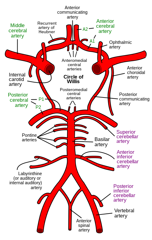
The circle of Willis (CoW) is the anastomotic arterial polygon at the base of the brain that connects the anterior and posterior circulations, providing the primary collateral pathway for cerebral perfusion [1]A1a[8]D5.
Also Called / Synonyms
- Circulus arteriosus cerebri (Latin), Willisian circle, cerebral arterial circle. Eponyms remain common in clinical discourse despite the push for standardized Terminologia Anatomica [20]D5.
Classification of Completeness and Variation
The CoW is conventionally described as a complete hexagon formed by the anterior communicating artery (ACoA), anterior cerebral arteries (A1 segments), internal carotid arteries (ICAs), posterior communicating arteries (PCoAs), and posterior cerebral arteries (P1 segments). However, only 30.8% of circles are structurally complete using strict diameter thresholds [16]C4. The following scheme, adapted from Saha et al. (2025), classifies circles by functional competence [16]C4:
Pearl: Five-group classification system (Ayre et al.) catalogues 82 variant configurations for clinical and research use [2]C4.
| Type | Definition | Presumed Functional Competence |
|---|---|---|
| I | All components present and ≥1 mm in diameter | Full collateral capacity |
| II | One hypoplastic segment (diameter <1 mm) | Partially reduced |
| III | One absent segment (aplasia) | Reduced in corresponding territory |
| IV | Multiple absent or hypoplastic segments | Severely limited collateral flow |
Data from Saha et al. [16]C4.
Gross Structure, Morphology & Function
- ▸The circle of Willis is complete in only 22-70% of individuals depending on population; the posterior arch (PCoA) is more frequently incomplete than the anterior arch.
- ▸The combined cross-sectional area of the connecting arteries (25.33 mm²) exceeds the outflow area (37.76 mm²), supporting a pressure-equalizing function.
From the definition of the Circle of Willis as a circular anastomosis, the structural arrangement of its component vessels determines its capacity to redistribute blood flow. The circle is formed by the bilateral anterior cerebral arteries (A1 segments), the anterior communicating artery (ACoA), the terminal segments of the bilateral internal carotid arteries (ICAs), the bilateral posterior communicating arteries (PCoAs), and the bilateral posterior cerebral arteries (P1 segments). This configuration creates a vascular ring at the base of the brain that interconnects the anterior and posterior circulations.
Component Vessels and Typical Configuration
In a textbook configuration, each A1 segment runs medially from the ICA bifurcation to join the ACoA, which connects the two anterior cerebral arteries. The PCoA arises from the ICA and runs posteriorly to join the P1 segment of the posterior cerebral artery (PCA), which itself originates from the basilar artery bifurcation. The circle is considered complete when all these vessels are present and patent. However, the typical symmetrical configuration is rare: in a Malawian cadaveric study, only 26.09% of circles were classified as typical [31]C4. A large CTA study of 511 patients found complete circles in 22.58% and nearly complete (one missing segment) in 28.6% [22]C4. The posterior arch is much more frequently incomplete than the anterior arch: 73.18% of posterior arches had at least one missing segment versus 18.4% for the anterior arch [22]C4. The left or right PCoA was unilaterally absent in 30.53% of patients, and both PCoAs were absent in 35.02% [22]C4.
Morphometric Dimensions
The diameters and cross-sectional areas of the circle's vessels are not uniform, and this asymmetry has functional implications. The combined cross-sectional area of the inflow arteries (bilateral ICAs and vertebral arteries) is 51.43 mm², which is significantly larger than the combined outflow area (37.76 mm²) but smaller than the combined area of outflow plus connecting arteries (25.33 mm² for connecting vessels alone) [26]C4. Table 1 summarizes key morphometric data from recent studies.
Table 1: Diameters and cross-sectional areas of circle of Willis vessels
| Vessel | Parameter | Value | Source |
|---|---|---|---|
| Inflow arteries (combined) | Cross-sectional area | 51.43 mm² | [26]C4 |
| Outflow arteries (combined) | Cross-sectional area | 37.76 mm² | [26]C4 |
| Connecting arteries (combined) | Cross-sectional area | 25.33 mm² | [26]C4 |
| PCA at origin | Median diameter | 3.20 mm (IQR 2.90-3.47) | [32]C4 |
| PCA at origin | Median cross-sectional area | 6.12 mm² (IQR 5.10-7.05) | [32]C4 |
| PCoA | Length (max) | 27.7 mm | [31]C4 |
| PCoA | Diameter (max) | 7.67 mm | [31]C4 |
| ACoA | Length (min) | 0.78 mm | [31]C4 |
| ACoA | Diameter (min) | 0.67 mm | [31]C4 |
The PCoA is the longest and widest vessel of the circle, while the ACoA is the shortest and narrowest [31]C4. The median PCA diameter at its origin is 3.20 mm [32]C4.
Completeness and Variability of the Circle
Completeness varies widely across populations. In Malawian cadavers, complete circles were found in 69.57% of specimens [31]C4, whereas in a European CTA study, complete circles were seen in only 22.58% [22]C4. This discrepancy may reflect differences in imaging technique, criteria for hypoplasia, or true population differences. The posterior communicating artery is the most variable vessel: 12 variations were identified in one study [31]C4. The PCoA is aplastic in 26.1% of sides [32]C4, and duplicated PCoA occurs in 2.8% [32]C4. The anterior communicating artery shows aplasia in 5.3% of cases, duplication in 8.92%, and fenestration in 1.78% [25]C4. The A1 segment of the anterior cerebral artery is hypoplastic in 21.43% of hemispheres [25]C4. These variations directly affect the circle's functional capacity.
Functional Significance of the Circle
The circle of Willis functions as a collateral network that can redistribute blood flow when one of the inflow arteries is stenotic or occluded. The cross-sectional area data support a pressure-equalizing role: the connecting arteries (combined area 25.33 mm²) provide a low-resistance pathway that attenuates peak pressure from the inflow vessels [26]C4. This mechanism is clinically critical: an incomplete circle reduces collateral reserve. In patients with middle cerebral artery (MCA) atherosclerosis, ACoA dysplasia or absence is associated with a 5.2-fold increase in the odds of symptomatic MCA plaques (OR 5.158) [33]B2b. Similarly, in patients, a severely compromised circle of Willis independently predicts ipsilateral brain infarcts and long-term mortality [34]B2b. The integrity of the circle is also central to the pathophysiology of disease, where progressive stenosis of the circle and terminal ICAs triggers the development of fragile collateral vessels [29]D5.
Pearl: A complete circle of Willis is present in fewer than 1 in 3 individuals; the posterior communicating arteries are the most commonly absent or hypoplastic vessels, making the posterior circulation dependent on the basilar artery alone during proximal occlusion of the ICA or vertebral artery.
Relations, Borders & Spaces
- ▸The circle of Willis resides within the suprasellar and interpeduncular cisterns; its relations to the optic chiasm, oculomotor nerves, and uncus govern surgical approach and risk.
- ▸Splitting the sylvian fissure to the anterior ascendant ramus optimizes exposure of the circle of Willis; extending dissection 2.0 cm distal offers no added benefit [38][39].
- ▸Infections of the retropharyngeal space can spread to the carotid sheath, threatening the ICA and relying on the circle of Willis for collateral supply [36].
Topographic Context Within the Basal Cisterns
Having described the constituent vessels, their topographic relations within the subarachnoid cisterns now define surgical corridors and predict pathologic spread. The entire circle of Willis is suspended in the , cradled primarily within the suprasellar cistern (chiasmatic cistern) and the interpeduncular cistern, with the posterior components reaching into the ambient cisterns. Anteriorly, the A1 segments and anterior communicating artery lie directly superior to the optic chiasm; the pituitary stalk descends between them. Posteriorly, the basilar bifurcation and P1 segments sit within the interpeduncular fossa, just ventral to the pons and between the cerebral peduncles. Laterally, the middle cerebral artery stems course into the sylvian fissure, with the uncus of the temporal lobe forming their lateral boundary. The posterior communicating arteries run above the oculomotor nerves (CN III), making these nerves vulnerable during aneurysm dissection.
Surgical Corridor: The Sylvian Fissure and Basal Cisterns
Access to the circle of Willis for aneurysm clipping or thrombectomy most commonly proceeds through the pterional-transsylvian approach. Splitting the opens the basal cisterns and progressively exposes the circle. A cadaveric study quantified this exposure in four steps: 1) dissection of the basal cisterns, 2) dissection of the sphenoidal compartment, 3) dissection of the operculoinsular compartment to the anterior ascendant ramus, and 4) dissection progressing 2.0 cm distal to that ramus [38]C4[39]C4. Exposure of the circle of Willis, the angle of approach to the carotid bifurcation, and the linear distance between the frontal lobe and skull base all increased significantly from Steps 1 to 2 and from Steps 2 to 3. Crucially, no significant difference was found between Steps 3 and 4 [38]C4[39]C4. The clinical implication is precise: splitting the sylvian fissure to the anterior ascendant ramus optimizes exposure; further distal dissection provides no additional gain [38]C4[39]C4.
Perivascular Space and Pathologic Relations
Beyond the macroscopic cisterns, penetrating branches of the circle of Willis course through the cerebral perivascular space (Virchow-Robin space), a pial-lined corridor that follows arterioles into the brain parenchyma. In the hyperacute phase after (SAH), neutrophils accumulate in this perivascular space, driven by the /RAGE axis, triggering cerebral vasospasm and impairing microarterial perfusion [37]D5. This perivascular compartment is thus not a passive cleft but an active immunologic and hemodynamic interface. In the neck, the internal carotid artery (ICA) travels within the carotid sheath, adjacent to the retropharyngeal space. Deep neck space infections (e.g., ) can erode into the ICA, producing a pseudoaneurysm that may stenose the vessel; in such cases, collateral flow through the circle of Willis can maintain hemispheric perfusion during childhood development and beyond [36]C4.
Relational Landmarks for Imaging Interpretation
On axial or , the circle of Willis appears as a pentagonal or heptagonal ring encircling the suprasellar cistern. Key relational landmarks: the optic chiasm sits anterior and midline, the pituitary stalk descends through the ring's center, the oculomotor nerves run lateral to the posterior communicating arteries, and the basilar apex lies posteriorly within the interpeduncular cistern. The uncus and parahippocampal gyri border the M1 segments laterally. These consistent relationships help distinguish aneurysms from infundibula and guide endovascular catheter navigation.
Pearl: When performing the pterional-transsylvian approach, stop splitting the sylvian fissure once the anterior ascendant ramus is reached, pushing further distal wastes time without improving exposure of the circle of Willis [38]C4[39]C4.
| Step | Dissection Target | Exposure & Angle Gain | Significance |
|---|---|---|---|
| 1 | Basal cisterns | Baseline | , |
| 2 | Sphenoidal compartment | Improved vs Step 1 | Significant [38]C4 |
| 3 | Operculoinsular to anterior ascendant ramus | Improved vs Step 2 | Significant [38]C4 |
| 4 | 2.0 cm distal to anterior ascendant ramus | No change vs Step 3 | Not significant [38]C4 |
Blood Supply, Innervation & Lymphatic Drainage
- ▸Only 22-31% of individuals have a complete Circle of Willis; the posterior communicating artery is the most frequently hypoplastic or absent segment, limiting collateral reserve.
- ▸Mean posterior communicating artery diameter is approximately 1.12 mm, approaching the threshold for effective collateral flow.
- ▸Combined non-contrast 4D-MRA achieves 95.2% sensitivity and 100% specificity for detecting collateral pathways through the CoW in ICA occlusion.
Having examined the spatial relationships of the Circle of Willis (CoW), its functional vascular anatomy, segmental diameters, collateral capacity, and hemodynamic properties, determines its role in cerebral perfusion. No direct innervation or lymphatic drainage of the CoW as a distinct structure is described in the available literature; the perivascular space of its component arteries participates in the glymphatic drainage of interstitial fluid.
Segmental Diameters and Flow Capacity
The eight constituent arteries of the CoW differ substantially in caliber, reflecting their respective contributions to cerebral blood flow. Table 1 lists mean diameters measured by magnetic resonance angiography in a Turkish population [18]C4.
Table 1. Mean diameters of CoW arteries (mm) [18]C4
| Artery | Mean diameter (mm) |
|---|---|
| Basilar artery | 2.85 |
| Right internal carotid artery (ICA) | 4.24 |
| Left ICA | 4.32 |
| Right A1 (precommunicating ACA) | 1.58 |
| Left A1 | 1.64 |
| Right M1 (middle cerebral artery) | 2.13 |
| Left M1 | 2.10 |
| Right P1 (precommunicating PCA) | 1.80 |
| Left P1 | 1.88 |
| Right posterior communicating artery (PCoA) | 1.12 |
| Left PCoA | 1.12 |
The PCoA is the narrowest and most variable segment; its mean diameter (1.12 mm) approaches the lower limit for effective collateral flow. In contrast, the ICAs are the largest-caliber vessels, supplying the majority of anterior circulation flow. The basilar artery (2.85 mm) provides the primary inflow for the posterior circulation. The anterior communicating artery (ACoA) is the shortest segment, measuring a mean of 0.78 mm in length [31]C4.
Collateral Circulation and Completeness
A complete CoW, all segments patent and ≥1 mm in diameter, is present in only 22.6% to 30.8% of individuals [16]C4[22]C4. An additional 28.6% have a nearly complete circle (only one missing segment) [22]C4. The posterior arch is far more often incomplete (73.18%) than the anterior arch (18.4%), with unilateral or bilateral PCoA absence or hypoplasia as the dominant cause [22]C4. Hypoplasia (diameter < 1 mm) is more common in cerebral arteries, whereas aplasia is more common in communicating arteries [16]C4. The right posterior quadrant has the highest incidence of incompleteness [16]C4.
A commonly used functional classification [16]C4 stratifies circles into:
- Type I: classical complete circle
- Type II: one hypoplastic or absent segment (most common variant)
- Type III: two nonadjacent hypoplastic or absent segments
- Type IV: three or more missing or severely hypoplastic segments
In a Malawian cadaveric study, 69.57% of circles were complete [31]C4, highlighting potential population differences.
Hemodynamic Consequences of Variant Configurations
An incomplete CoW limits collateral reserve during acute occlusion. Full separation of the carotid and vertebrobasilar territories (i.e., absence of any PCoA) occurs in 40.1% of individuals, and complete isolation of the middle cerebral artery (MCA) in 1.4% [22]C4. These configurations eliminate the protective shunt capacity of the circle, increasing infarct risk in large vessel occlusion. In patients with internal carotid artery occlusion (ICAO), non-contrast 4D-MRA (combined 4D-PACK and 4D-S-PACK) provides 95.2% sensitivity and 100% specificity for assessing collateral pathway patterns through the CoW, superior to standard TOF-MRA [54]B2b.
Pearl: A mean arterial-venous pressure gradient ≥ 20 mmHg during cardiopulmonary resuscitation is associated with complete CoW contrast enhancement on CT, suggesting a hemodynamic threshold for antegrade cerebral perfusion [52]B2b.
Influence of Vessel Caliber on Pathology
Larger CoW arteries accumulate more calcification: calcification percentage correlates positively with vessel diameter, male sex, and age, but not with coronary artery disease as a cause of death [35]C4[43]C4. Increased luminal surface roughness secondary to calcification alters wall shear stress and vortex formation [35]C4. and estrogen deficiency lower the threshold for flow-induced aneurysmal remodeling in the CoW, producing greater vessel tortuosity and more widespread saccular changes after bilateral common carotid ligation in a rabbit model [47]D5.
Lymphatic Drainage
The brain lacks conventional lymphatic vessels; interstitial fluid and solutes drain via the glymphatic system along perivascular spaces of the CoW arteries and their penetrating branches. The walls of the larger CoW arteries (ICA, MCA, ACA) are surrounded by a pial-glial sheath that forms a conduit for fluid exchange, though this system is not unique to the CoW and is not separately innervated.
| Artery | Mean diameter (mm) |
|---|---|
| Basilar artery | 2.85 |
| Right internal carotid artery (ICA) | 4.24 |
| Left ICA | 4.32 |
| Right A1 (precommunicating ACA) | 1.58 |
| Left A1 | 1.64 |
| Right M1 (middle cerebral artery) | 2.13 |
| Left M1 | 2.10 |
| Right P1 (precommunicating PCA) | 1.80 |
| Left P1 | 1.88 |
| Right posterior communicating artery (PCoA) | 1.12 |
| Left PCoA | 1.12 |
Microscopic & Histological Notes
- ▸Vessel wall thickness ranges from 0.31 mm (minimum) to 0.86 mm (maximum) and is greater in symptomatic patients [[63]].
- ▸The recurrent artery of Heubner has a mean diameter of 1 mm and 2-30 branches; its number negatively correlates with lenticulostriate artery count [[57]].
- ▸Decreased endothelial tight junction proteins (ZO-1, claudin-5) are found in PKD-related IAs and human ruptured aneurysms [[62]].
- ▸Distinct histopathologic patterns differentiate FMD, Ehlers-Danlos type IV, NF1, and moyamoya [[59]].
Having traced the blood supply and innervation, the tissue-level architecture of the circle of Willis reveals a muscular artery system adapted to intracranial hemodynamics. The vessel wall comprises three layers: a thin intima lined by endothelial cells, a prominent internal elastic lamina, a thick media of circumferentially oriented smooth muscle cells interspersed with elastin and collagens (types I and III), and a thin adventitia lacking an external elastic lamina. This intimal barrier is central to the blood-brain barrier; endothelial tight junction proteins, zonula occludens-1 and claudin-5, are reduced in intracranial aneurysms (IAs) from both rat models and human ruptured specimens, suggesting that endothelial dysfunction precedes wall weakening .
Vessel Wall Thickness and Regional Variation
Ex vivo 7T MRI validated against histology demonstrates that mean wall thickness varies from 0.45 to 0.66 mm across major segments, with minimum values of 0.31 mm and maxima of 0.86 mm; the normalized wall index ranges 0.64-0.75 . Vessel walls are consistently thicker in patients with symptomatic cerebrovascular disease compared with asymptomatic controls . The recurrent artery of Heubner, a key perforator from the anterior cerebral artery, has a mean length of 25.2 mm and an origin diameter of 1 mm, giving off 2 to 30 branches (mean 9.4) that penetrate the striatum and internal capsule . Its number correlates negatively with lenticulostriate arteries from the MCA (R = -0.62; p < 0.0001), reflecting an inverse embryologic relationship.
| Parameter | Value (mean ± SD or range) |
|---|---|
| Mean wall thickness | 0.45-0.66 mm |
| Minimum wall thickness | 0.31-0.42 mm |
| Maximum wall thickness | 0.52-0.86 mm |
| Normalized wall index | 0.64-0.75 |
| RAH diameter at origin | 1 mm |
| RAH branches per vessel | 2-30 (mean 9.4) |
Histopathology of Selected Arteriopathies
Distinct histologic changes distinguish intrinsic arteriopathies that involve the circle of Willis . In , disorganized collagen within the muscularis, highlighted by picrosirius red staining, is confined to the intracranial circulation. syndrome shows fibrocellular smooth muscle intimal proliferation confirmed by smooth muscle actin immunohistochemistry. Ehlers-Danlos type IV (vascular type) presents with eccentric intimal thickening of circle vessels but no overt muscularis abnormality, alongside aneurysm formation. Neurofibromatosis I preferentially affects smaller leptomeningeal arteries, particularly around the spinal cord. These patterns require extensive sampling, including multiple sections of the entire circle of Willis, to avoid underdiagnosis .
Perforating Arteries and the Basilar Tip
The basilar bifurcation gives rise to a variable set of mesencephalic and thalamoperforating arteries critical to midbrain and thalamic perfusion. A reported case of basilar tip fenestration gave origin to five perforating branches, one artery of Percheron and four mesencephalic arteries, underscoring that fenestration, if misdiagnosed as a saccular aneurysm, would risk catastrophic sequelae (consciousness disturbances, quadriplegia, sensory loss) during endovascular treatment .
Pearl: Disruption of the internal elastic lamina, not medial thinning, is the earliest histologic event in intracranial aneurysm formation; its preservation on histology argues against a congenital saccular origin.
Development (Brief Embryology)
- ▸The Circle of Willis develops between weeks 4-8 of gestation from the aortic arches (ICAs), longitudinal neural arteries (basilar), and their connecting vessels (PComA, AComA).
- ▸Pediatric development follows a biphasic growth pattern: rapid infant expansion then slower growth; children exhibit greater symmetry and fewer anomalies than adults [44].
- ▸Variations arise from persistence, disappearance, or new sprouting of embryonic vessels driven by hemodynamic and genetic factors, explaining common variants like fetal PCA [68][65].
From the microscopic organization of the arterial wall to its origin, the Circle of Willis emerges during the fourth to eighth weeks of gestation through a tightly choreographed sequence of vessel formation, fusion, and regression that explains its adult configuration and many common anomalies.
Formation of the Primary Arterial Network
The internal carotid arteries arise from the third aortic arch and the dorsal aortae. The posterior communicating arteries develop as connections between the internal carotid and the developing vertebrobasilar system. The basilar artery forms from fusion of the paired longitudinal neural arteries, which are supplied by the vertebral arteries developing from cervical intersegmental arteries. The anterior cerebral arteries sprout from the internal carotid arteries and are connected by the anterior communicating artery, completing the anastomotic ring [68]C4.
Biphasic Growth and Pediatric Maturation
Pediatric development follows a biphasic growth pattern: rapid arterial expansion during infancy, followed by slower growth, with strong correlations between vessel dimensions and circumference [44]B2a. Children exhibit greater Circle of Willis symmetry and fewer anatomical anomalies than adults, challenging the assumption that vascular configurations stabilize only with age [44]B2a. This suggests that environmental, hemodynamic, or structural factors drive asymmetry in adulthood.
Embryological Basis of Variations
Variations arise from persistence of embryonic vessels that normally disappear, disappearance of vessels that would normally persist, or sprouting of new vessels due to hemodynamic and genetic factors [68]C4. For example, fetal origin of the posterior cerebral artery (a common variant, seen in ~10-30%) results from persistence of the embryonic posterior communicating artery as the dominant supply [65]C4. Internal carotid artery agenesis (228 cases reviewed) can be classified by the type of flow compensation (Type I-IV) based on which embryonic arteries regress or persist [3]D5. The accessory middle cerebral artery and fenestrations of the anterior cerebral artery also derive from embryological development [65]C4[41]C4.
Clinical Relevance of Developmental Anatomy
Understanding the embryological timetable aids in predicting which anomalies may be associated with aneurysms (e.g., MCA duplication, A1 hypoplasia) [65]C4 and guides interpretation of pediatric neuroimaging [44]B2a. disease involves progressive stenosis of the terminal internal carotid and Circle of Willis, likely with a developmental component [29]D5.
Pearl: The Circle of Willis forms by the 8th gestational week, and any deviation from the standard regression/persistence pattern during embryogenesis predicts the specific variant seen in adulthood, knowledge that helps anticipate associated risks (aneurysm, stroke) [68]C4[65]C4.
Variations & Anomalies
- ▸A complete circle is present in only ~30% of individuals; the most common variant is PCoA hypoplasia/aplasia.
- ▸Fetal PCoA (PCA supplied by ICA) is the single most frequent variant in the posterior circulation.
From this embryological template, the mature circle of Willis is complete in only about one‑third of individuals - 30.8% in cadaveric series and 30.9% on MRA [16]C4[74]C4. A meta‑analysis of 33 studies reported a 68.2% prevalence of general variation), with unilateral posterior communicating artery (PCoA) hypoplasia/aplasia in 19.45% and bilateral in 22.83% [1]A1a. A systematic review of 42 studies catalogued 82 distinct variants organized into five groups: (1) hypoplastic segments only, (2) absent segments only, (3) mixed hypoplastic/absent, (4) accessory segments, (5) other anomalies [2]C4.
Anterior Circulation Variations
Unilateral absence of the A1 segment of the anterior cerebral artery (ACA) is the most common anterior variant (13.5% in one CTA study [75]C4). Absence of the anterior communicating artery (ACoA) occurs in 22.8% and is associated with symptomatic middle cerebral artery (MCA) atherosclerosis (for symptomatic status; 95% CI 1.74‑15.25) and non‑positive remodeling [33]B2b. A single A2 segment supplying both hemispheres (azygos ACA) is rare but critical because a unilateral lesion can cause bihemispheric ACA territory infarction [73]C4. Infra‑optic or inter‑optic ACA courses, often with associated A1 aplasia and carotid‑ACA anastomoses, are exceedingly rare but carry a high risk of aneurysm [23]C4[45]C4[70]C4.
MCA variations are also frequent: bifurcation is the most common configuration (86.2%), trifurcation in 13.8%, with upper‑trunk dominance in 26% and lower‑trunk in 25.4% [9]C4. An accessory MCA, defined as an additional vessel arising from the A1 segment, is rare; bilateral occurrence has been reported in association with anterior circulation aneurysms [72]C4[65]C4.
Posterior Circulation Variations
Fetal‑type PCoA - in which the PCoA supplies the posterior cerebral artery (PCA) territory - is the most common posterior variant, found unilaterally in 11.1-14.3% and bilaterally in 4.8-5.6% of individuals [16]C4[74]C4. Unilateral absence of the PCoA occurs in 22.2% and bilateral absence in 29.2% [75]C4. Basilar tip fenestration is a rare variant that can give rise to the artery of Percheron and other perforating arteries; misdiagnosis as an aneurysm during embolization would be catastrophic [60]C4.
Rare Variants
Internal carotid artery (ICA) agenesis or hypoplasia occurs in approximately 0.16% of individuals and is usually compensated by collateral flow through the circle of Willis, intercavernous anastomoses, or persistent embryologic arteries [69]C4. Carotid‑ACA anastomoses, where an anomalous vessel takes an infra‑optic course from the ICA to the contralateral A2 segment, have been described with concurrent A1 aplasia and ipsilateral ACA fenestration [23]C4.
Clinical Implications
An incomplete circle reduces collateral reserve. During with cross‑clamping, contralateral carotid stenosis >70% or occlusion - but not CoW completeness alone - predicted neurologic deficit (positive awake test in 5.8-45.7%), yet patients with incomplete anterior circulation had positive awake tests in 5-91% of cases [4]B2a. ACoA absence independently predicts symptomatic MCA atherosclerotic plaques (, p=0.003) [33]B2b. Recognition of variants prevents misinterpretation of CT perfusion “false penumbras” caused by upstream flow restriction due to variant anatomy [7]D5.
Pearl: When interpreting CTA or MRA for stroke workup, always assess the A1‑ACoA complex: its absence increases the odds that a contralateral MCA stenosis will cause symptoms (OR >5), and its presence may supply crucial collateral flow that alters the ischemic penumbra.
| Group | Description | Variants (n) | Key Examples |
|---|---|---|---|
| 1 | Hypoplastic segments only | 24 | Hypoplastic A1, hypoplastic PCoA |
| 2 | Absent segments only | 11 | ACoA aplasia, A1 agenesis |
| 3 | Hypoplastic + absent segments | 6 | Hypoplastic A1 + absent PCoA |
| 4 | Accessory segments only | 26 | Duplicate ACoA, accessory MCA |
| 5 | Other variations | 15 | Fenestrations, azygos ACA, infra‑optic ACA |
Surface Anatomy & Imaging Correlation
- ▸The Circle of Willis resides in the suprasellar cistern; familiarity with this location enables rapid identification on axial, coronal, and sagittal imaging.
- ▸The Vascular Asymmetry Coefficient (VAC) standardizes the diagnosis of asymmetry (>10%) and hypoplasia (>40%) from CTA, essential for interpreting perfusion studies.
- ▸Fractal analysis (Df[W]) from 3D TOF-MRA outperforms traditional risk scores in detecting silent cerebral small vessel disease (AUC 0.928), and combined biomarker-imaging models achieve near-perfect accuracy (AUC 0.952).
Recognition of these anatomic variants is essential during imaging interpretation, because an incomplete or asymmetric Circle of Willis (CoW) can mimic pathology, alter hemodynamic patterns, and confound perfusion parameters. The CoW lies within the suprasellar cistern, a CSF-filled space at the base of the brain that is consistently identified on axial, coronal, and sagittal imaging. The carotid and basilar systems join here, and the key surface landmarks, the optic chiasm anteriorly, the interpeduncular fossa posteriorly, and the internal carotid artery (ICA) termini laterally, provide reliable radiographic coordinates for localizing aneurysm and stroke lesions.
Imaging Modalities and Anatomic Landmarks
Modern neuroimaging visualizes the CoW with high spatial resolution, but each modality has distinct advantages and pitfalls. Computed tomography angiography (CTA) offers rapid, widespread availability and is the first-line test for acute ; the bony carotid canal is visible on skull-base CT, and its absence helps distinguish ICA agenesis from aplasia or hypoplasia [77]C4. Magnetic resonance angiography (MRA), especially 3D time-of-flight (TOF) sequence, provides superior soft-tissue contrast without ionizing radiation and allows calculation of the fractal dimension of the CoW (Df [W]), a novel biomarker for cerebrovascular network complexity that correlates with white matter hyperintensity burden [76]C4. Digital subtraction angiography (DSA) remains the gold standard for dynamic flow assessment and detection of small aneurysms, particularly in the posterior circulation, and can identify rare anomalies such as basilar tip fenestration giving rise to multiple perforating arteries (artery of Percheron and mesencephalic arteries) that would be missed on non-invasive imaging [60]C4.
| Modality | Key Feature | Diagnostic Utility | Limitation |
|---|---|---|---|
| CTA | Rapid acquisition, bone detail | First-line for acute SAH; ICA agenesis (absence of carotid canal) [77]C4 | Radiation, iodinated contrast |
| TOF-MRA | No contrast, flow-sensitive | Fractal analysis (Df[W]) for silent CSVD [76]C4; pediatric CoW growth [44]B2a | Overestimates stenosis; time-consuming |
| DSA | Dynamic flow, high resolution | Detection of fenestrations and perforator anatomy [60]C4; puff-of-smoke [29]D5 | Invasive, radiation, contrast |
| Transcranial Doppler (TCD) | Bedside, portable | Screening for vasospasm; collateral flow direction | Operator-dependent; limited windows |
Interpretation of Variants and Asymmetry
Quantitative assessment of vessel diameter asymmetry is critical for distinguishing normal variants from pathology. The Vascular Asymmetry Coefficient (VAC), calculated from CT angiography datasets, classifies asymmetry when the diameter difference between paired segments exceeds 10% and hypoplasia when the difference exceeds 40% [82]C4. Pediatric CoW development follows a biphasic growth pattern, rapid arterial expansion in infancy then slower growth, that correlates strongly with circumference, and children exhibit greater symmetry and fewer anomalies than adults, suggesting that asymmetry in adulthood reflects acquired hemodynamic or environmental factors [44]B2a.
Fractal analysis of the CoW from 3D TOF-MRA outperforms conventional cardiovascular risk scores (QRISK3) in discriminating silent cerebral small vessel disease (AUC 0.928 vs. ~0.75). A combined model integrating circulating microparticles and Df(W) achieved near-perfect diagnostic accuracy (AUC 0.952) [76]C4. These quantitative imaging tools are moving from research into clinical practice, enabling biologically grounded detection of preclinical cerebrovascular injury.
Impact on Perfusion Imaging Interpretation
CoW variants can produce false ischemic penumbras on CT perfusion studies, regions with prolonged mean transit time and normal cerebral blood volume that mimic salvageable penumbra but actually represent benign oligemia due to variant anatomy rather than true ischemia [7]D5. Five principal causes have been identified: upstream flow restriction, evolving infarction, vascular dysregulation, head positioning artifacts, and CoW variant anatomy (e.g., absent A1 segment, fetal posterior communicating artery). Radiologists must correlate CT perfusion parameters with unenhanced head CT, CTA, and diffusion-weighted MRI to avoid inappropriate thrombolysis [7]D5.
Special Clinical Scenarios
- ICA agenesis/hypoplasia: Most patients are asymptomatic due to collateral circulation via the CoW, intercavernous anastomosis, or persistent embryologic arteries, but these anomalies carry a risk of associated cerebral aneurysms and must be recognized before carotid or transsphenoidal surgery [69]C4. The definitive imaging sign is absence of the bony carotid canal on skull-base CT [77]C4.
- Moyamoya disease: Progressive stenosis of the terminal ICA and CoW produces a classic "puff-of-smoke" collateral network on DSA, best appreciated on lateral projections. Ischemic symptoms predominate, but adults are twice as likely to present with hemorrhage [29]D5.
- AI-assisted detection: Machine learning algorithms now achieve 83% sensitivity and specificity for aneurysm rupture prediction, and the RAPID Aneurysm software demonstrates high accuracy on CTA [80]D5. Deep learning architectures (e.g., PointNet++) yield an AUC of 0.85 for rupture risk [80]D5.
Pearl: The suprasellar cistern is the single most important radiographic landmark for the Circle of Willis; any asymmetry in vessel caliber or flow on CTA/MRA should prompt calculation of the VAC coefficient, when the difference exceeds 10%, the vascular segment is asymmetric, and when it exceeds 40%, hypoplasia should be documented, because these variations directly affect the interpretation of CT perfusion maps and the risk of false penumbras [7]D5[82]C4.
Clinical Correlations
- ▸CoW variations affect collateral reserve; the most consequential is bilateral PCoA absence (35%), which eliminates anterior-posterior cross-flow and increases hemispheric stroke risk with ipsilateral carotid occlusion [22].
- ▸Aneurysm formation in the anterior circulation is associated with specific CoW variants (e.g., accessory MCA, A1 agenesis), and these variants must be recognized during preprocedural planning to avoid complications [72][45].
- ▸CT perfusion false penumbras can be caused by fetal PCA variants, leading to misdiagnosis of salvageable tissue; knowledge of CoW anatomy is essential for accurate interpretation [7].
From the imaging patterns detailed above, the clinical implications of Circle of Willis (CoW) anatomy manifest at the bedside through collateral circulation failure, ischemic stroke syndromes, and aneurysm-related presentations. The morphology, complete versus incomplete, fetal versus adult, determines vulnerability to hemodynamic stress and determines the clinical phenotype when proximal vessels become diseased or occluded.
Presenting Symptoms
Most individuals with CoW variations remain asymptomatic throughout life; the anastomotic network is sufficiently robust under resting conditions. Symptoms emerge when a variation compromises collateral reserve and a second insult, thrombotic occlusion, embolus, hypotension, or vasospasm, overwhelms the remaining channels.
Transient ischemic attack (TIA) and ischemic stroke are the most common clinical presentations attributable to CoW variants. In a series of 511 patients undergoing CTA, 40.1% had full separation of the carotid and vertebrobasilar territories (absent or hypoplastic posterior communicating arteries bilaterally), and 8.6% had separation of all three main arterial axes (both ICAs and VB) [22]C4. Such configurations eliminate cross-flow from the anterior to posterior circulation (or vice versa), rendering the hemisphere dependent on ipsilateral carotid inflow. When the internal carotid artery becomes stenotic or occluded, these patients present with territorial infarction in the middle cerebral artery (MCA) or watershed infarcts. Absence of at least one posterior communicating artery (PCoA) is linked to lower occipital lobe cerebral blood flow (p = 0.001) and higher global perfusion heterogeneity (p = 0.003) in 390 community-dwelling adults free of clinical stroke [87]B2b. Bilateral PCoA absence is associated with even more pronounced reductions in global (p = 0.01) and occipital lobe CBF (p = 0.0001) [87]B2b.
(SAH) from ruptured intracranial aneurysm is the second major symptom complex. CoW variations predispose to aneurysm formation by altering hemodynamic shear stress at branching points. Anomalies such as accessory middle cerebral arteries [72]C4, infra-optic course of the anterior cerebral artery (ACA) [45]C4, and inter-optic course of a unique A1 segment [70]C4 are each reported in association with anterior communicating artery (ACoA) complex aneurysms. Aneurysm rupture presents with the classic “thunderclap” headache, nausea, vomiting, and transient loss of consciousness. In the hyperacute phase, cerebral vasospasm driven by neutrophil RAGE signaling further compromises perfusion of the CoW and can lead to delayed ischemic neurologic deficits [37]D5.
Cognitive decline may be the only symptom in patients with silent cerebral small vessel disease (CSVD) related to reduced cerebrovascular complexity. In one study of 60 asymptomatic adults, reduced fractal dimension of the CoW (Df[W]), reflecting diminished network complexity, was strongly associated with white matter hyperintensity burden on MRI (p < 0.001), yet neurocognitive performance remained normal, consistent with network reserve [76]C4.
Mass effect from large CoW aneurysms can compress adjacent cranial nerves. A posterior communicating artery aneurysm may present with ipsilateral third nerve palsy (ptosis, mydriasis, “down and out” gaze), whereas a basilar tip aneurysm can cause oculomotor or abducens deficits.
Neurological Examination Findings
The examination in a patient with a symptomatic CoW variation is driven by the territory at risk:
- Anterior circulation (ICA/MCA): Contralateral hemiparesis (face > arm > leg), hemisensory loss, homonymous hemianopia, and (if dominant hemisphere) aphasia. A hypoplastic A1 segment or absent ACoA eliminates the potential for cross-filling from the contralateral ICA, so internal borderzone infarcts between the ACA and MCA territories are more likely.
- Anterior cerebral artery territory: Contralateral leg-predominant weakness and sensory loss, plus urinary incontinence and abulia. The rare variant of a single A2 segment supplying both medial frontal lobes (seen in 2.1-2.5% [19]C4) places both medial frontal lobes at risk from a single embolic event, producing bilateral leg weakness and mutism [6]C4.
- Posterior circulation (vertebrobasilar): Visual field defects (occipital lobe), ataxia, dysarthria, diplopia, and cranial nerve deficits. A fetal-type posterior cerebral artery (PCA) (unilateral 9.4%, bilateral 2.54% [22]C4) means the PCA is supplied predominantly by the ICA; occlusion of the ICA thus produces both anterior and posterior territory signs. Percheron artery infarction (from basilar tip fenestration [60]C4) presents with bilateral thalamic and midbrain signs: altered consciousness, vertical gaze palsy, and amnesia.
- Cranial nerve examination: Ptosis, mydriasis, and extraocular movement restriction (CN III) suggest a PCoA aneurysm. Bilateral optic atrophy or visual field cuts may occur from an inter-optic ACA compressing the optic chiasm [70]C4.
- Cognitive assessment: In silent CSVD, bedside tests (Montreal Cognitive Assessment) may be normal until significant progression, but processing speed shows weak association with Df(W) [76]C4.
Phenotypic Variants, Clinical Significance
The following table summarizes the most clinically relevant CoW configurations encountered in practice, their frequency (as reported in imaging or cadaveric series), and the clinical syndrome they predispose to.
| Variant | Key Features | Frequency (pooled ranges) | Clinical Correlation |
|---|---|---|---|
| Complete CoW | All segments patent, normal caliber | 22.5-27% [22]C4[85]C4 | Optimal collateral reserve; stroke risk approximates general population |
| Incomplete CoW (any segment absent) | At least one missing segment | 73-78% (posterior arch most common) [22]C4[85]C4 | Reduced collateral capacity; 40.1% have full carotid-vertebrobasilar separation [22]C4 |
| Bilateral absent PCoA | Both posterior communicating arteries aplastic | 35% [22]C4 | Lower global and occipital CBF; higher perfusion heterogeneity; increased watershed infarct risk [87]B2b |
| Fetal-type PCA (full) | PCA origin from ICA via PCoA; basilar contribution absent | Unilateral 9.4%, bilateral 2.54% [22]C4 | Posterior circulation stroke with ipsilateral ICA occlusion; false penumbra on CT perfusion [7]D5 |
| Basilar artery non-furcation | No bifurcation; single trunk continues as PCA | 8.95% [83]A1a | May mimic aneurysm on angiography; risk of misdiagnosis during intervention [83]A1a |
| Basilar tip trifurcation/quadrifurcation | Three or more PCA branches from basilar tip | 7.05% / 5.30% [83]A1a | Altered flow dynamics; potential for perforator (Percheron) infarction [60]C4 |
| A1 segment agenesis/hypoplasia | Absent or <1.5 mm A1 segment | ~5.65% [19]C4 | Association with ACoA aneurysm (contralateral A1 supplies both A2s); risk of bilateral medial frontal ischemia [6]C4 |
| Accessory MCA | Duplicated MCA from ACA | Rare (bilateral extremely rare) [72]C4 | Increased aneurysm risk in anterior circulation [72]C4 |
| Median pericallosal artery (triple A2) | Unpaired third A2 from ACoA complex | 2.1% [19]C4 | Risk of intraoperative avulsion during ACoA aneurysm clipping [78]C4 |
| Persistent fetal carotid-vertebrobasilar anastomosis (e.g., persistent hypoglossal artery) | Embryonic vessel connecting carotid to vertebrobasilar system | Rare (<0.1%) [84]C4 | May be sole supply to posterior circulation; associated with hypoplastic vertebral arteries and absent PCoA; aneurysm risk [84]C4 |
Red Flags, Symptoms Requiring Urgent Action
Certain presentations demand immediate evaluation because they herald catastrophic loss of collateral reserve or aneurysm rupture:
- Sudden severe (“thunderclap”) headache with neck stiffness, suspect SAH from a ruptured CoW aneurysm. Perform noncontrast CT followed by CTA to identify the source.
- Acute onset of third nerve palsy (ptosis, mydriasis, diplopia), ipsilateral PCoA aneurysm until proven otherwise.
- New bilateral leg weakness or mutism, consider the variant of single A2 segment supplying both medial frontal lobes (especially if carotid disease is present) [6]C4.
- Hemodynamic collapse after carotid occlusion (e.g., during CEA), patients with complete separation of anterior and posterior circulation (40% [22]C4) cannot recruit cross-flow and develop massive MCA territory infarction. Preoperative CoW assessment with CTA or MRA is mandatory [22]C4.
- Delayed neurological deterioration after SAH, cerebral vasospasm can reduce CoW caliber by 50% or more. The HMGB1/RAGE axis on neutrophils drives this vasospasm; plasma soluble RAGE levels may predict symptomatic vasospasm [37]D5.
Atypical Presentations
CoW variants can mimic other conditions or present with subtle, non-focal symptoms:
- False ischemic penumbra on CT perfusion: A fetal-type PCA can produce a delay in contrast bolus arrival in the occipital lobe, mimicking the prolonged mean transit time and reduced cerebral blood volume of salvageable penumbra. This variant anatomy must be recognized to avoid inappropriate thrombolysis [7]D5. Similarly, upstream flow restriction from a hypoplastic A1 segment can produce an apparent penumbra in the ACA territory.
- Silent cerebral small vessel disease: In heart-healthy middle-aged and older adults, missing PCoAs correlate with lower occipital CBF and higher perfusion heterogeneity in the absence of any clinical symptoms [87]B2b. Fractal analysis of CoW complexity can detect subclinical CSVD with high accuracy (AUC 0.928) before cognitive decline emerges [76]C4.
- Cognitive impairment without stroke: Long-standing hemodynamic stress from an incomplete posterior circle may contribute to white matter disease and vascular dementia, though this remains an area of active investigation [76]C4[87]B2b.
- Headache and syncope: Rarely, significant steal phenomena through persistent fetal anastomoses (e.g., persistent trigeminal artery) can produce vertebrobasilar insufficiency symptoms with head turning or hypotension.
- Incidental finding during cardiac or subclavian artery surgery: In patients with a dominant left vertebral artery and an incomplete CoW, subclavian artery aneurysm repair carries risk of posterior circulation ischemia if the vertebral is compromised [12]D5.
Controversies and Guideline Disagreement
| Question | Position A | Position B | Strength | Implication |
|---|---|---|---|---|
| Should all patients with TIA undergo CTA/MRA to assess CoW completeness? | AHA/ASA guidelines recommend vascular imaging, but do not require full CoW characterization for routine TIA [implied by standard care]. | Some interventionalists argue that CoW status should be documented before any carotid intervention (CEA or stenting) to stratify hemodynamic risk [22]C4. | Class IIa (reasonable) | Preoperative CTA with 3D volume rendering can identify high-risk configurations (40% with complete anterior-posterior separation) and alter surgical planning (e.g., shunt use) [22]C4. |
| Should CoW screening be performed in patients with Ehlers-Danlos syndrome? | hEDS has generally been considered low-risk for vascular rupture. | A retrospective cohort found intracranial aneurysms in 7.3% of hEDS patients, suggesting a need for screening CTA [86]C4. | Low evidence (single cohort, small N) | Until larger studies confirm, screening is not mandated but may be considered on a case-by-case basis, especially if other risk factors are present. |
Pearl: A missing posterior communicating artery is the single most important CoW variation for the clinician: present in roughly one in three patients, it eliminates cross-flow between the anterior and posterior circulations, elevates perioperative risk during carotid interventions, and is an independent predictor of reduced occipital perfusion and increased stroke susceptibility in aging adults [22]C4[87]B2b. When planning any ipsilateral carotid or subclavian procedure, verify patency of both PCoAs on the preoperative angiogram.
| Variant | Key Features | Frequency (pooled ranges) | Clinical Correlation |
|---|---|---|---|
| Complete CoW | All segments patent | 22.5-27% [22]C4[85]C4 | Optimal collateral reserve; stroke risk approximates general population |
| Incomplete CoW (any segment absent) | At least one missing segment | 73-78% [22]C4[85]C4 | Reduced collateral capacity; 40.1% have full carotid-vertebrobasilar separation [22]C4 |
| Bilateral absent PCoA | Both PCoAs aplastic | 35% [22]C4 | Lower global and occipital CBF; higher perfusion heterogeneity [87]B2b |
| Fetal-type PCA (full) | PCA from ICA via PCoA | Unilateral 9.4%, bilateral 2.54% [22]C4 | Posterior circulation stroke with ICA occlusion; false CT penumbra [7]D5 |
| Basilar non-furcation | No bifurcation; single trunk | 8.95% [83]A1a | Mimics aneurysm on angiography; intervention risk [83]A1a |
| A1 agenesis/hypoplasia | A1 <1.5 mm or absent | 5.65% [19]C4 | Associated with ACoA aneurysm; bilateral medial frontal stroke risk [6]C4 |
| Accessory MCA | Duplicated MCA from ACA | Rare (bilateral extremely rare) [72]C4 | Increased anterior circulation aneurysm risk [72]C4 |
Eponyms & Nomenclature
- ▸The Terminologia Anatomica term for the circle of Willis is circulus arteriosus cerebri, but the eponym dominates clinical usage.
- ▸Thomas Willis described the circle in 1664, recognizing its collateral function; he also coined 'neurologie'.
- ▸Eponyms like recurrent artery of Heubner lack standardized anatomica terms but persist in operative literature.
Given the clinical significance of the circle's variant anatomy in stroke and aneurysm , precise terminology ensures clear communication across disciplines.
The Eponym and Its History
Thomas Willis first described the circular arterial anastomosis at the base of the brain in his 1664 work Cerebri anatome, recognizing its functional importance in maintaining cerebral perfusion when one feeding vessel fails [8]D5. Willis also coined the term "neurologie," marking a transition between mediaeval and modern concepts of brain function [8]D5. The structure has borne his name ever since, though its official Terminologia Anatomica designation is circulus arteriosus cerebri [20]D5.
Terminologia Anatomica and Synonyms
Eponyms "bring colour to medicine" but are criticized for lacking accuracy, leading to confusion and hampering scientific discussion [20]D5. Despite this, the eponym "circle of Willis" remains overwhelmingly dominant in clinical practice, operative reports, and the literature.
Other Associated Eponyms
The recurrent artery of Heubner (RAH), a key branch of the anterior cerebral artery supplying the of the caudate nucleus and anterior limb of the internal capsule, is frequently referenced in aneurysm surgery involving the anterior communicating artery complex [50]C4. No universally accepted Terminologia Anatomica synonym exists for this vessel, but it is consistently called by its eponym in neurosurgical contexts. The fetal posterior cerebral artery (fetal PCA) is a common variant but is not eponymous.
Pearl: When documenting or dictating, use "circle of Willis" for clinical communication but be prepared to reconcile it with the official Terminologia Anatomica term "circulus arteriosus cerebri" in formal anatomical reports.
| Eponym | Terminologia Anatomica | Clinical Context |
|---|---|---|
| Circle of Willis | Circulus arteriosus cerebri [20]D5 | Universal clinical term for the basal arterial anastomosis |
| Recurrent artery of Heubner | No standardized TA synonym; often called 'recurrent branch of ACA' | Supplies head of caudate and anterior limb of internal capsule; important in ACom aneurysm surgery [50]C4 |
| Fetal posterior cerebral artery | Not an eponym; variant of PCA | Common variant where PCA origin is from ICA; increases risk of TIA/stroke [65]C4 |
References
- [1]
Jones JD, Castanho P, Bazira P et al.. “Anatomical variations of the circle of Willis and their prevalence, with a focus on the posterior communicating artery: A literature review and meta-analysis.” Clinical anatomy (New York, N.Y.) (2020). PMID: 32713011 ↗
L1SR_OBSCited in: Definition & Classification, Blood Supply, Innervation & Lymphatic Drainage, Variations & Anomalies - [2]
Ayre JR, Bazira PJ, Abumattar M et al.. “A new classification system for the anatomical variations of the human circle of Willis: A systematic review.” Journal of anatomy (2021). PMID: 34936097 ↗
L4SR_OBSCited in: Definition & Classification, Variations & Anomalies, Clinical Correlations - [3]
Bonasia S, Smajda S, Ciccio G et al.. “Proposed new classification for internal carotid artery segmental agenesis based on embryologic and angiographic correlation.” Surgical and radiologic anatomy : SRA (2023). PMID: 36786934 ↗
L5REVIEW_NARRATIVECited in: Definition & Classification, Blood Supply, Innervation & Lymphatic Drainage, Development (Brief Embryology) - [4]
Myrcha P, Pinheiro F, Rocha-Neves J et al.. “The effect of the collateral cerebrovascular circulation on tolerance to carotid artery cross-clamping and on early outcome after carotid endarterectomy.” Journal of vascular surgery (2024). PMID: 38679219 ↗
L2SR_OBSCited in: Definition & Classification, Variations & Anomalies - [5]
Chen Q, Lu K, Tang Y et al.. “Isolated left common carotid artery: steal syndrome and neurological complications.” Surgical and radiologic anatomy : SRA (2026). PMID: 41838163 ↗
L4CASE_REPORTCited in: Definition & Classification, Blood Supply, Innervation & Lymphatic Drainage - [6]
Ozturk S, Saglam L, Barburoğlu M et al.. “An unusual variant of the anterior cerebral and anterior communicating arterial system: anatomical description with clinical and radiological implications.” Surgical and radiologic anatomy : SRA (2026). PMID: 41886088 ↗
L4CASE_REPORTCited in: Definition & Classification, Blood Supply, Innervation & Lymphatic Drainage, Variations & Anomalies, Clinical Correlations - [7]
Best AC, Acosta NR, Fraser JE et al.. “Recognizing false ischemic penumbras in CT brain perfusion studies.” Radiographics : a review publication of the Radiological Society of North America, Inc (2012). PMID: 22787001 ↗
L5OTHERCited in: Definition & Classification, Variations & Anomalies, Surface Anatomy & Imaging Correlation, Clinical Correlations, Eponyms & Nomenclature - [8]
Arráez-Aybar LA, Navia-Álvarez P, Fuentes-Redondo T et al.. “Thomas Willis, a pioneer in translational research in anatomy (on the 350th anniversary of Cerebri anatome).” Journal of anatomy (2015). PMID: 25688933 ↗
L5OTHERCited in: Definition & Classification, Gross Structure, Morphology & Function, Development (Brief Embryology), Variations & Anomalies, Clinical Correlations, Eponyms & Nomenclature - [9]
Brzegowy P, Polak J, Wnuk J et al.. “Middle cerebral artery anatomical variations and aneurysms: a retrospective study based on computed tomography angiography findings.” Folia morphologica (2017). PMID: 29235088 ↗
L4COHORTCited in: Definition & Classification, Variations & Anomalies, Clinical Correlations - [10]
Osiowski A, Stolarz K, Osiowski M et al.. “The prevalence and anatomy of recurrent artery of Heubner: a meta analysis with neurosurgical considerations.” Acta neurochirurgica (2024). PMID: 39472321 ↗
L1SR_OBSCited in: Definition & Classification - [11]
Jeon YS, Park JJ, Chun YI et al.. “Lateral Spinal Artery Aneurysm Causing Subarachnoid Hemorrhage: Literature Review and Case Report.” Journal of clinical medicine (2024). PMID: 39201057 ↗
L5CASE_REPORTCited in: Definition & Classification, Gross Structure, Morphology & Function, Surface Anatomy & Imaging Correlation - [12]
Zhang K, Gu Y, Zhang H et al.. “Subclavian artery aneurysms: Management strategies by etiology and anatomical classification.” Vascular medicine (London, England) (2026). PMID: 42273880 ↗
L5REVIEW_NARRATIVECited in: Definition & Classification, Clinical Correlations - [13]
Kumar N, Lee JJ, Perlmutter JS et al.. “Cervical carotid and circle of willis arterial anatomy of macaque monkeys: a comparative anatomy study.” Anatomical record (Hoboken, N.J. : 2007) (2009). PMID: 19434671 ↗
L4OTHERCited in: Definition & Classification, Blood Supply, Innervation & Lymphatic Drainage - [14]
Kim YY, Chao JR, Kim C et al.. “Comparing the Superficial Vasculature of the Central Nervous System in Six Laboratory Animals: A Hypothesis About the Role of the "Circle of Willis".” Anatomical record (Hoboken, N.J. : 2007) (2019). PMID: 31087813 ↗
L4OTHERCited in: Definition & Classification, Gross Structure, Morphology & Function, Clinical Correlations - [15]
Schmidt AR, Inserra PIF, Giacchino M et al.. “Neurovascular description in the South American plains vizcacha, Lagostomus maximus (Chinchilloidea, Caviomorpha). A study involving evolutionarily related species of Caviomorpha and Muroidea.” Anatomical record (Hoboken, N.J. : 2007) (2023). PMID: 38328879 ↗
L4OTHERCited in: Definition & Classification, Gross Structure, Morphology & Function, Blood Supply, Innervation & Lymphatic Drainage - [16]
Saha A, Bhattacharya A, Mullick A. “Morphological evaluation of completeness of Circle of Willis.” Anatomical science international (2025). PMID: 40032796 ↗
L4OTHERCited in: Definition & Classification, Blood Supply, Innervation & Lymphatic Drainage, Variations & Anomalies - [17]
On TJ, Xu Y, Tayebi Meybodi A et al.. “Historical Roots of Modern Neurosurgical Cadaveric Research Practices: Dissection, Preservation, and Vascular Injection Techniques.” World neurosurgery (2024). PMID: 39216723 ↗
L5REVIEW_NARRATIVECited in: Definition & Classification - [18]
Yeniçeri IÖ, Çullu N, Deveer M et al.. “Circle of Willis variations and artery diameter measurements in the Turkish population.” Folia morphologica (2017). PMID: 28150270 ↗
L4OTHERCited in: Definition & Classification, Blood Supply, Innervation & Lymphatic Drainage - [19]
Shatri J, Cerkezi S, Ademi V et al.. “Anatomical variations and dimensions of arteries in the anterior part of the circle of Willis.” Folia morphologica (2018). PMID: 30311936 ↗
L4OTHERCited in: Definition & Classification, Clinical Correlations - [20]
Burdan F, Dworzański W, Cendrowska-Pinkosz M et al.. “Anatomical eponyms - unloved names in medical terminology.” Folia morphologica (2016). PMID: 27830870 ↗
L5OTHERCited in: Definition & Classification, Gross Structure, Morphology & Function, Eponyms & Nomenclature - [21]
Dubrowski A, Stachera B, Dziedzic M et al.. “Anatomical variations of the anterior inferior cerebellar artery.” Folia morphologica (2025). PMID: 40611725 ↗
L4OTHERCited in: Definition & Classification, Development (Brief Embryology), Clinical Correlations - [22]
Coulier B. “Morphologic variants of the Cerebral Arterial Circle on computed tomographic angiography (CTA): a large retrospective study.” Surgical and radiologic anatomy : SRA (2021). PMID: 33483832 ↗
L4COHORTCited in: Gross Structure, Morphology & Function, Blood Supply, Innervation & Lymphatic Drainage, Clinical Correlations - [23]
Fuentes-Redondo T, Arráez-Aybar LA, Navia-Álvarez P. “Left anterior cerebral artery arising from a right internal carotid artery: a rare case of carotid-anterior cerebral artery anastomosis.” Surgical and radiologic anatomy : SRA (2023). PMID: 37572147 ↗
L4CASE_REPORTCited in: Gross Structure, Morphology & Function, Variations & Anomalies - [24]
Rusu MC, Vrapciu AD, Lazăr M. “A rare variant of accessory posterior cerebral artery.” Surgical and radiologic anatomy : SRA (2023). PMID: 36991211 ↗
L4OTHERCited in: Gross Structure, Morphology & Function - [25]
Saha A, Bhattacharya A, Ghosh SP et al.. “Morphology and morphometry of the anterior cerebral-anterior communicating artery complex.” Surgical and radiologic anatomy : SRA (2024). PMID: 39103573 ↗
L4OTHERCited in: Gross Structure, Morphology & Function - [26]
Burlakoti A, Kumaratilake J, Taylor J et al.. “The cerebral basal arterial network: morphometry of inflow and outflow components.” Journal of anatomy (2017). PMID: 28370065 ↗
L4OTHERCited in: Gross Structure, Morphology & Function - [27]
Deshpande A, Zhang LQ, Balu R et al.. “Cerebrovascular morphology: Insights into normal variations, aging effects and disease implications.” Journal of cerebral blood flow and metabolism : official journal of the International Society of Cerebral Blood Flow and Metabolism (2025). PMID: 40314210 ↗
L5REVIEW_NARRATIVECited in: Gross Structure, Morphology & Function, Development (Brief Embryology) - [28]
Lengyel B, Magyar-Stang R, Pál H et al.. “Non-Invasive Tools in Perioperative Stroke Risk Assessment for Asymptomatic Carotid Artery Stenosis with a Focus on the Circle of Willis.” Journal of clinical medicine (2024). PMID: 38731014 ↗
L5REVIEW_NARRATIVECited in: Gross Structure, Morphology & Function - [29]
Kappel AD, Feroze AH, Torio E et al.. “Management of moyamoya disease: a review of current and future therapeutic strategies.” Journal of neurosurgery (2024). PMID: 38626477 ↗
L5REVIEW_NARRATIVECited in: Gross Structure, Morphology & Function, Development (Brief Embryology), Surface Anatomy & Imaging Correlation - [30]
Kuchinka J. “Morphometry and Variability of the Brain Arterial Circle in Chinchilla (Chinchilla laniger, Molina).” Anatomical record (Hoboken, N.J. : 2007) (2017). PMID: 28181413 ↗
L4OTHERCited in: Gross Structure, Morphology & Function, Blood Supply, Innervation & Lymphatic Drainage - [31]
Nyasa C, Mwakikunga A, Tembo LH et al.. “Anatomical variations and morphometric properties of the circulus arteriosus cerebri in a cadaveric Malawian population.” Folia morphologica (2020). PMID: 33330970 ↗
L4OTHERCited in: Gross Structure, Morphology & Function, Blood Supply, Innervation & Lymphatic Drainage, Clinical Correlations - [32]
Błaszczyk M, Stachera B, Gliwa J et al.. “The anatomy of the posterior cerebral artery - a detailed analysis.” Folia morphologica (2026). PMID: 41878813 ↗
L4OTHERCited in: Gross Structure, Morphology & Function, Clinical Correlations - [33]
Li J, Yang W, Zheng L et al.. “Lack of Anterior Communicating Artery Is Associated with Symptomatic Middle Cerebral Artery Atherosclerosis.” Biomedicines (2026). PMID: 42193446 ↗
L2OTHERCited in: Gross Structure, Morphology & Function, Variations & Anomalies - [34]
Varga A, Vecsey-Nagy M, Péter C et al.. “Carotid plaque vulnerability and circle of Willis anatomy predict ipsilateral brain infarcts and long-term mortality in carotid endarterectomy patients.” GeroScience (2026). PMID: 42174347 ↗
L2OTHERCited in: Gross Structure, Morphology & Function - [35]
Weiner J, Akhavan M, Raja M et al.. “Arterial Calcification Alters Lumen Surface Roughness of Branches of the Circle of Willis.” Clinical anatomy (New York, N.Y.) (2026). PMID: 41999085 ↗
L4OTHERCited in: Relations, Borders & Spaces, Blood Supply, Innervation & Lymphatic Drainage - [36]
Sankararaman S, Velayuthan S, Gonzalez-Toledo E. “Internal carotid artery stenosis as the sequela of a pseudoaneurysm after methicillin-resistant Staphylococcus aureus infection.” Pediatric neurology (2012). PMID: 22964450 ↗
L4CASE_REPORTCited in: Relations, Borders & Spaces - [37]
Ishii H, Demura M, Hattori T et al.. “EXPRESS: RAGE in neutrophils triggers cerebral vasospasm in the hyperacute phase after subarachnoid hemorrhage.” Journal of cerebral blood flow and metabolism : official journal of the International Society of Cerebral Blood Flow and Metabolism (2026). PMID: 42220272 ↗
L5OTHERCited in: Relations, Borders & Spaces, Clinical Correlations - [38]
Figueiredo EG, Deshmukh P, Zabramski JM et al.. “The pterional-transsylvian approach: an analytical study.” Neurosurgery (2006). PMID: 17041496 ↗
L4OTHERCited in: Relations, Borders & Spaces - [39]
Figueiredo EG, Deshmukh P, Zabramski JM et al.. “The pterional-transsylvian approach: an analytical study.” Neurosurgery (2008). PMID: 18695556 ↗
L4OTHERCited in: Relations, Borders & Spaces - [40]
Thirugnanachandran T, Beare R, Mitchell M et al.. “Anterior Cerebral Artery Stroke: Role of Collateral Systems on Infarct Topography.” Stroke (2021). PMID: 34015938 ↗
L2OTHERCited in: Relations, Borders & Spaces - [41]
Vuksanovic-Bozaric A, Vukcevic B, Abramovic M et al.. “Duplication of the middle cerebral artery in a neonate: case report.” Surgical and radiologic anatomy : SRA (2018). PMID: 30069575 ↗
L4CASE_REPORTCited in: Blood Supply, Innervation & Lymphatic Drainage, Development (Brief Embryology), Variations & Anomalies - [42]
Sahin H, Cinar C, Oran I. “Carotid and vertebrobasilar rete mirabile: a case report.” Surgical and radiologic anatomy : SRA (2009). PMID: 19603132 ↗
L4CASE_REPORTCited in: Blood Supply, Innervation & Lymphatic Drainage - [43]
Akhavan M, Weiner J, Raja M et al.. “Arterial Calcification of the Circle of Willis is Correlated With Age, Sex, and Vessel Size, but Not Coronary Artery Disease as a Cause of Death.” Clinical anatomy (New York, N.Y.) (2026). PMID: 41708926 ↗
L4OTHERCited in: Blood Supply, Innervation & Lymphatic Drainage, Development (Brief Embryology) - [44]
Arrighi M, Peltier J, Berton Q et al.. “Pediatric Growth Patterns of the Circle of Willis: Systematic Review and Implications for Clinical Neurovascular Applications.” Neurosurgery (2025). PMID: 41460071 ↗
L2SR_OBSCited in: Blood Supply, Innervation & Lymphatic Drainage, Development (Brief Embryology), Surface Anatomy & Imaging Correlation, Clinical Correlations - [45]
Peltier J, Fichten A, Havet E et al.. “The infra-optic course of the anterior cerebral arteries: an anatomic case report.” Surgical and radiologic anatomy : SRA (2007). PMID: 17599237 ↗
L4CASE_REPORTCited in: Blood Supply, Innervation & Lymphatic Drainage, Variations & Anomalies, Clinical Correlations - [46]
Furuichi K, Ishikawa A, Uwabe C et al.. “Variations of the Circle of Willis at the End of the Human Embryonic Period.” Anatomical record (Hoboken, N.J. : 2007) (2018). PMID: 29457875 ↗
L4OTHERCited in: Blood Supply, Innervation & Lymphatic Drainage - [47]
Tutino VM, Mandelbaum M, Takahashi A et al.. “Hypertension and Estrogen Deficiency Augment Aneurysmal Remodeling in the Rabbit Circle of Willis in Response to Carotid Ligation.” Anatomical record (Hoboken, N.J. : 2007) (2015). PMID: 26248728 ↗
L5OTHERCited in: Blood Supply, Innervation & Lymphatic Drainage - [48]
Ding Y, Shao B, Wang J. “The arterial supply to the brain of the yak (Bos grunniens).” Annals of anatomy = Anatomischer Anzeiger : official organ of the Anatomische Gesellschaft (2007). PMID: 17319606 ↗
L4OTHERCited in: Blood Supply, Innervation & Lymphatic Drainage - [49]
Polguj M, Majos M, Topol M et al.. “An asymmetrical fenestration of the basilar artery coexisting with two aneurysms in a patient with subarachnoid haemorrhage: case report and review of the literature.” Folia morphologica (2014). PMID: 24902104 ↗
L4CASE_REPORTCited in: Blood Supply, Innervation & Lymphatic Drainage - [50]
Maga P, Tomaszewski KA, Krzyżewski RM et al.. “Branches and arterial supply of the recurrent artery of Heubner.” Anatomical science international (2013). PMID: 23860669 ↗
L4OTHERCited in: Blood Supply, Innervation & Lymphatic Drainage, Eponyms & Nomenclature - [51]
Rusu MC, Jianu AM, Monea MD et al.. “Two cases of combined anatomical variations: maxillofacial trunk, vertebral, posterior communicating and anterior cerebral atresia, linguofacial and labiomental trunks.” Folia morphologica (2021). PMID: 33559115 ↗
L4OTHERCited in: Blood Supply, Innervation & Lymphatic Drainage, Clinical Correlations - [52]
Koyama Y, Matsuyama T, Nakada K et al.. “Association between haemodynamics during cardiopulmonary resuscitation and cerebral arterial enhancement.” Resuscitation plus (2026). PMID: 42179801 ↗
L2OTHERCited in: Blood Supply, Innervation & Lymphatic Drainage - [53]
Zheng J, Wang Z, Zhang J et al.. “GA-TAN: a graph aggregation and topology-aware network for unified 3D ocular-cerebral vascular segmentation.” Frontiers in medicine (2026). PMID: 42006873 ↗
L5OTHERCited in: Blood Supply, Innervation & Lymphatic Drainage, Variations & Anomalies - [54]
Zhang J, Wang W, Chen Z et al.. “Evaluation of collateral circulation through the Circle of Willis in internal carotid artery occlusion using non-contrast 4D-MRA: a comparative analysis of 4D-PACK, 4D-S-PACK, TOF-MRA, CE-MRA, and DSA.” The British journal of radiology (2026). PMID: 41984791 ↗
L2OTHERCited in: Blood Supply, Innervation & Lymphatic Drainage - [55]
Rallios D, Hilbert A, Majoie C et al.. “Cerebrovascular CTA radiomics for objective collateral grading in acute ischemic stroke.” European radiology experimental (2026). PMID: 41838258 ↗
L2OTHERCited in: Blood Supply, Innervation & Lymphatic Drainage - [56]
Bakker MK, Groenheide PJ, Vos IN et al.. “Genetic basis of the circle of Willis characteristics in the healthy and intracranial aneurysm population.” European journal of human genetics : EJHG (2026). PMID: 41826712 ↗
L4OTHERCited in: Blood Supply, Innervation & Lymphatic Drainage - [57]
Maga P, Tomaszewski KA, Skrzat J et al.. “Microanatomical study of the recurrent artery of Heubner.” Annals of anatomy = Anatomischer Anzeiger : official organ of the Anatomische Gesellschaft (2013). PMID: 23701902 ↗
L4OTHERCited in: Microscopic & Histological Notes - [58]
Gleeson H, Amin R, Maghnie M. “'Do no harm': management of craniopharyngioma.” European journal of endocrinology (2008). PMID: 18775978 ↗
L4CASE_REPORTCited in: Microscopic & Histological Notes - [59]
Lummus S, Breeze R, Lucia MS et al.. “Histopathologic features of intracranial vascular involvement in fibromuscular dysplasia, ehlers-danlos type IV, and neurofibromatosis I.” Journal of neuropathology and experimental neurology (2014). PMID: 25192048 ↗
L4CASE_REPORTCited in: Microscopic & Histological Notes - [60]
Rzepliński R, Sługocki M, Tomaszewski M et al.. “Basilar tip fenestration giving rise to Percheron's and mesencephalic arteries.” Folia morphologica (2023). PMID: 37622393 ↗
L4OTHERCited in: Microscopic & Histological Notes, Variations & Anomalies, Surface Anatomy & Imaging Correlation, Clinical Correlations - [61]
Slayden A, Mysiewicz S, North K et al.. “Cerebrovascular Effects of Alcohol Combined with Tetrahydrocannabinol.” Cannabis and cannabinoid research (2022). PMID: 36108317 ↗
L5OTHERCited in: Microscopic & Histological Notes - [62]
Cayron AF, Morel S, Azam M et al.. “Enhanced intracranial aneurysm development in a rat model of polycystic kidney disease.” Cardiovascular research (2025). PMID: 40249101 ↗
L5OTHERCited in: Microscopic & Histological Notes - [63]
Harteveld AA, Denswil NP, Van Hecke W et al.. “Ex vivo vessel wall thickness measurements of the human circle of Willis using 7T MRI.” Atherosclerosis (2018). PMID: 29715587 ↗
L4OTHERCited in: Microscopic & Histological Notes - [64]
Leng X, Leung TW. “Collateral Flow in Intracranial Atherosclerotic Disease.” Translational stroke research (2022). PMID: 35672561 ↗
L5SR_OBSCited in: Development (Brief Embryology) - [65]
Kowalewska KK, Pilawska SA, Łasocha B et al.. “Accessory middle cerebral artery, hypoplasia of the A1 segment of the left anterior cerebral artery fenestration of the A2 segment of the left anterior cerebral artery and fetal variant of posterior cerebral artery.” Surgical and radiologic anatomy : SRA (2025). PMID: 40775558 ↗
L4CASE_REPORTCited in: Development (Brief Embryology), Variations & Anomalies, Eponyms & Nomenclature - [66]
Shen Y, Molenberg R, Bokkers RPH et al.. “The Role of Hemodynamics through the Circle of Willis in the Development of Intracranial Aneurysm: A Systematic Review of Numerical Models.” Journal of personalized medicine (2022). PMID: 35743791 ↗
L5SR_OBSCited in: Development (Brief Embryology) - [67]
Llopis G, Quinones S, Konschake M et al.. “Atheromatosis of the brain-supplying arteries: Circle of Willis, basilar, vertebral and their branches.” Annals of anatomy = Anatomischer Anzeiger : official organ of the Anatomische Gesellschaft (2022). PMID: 35378255 ↗
L4OTHERCited in: Development (Brief Embryology) - [68]
Kapoor K, Singh B, Dewan LI. “Variations in the configuration of the circle of Willis.” Anatomical science international (2008). PMID: 18507619 ↗
L4OTHERCited in: Development (Brief Embryology) - [69]
Akdemir Aktaş H, Farımaz M, Fırat A et al.. “Rare congenital anomalies of the internal carotid artery: anatomic and radiologic aspects of three cases and review of the literature.” Surgical and radiologic anatomy : SRA (2020). PMID: 32814983 ↗
L4CASE_REPORTCited in: Variations & Anomalies, Surface Anatomy & Imaging Correlation - [70]
Hannequin P, Peltier J, Destrieux C et al.. “The inter-optic course of a unique precommunicating anterior cerebral artery with aberrant origin of an ophthalmic artery: an anatomic case report.” Surgical and radiologic anatomy : SRA (2012). PMID: 23053121 ↗
L4CASE_REPORTCited in: Variations & Anomalies, Clinical Correlations - [71]
Boscolo-Berto R, Macchi V, Porzionato A et al.. “Post-traumatic aneurysmal rupture involving the circle of Willis affected by fibromuscular dysplasia. A case report and systematic review.” Legal medicine (Tokyo, Japan) (2020). PMID: 32653857 ↗
L4SR_OBSCited in: Variations & Anomalies - [72]
Daghigi MH, Tubbs RS, Shoja MM et al.. “Bilateral accessory middle cerebral arteries associated with an aneurysm of the anterior circulation.” Folia morphologica (2006). PMID: 16773607 ↗
L4CASE_REPORTCited in: Variations & Anomalies, Clinical Correlations - [73]
Wadi LC, Nnodim S, Pack J et al.. “Bihemisphere Ischemia Due to a Unilateral Lesion: A Case Report.” Journal of stroke and cerebrovascular diseases : the official journal of National Stroke Association (2019). PMID: 31883780 ↗
L4CASE_REPORTCited in: Variations & Anomalies - [74]
Enyedi M, Scheau C, Baz RO et al.. “Circle of Willis: anatomical variations of configuration. A magnetic resonance angiography study.” Folia morphologica (2021). PMID: 34966998 ↗
L4OTHERCited in: Variations & Anomalies - [75]
Klimek-Piotrowska W, Kopeć M, Kochana M et al.. “Configurations of the circle of Willis: a computed tomography angiography based study on a Polish population.” Folia morphologica (2013). PMID: 24402749 ↗
L4OTHERCited in: Variations & Anomalies - [76]
Hein ZM, Che Mohd Nassir CMN. “Fractal Complexity of the Circle of Willis Links Circulating Microparticles to Silent Cerebral Small Vessel Disease.” Clinical anatomy (New York, N.Y.) (2026). PMID: 42365442 ↗
L4OTHERCited in: Surface Anatomy & Imaging Correlation, Clinical Correlations - [77]
Hebant B, Gerardin E, Delpierre C et al.. “A case of right internal carotid agenesis with associated transcavernous anastomosis revealed by transient ischemic attack.” Surgical and radiologic anatomy : SRA (2019). PMID: 31535194 ↗
L4CASE_REPORTCited in: Surface Anatomy & Imaging Correlation - [78]
Kornieieva MA, Hadidy AM, Hinno SH. “Median pericallosal artery with double proximal origin: case report and clinical consideration.” Surgical and radiologic anatomy : SRA (2017). PMID: 28396980 ↗
L4CASE_REPORTCited in: Surface Anatomy & Imaging Correlation, Clinical Correlations - [79]
Dreizin D, Munera F. “Blunt polytrauma: evaluation with 64-section whole-body CT angiography.” Radiographics : a review publication of the Radiological Society of North America, Inc (2012). PMID: 22582350 ↗
L5OTHERCited in: Surface Anatomy & Imaging Correlation, Clinical Correlations - [80]
Akhtar M, Farooqi HA, Nabi R. “Revolutionizing Aneurysm detection: The role of artificial intelligence in reducing rupture rates.” Neurosurgical review (2024). PMID: 39088154 ↗
L5REVIEW_NARRATIVECited in: Surface Anatomy & Imaging Correlation - [81]
Széphelyi K, Kóra S, Orsi G et al.. “Structural Brain Abnormalities, Diagnostic Approaches, and Treatment Strategies in Vertigo: A Case-Control Study.” Neurology international (2025). PMID: 41002934 ↗
L3CASE_CONTROLCited in: Surface Anatomy & Imaging Correlation - [82]
Zurada A, Gielecki JS. “A novel formula for the classification of blood vessels according to symmetry, asymmetry and hypoplasia.” Folia morphologica (2007). PMID: 18058758 ↗
L4OTHERCited in: Surface Anatomy & Imaging Correlation - [83]
Mathangasinghe Y, Jayapala D, Narayan A et al.. “Anatomic variations in basilar artery termination: a systematic review and meta-analysis with presentation of an illustrative case.” Surgical and radiologic anatomy : SRA (2026). PMID: 42390637 ↗
L1SR_OBSCited in: Clinical Correlations - [84]
Guerri-Guttenberg RA. “Fetal carotid-vertebrobasilar anastomoses: persistent hypoglossal artery associated with further variations of the circle of Willis.” Surgical and radiologic anatomy : SRA (2008). PMID: 18762856 ↗
L4CASE_REPORTCited in: Clinical Correlations - [85]
Klimek-Piotrowska W, Rybicka M, Wojnarska A et al.. “A multitude of variations in the configuration of the circle of Willis: an autopsy study.” Anatomical science international (2015). PMID: 26439730 ↗
L4OTHERCited in: Clinical Correlations - [86]
Gehin T, Foy M, Carlier R et al.. “Vascular Abnormalities in Hypermobile Ehlers-Danlos Syndrome: A Retrospective Cohort Study.” American journal of medical genetics. Part A (2026). PMID: 42057351 ↗
L4COHORTCited in: Clinical Correlations - [87]
Cambronero FE, Zhang P, Robb WH et al.. “Posterior communicating artery variation is linked to compromised cerebral hemodynamics in middle-aged and older adults.” Journal of cerebral blood flow and metabolism : official journal of the International Society of Cerebral Blood Flow and Metabolism (2026). PMID: 41816815 ↗
L2OTHERCited in: Clinical Correlations - [88]
Royo-Villanova M, Domínguez-Gil B, Moya J et al.. “Clinical evidence of the absence of brain perfusion during thoraco-abdominal normothermic regional perfusion in donation after circulatory determination of death.” American journal of transplantation : official journal of the American Society of Transplantation and the American Society of Transplant Surgeons (2026). PMID: 41759935 ↗
L2OTHERCited in: Clinical Correlations - [89]
Duma SR, Ghattas S, Chang FCF. “Internal Carotid Artery Occlusion Causing Acute Cranial Neuropathies.” Journal of stroke and cerebrovascular diseases : the official journal of National Stroke Association (2019). PMID: 30638944 ↗
L4CASE_REPORTCited in: Eponyms & Nomenclature - [90]
Hosainey SA, Meling TR. “A 54-year-old man with 12 intracranial aneurysms and familial subarachnoid hemorrhage: case report.” Neurosurgical review (2016). PMID: 27452953 ↗
L4CASE_REPORTCited in: Eponyms & Nomenclature - [91]
Puri AS, Massari F, Asai T et al.. “Safety, efficacy, and short-term follow-up of the use of Pipeline Embolization Device in small (<2.5 mm) cerebral vessels for aneurysm treatment: single institution experience.” Neuroradiology (2015). PMID: 26700827 ↗
L4CASE_REPORTCited in: Eponyms & Nomenclature - [92]
Helle M, Rüfer S, van Osch MJ et al.. “Superselective arterial spin labeling applied for flow territory mapping in various cerebrovascular diseases.” Journal of magnetic resonance imaging : JMRI (2013). PMID: 23526786 ↗
L4CASE_REPORTCited in: Eponyms & Nomenclature