On this page
Quick Reference
Overview and Recommendations
Background
- •Glaucoma is a chronic, progressive optic neuropathy defined by the degeneration of retinal ganglion cells and their axons, leading to characteristic optic nerve cupping and corresponding visual field defects. It is the leading cause of irreversible blindness globally, affecting 76 million people in 2020, projected to reach 112 million by 2040.
- •The disease is not defined by intraocular pressure (IOP) alone; while elevated IOP is the only proven modifiable risk factor, approximately 30-50% of patients have IOP in the normal range at diagnosis (normal-tension glaucoma). The Ocular Hypertension Treatment Study (OHTS) showed that a 20% reduction in IOP reduces the 5-year risk of developing primary open-angle glaucoma (POAG) from 9.5% to 4.4%.
- •Glaucoma is classified anatomically by the appearance of the anterior chamber angle (open-angle vs. angle-closure) and by etiology (primary, with no identifiable cause, vs. secondary, due to another ocular or systemic condition). Primary open-angle glaucoma (POAG) accounts for ~74% of cases globally, while primary angle-closure glaucoma (PACG) causes a disproportionate share of bilateral blindness, especially in Asia.
- •The pathophysiology involves three integrated domains: increased resistance to aqueous humor outflow through the trabecular meshwork (the primary driver of IOP elevation in POAG), biomechanical strain on the lamina cribrosa leading to axonal transport failure, and a cascade of mitochondrial dysfunction, oxidative stress, and neuroinflammation that triggers retinal ganglion cell death. The Rho kinase (ROCK) pathway is central to trabecular meshwork dysfunction, and ROCK inhibitors like netarsudil are a novel therapeutic class.
- •Major risk factors include age (prevalence rises steeply after 40, reaching >10% in those aged ≥80), family history (2- to 4-fold increased risk), African ancestry (3.5-fold higher POAG prevalence vs. White individuals), myopia (each diopter increases risk by ~20%), and corticosteroid use. Protective factors include statin use (12-20% risk reduction) and metformin use (28% risk reduction vs. other antidiabetic agents).
- •The disease is asymptomatic until moderate-to-severe stages; by the time a patient notices a blind spot, approximately 40% of retinal ganglion cells have been lost. This highlights the critical importance of screening in at-risk populations and the need for lifelong monitoring and treatment.
Evaluation
- •Suspect glaucoma in any patient aged >40 with risk factors: family history of glaucoma, African or Asian ancestry, high myopia (> -3 D), corticosteroid use, or a history of ocular trauma. Also suspect in patients with unexplained visual field loss, difficulty with night vision, or a sensation of missing steps.
- •Ask about the onset and duration of symptoms: most patients with POAG are asymptomatic; acute angle-closure glaucoma (AACG) presents with sudden, severe unilateral eye pain, headache, nausea/vomiting, blurred vision, and colored halos around lights. Ask about a history of corticosteroid use (topical, periocular, systemic, inhaled).
- •Examine visual acuity: central acuity is preserved until advanced disease. Assess pupillary function: a relative afferent pupillary defect (RAPD) suggests asymmetric or unilateral disease, quantifiable with neutral density filters.
- •Measure intraocular pressure (IOP) by Goldmann applanation tonometry: normal range is 10-21 mmHg, but glaucoma can occur at any IOP. A single normal reading does not rule out glaucoma; consider a diurnal curve with multiple measurements if suspicion is high.
- •Perform gonioscopy to classify the anterior chamber angle as open, narrow, or closed. Use a four-mirror lens; grade the angle using the Shaffer or Spaeth systems. Gonioscopy is mandatory to differentiate open-angle from angle-closure glaucoma and to identify secondary causes like neovascularization or pigment dispersion.
- •Evaluate the optic nerve head (ONH) stereoscopically: look for vertical cup-to-disc ratio (CDR) >0.5, asymmetry of CDR >0.2 between eyes, focal or generalized neuroretinal rim thinning (notching), optic disc hemorrhages (small splinter hemorrhages at the rim), and beta-zone peripapillary atrophy.
- •Assess the retinal nerve fiber layer (RNFL) with red-free light or spectral-domain optical coherence tomography (SD-OCT): look for dark wedge-shaped RNFL defects emanating from the disc margin. SD-OCT provides quantitative RNFL and ganglion cell-inner plexiform layer (GCIPL) thickness; abnormal if below the 1st percentile of age-matched normative data.
- •Order standard automated perimetry (SAP), typically Humphrey 24-2 SITA-Standard, to assess functional loss. Abnormal if the Glaucoma Hemifield Test (GHT) is outside normal limits and pattern standard deviation (PSD) has p <0.05, or if there is a cluster of ≥3 points at p <0.05 on the pattern deviation plot (Hodapp-Parrish-Anderson criteria). Repeat testing within 1-3 months to confirm persistence; single unreliable tests (false positives >15%, fixation losses >20%) should be repeated.
- •Integrate structural and functional data for definitive diagnosis: definite glaucoma requires concordant structural damage (RNFL or GCIPL below 1st percentile) and functional loss (SAP abnormality) in corresponding regions. Preperimetric glaucoma: structural defect with normal SAP. Glaucoma suspect: elevated IOP, suspicious disc, or family history without confirmatory OCT or SAP defect.
- •Consider secondary causes if glaucoma is unilateral, asymmetric, presents at an unusual age (juvenile or very elderly), or if anterior segment findings are suggestive (pigment dispersion, pseudoexfoliation, neovascularization). Order targeted history, gonioscopy, and serology (syphilis, herpes, HLA-B27) as indicated.
- •In suspected acute angle-closure glaucoma (AACG), recognize the emergency: fixed, mid-dilated pupil, corneal edema, IOP > 40 mmHg, and severe pain. Immediate management (see below) takes precedence over further diagnostic testing. Do not dilate the pupil in a patient with a shallow anterior chamber.
- •In children, suspect congenital glaucoma if there is epiphora, photophobia, blepharospasm, corneal enlargement (buphthalmos, >12 mm in first year), or Haab striae. Examination under anesthesia is often required; measure IOP with a handheld tonometer and assess the optic nerve.
Management
- •Set a target intraocular pressure (IOP) individualized to disease severity and rate of progression. For mild disease (visual field MD > -6 dB), target IOP 18-21 mmHg. For moderate disease (MD -6 to -12 dB), target 15-18 mmHg. For severe disease (MD < -12 dB or advanced structural damage), target ≤ 14 mmHg. For normal-tension glaucoma, a 30% reduction from baseline IOP is a common initial goal.
- •Initiate first-line medical therapy with a prostaglandin analog (latanoprost 0.005%, travoprost 0.004%, or bimatoprost 0.01%): one drop in the affected eye(s) once daily at bedtime. These provide the greatest IOP reduction of any monotherapy class (mean 25-33% from baseline). Bimatoprost may have slightly greater efficacy but causes more conjunctival hyperemia.
- •Alternatively, offer primary selective laser trabeculoplasty (SLT) as a first-line treatment for mild-to-moderate open-angle glaucoma or ocular hypertension. The LiGHT trial demonstrated SLT is noninferior to drops for IOP control and quality of life at 6 years, with 71% of SLT-treated eyes remaining drop-free. SLT is also cost-effective.
- •If monotherapy (prostaglandin or SLT) fails to achieve target IOP, add a second agent. Fixed combinations (e.g., latanoprost/timolol, dorzolamide/timolol, brimonidine/brinzolamide) improve adherence. Beta-blockers (timolol 0.5% one drop BID) or alpha-2 agonists (brimonidine 0.2% one drop TID) are common second-line agents. Rho-kinase inhibitors (netarsudil 0.02% one drop QHS) can be added for an additional 3-5 mmHg reduction.
- •For acute angle-closure glaucoma, administer immediate medical therapy to break the attack: one drop of topical beta-blocker (timolol 0.5%), alpha-2 agonist (brimonidine or apraclonidine), and carbonic anhydrase inhibitor (dorzolamide 2%) immediately. Give systemic acetazolamide 500 mg IV or 500-1000 mg PO as a single dose. If IOP remains >40 mmHg, add osmotic agents: mannitol 20% 1-2 g/kg IV over 30-60 minutes or glycerin 50% PO 1-1.5 g/kg (avoid in diabetics). Do not use pilocarpine until IOP is <40 mmHg.
- •Perform laser peripheral iridotomy (LPI) as soon as the cornea clears, using argon and/or Nd:YAG laser. LPI creates a full-thickness hole in the peripheral iris to relieve pupillary block. Prophylactic LPI in primary angle-closure suspects reduces the risk of acute attack by 50% over 14 years (NNT=18). If LPI fails, proceed to laser peripheral iridoplasty (LPIp) or surgical intervention (phacoemulsification with goniosynechialysis, or trabeculectomy).
- •For neovascular glaucoma (NVG), give urgent intravitreal anti-VEGF injection (bevacizumab 1.25 mg or ranibizumab 0.5 mg) to induce regression of iris vessels within 24-72 hours. Perform panretinal photocoagulation (PRP) as soon as media clears to reduce VEGF drive. Add topical IOP-lowering agents (beta-blocker, alpha-agonist, dorzolamide); avoid pilocarpine. If IOP remains >30 mmHg, consider glaucoma drainage device (e.g., Ahmed valve) or cyclophotocoagulation.
- •Indications for incisional surgery: failure of medical and laser therapy, advanced disease at presentation, intolerance to medications, or pediatric glaucoma. Trabeculectomy with mitomycin C (MMC 0.2-0.4 mg/mL for 2-4 minutes) is the gold-standard procedure. The TAGS trial showed primary trabeculectomy achieves lower mean IOP (12.3 vs 14.8 mmHg) and better visual field preservation than medical therapy at 5 years.
- •Tube shunts (Baerveldt or Ahmed) are preferred when trabeculectomy is high-risk (e.g., failed prior surgery, uveitic glaucoma, neovascular glaucoma). The PTVT study found similar 5-year failure rates for tube shunt (38%) and trabeculectomy (46%) in virgin eyes. For uveitic glaucoma, glaucoma drainage devices have better long-term success than trabeculectomy.
- •Minimally invasive glaucoma surgery (MIGS) is reserved for mild-to-moderate disease, often combined with cataract surgery. The HORIZON trial showed cataract surgery with Hydrus microstent reduced IOP (14.6 vs 15.4 mmHg) and slowed visual field progression compared to cataract surgery alone at 5 years. Phacogoniotomy (phaco + goniosynechialysis + goniotomy) is noninferior to phacotrabeculectomy for angle-closure glaucoma with cataract.
- •Monitor patients lifelong: IOP at 3-6 month intervals, visual field testing annually (or semiannually for advanced disease), and OCT of RNFL and GCIPL every 6-12 months. Assess adherence at every visit; nonadherence is a leading cause of progression. Escalate therapy if IOP exceeds target, visual field progression is confirmed (≥2 dB MD loss over 2 years), or structural loss accelerates.
- •What NOT to do: Do not use beta-blockers (timolol) in patients with asthma, COPD, bradycardia, or heart block. Do not use brimonidine in children <6 years (risk of CNS depression and apnea). Do not perform laser iridotomy alone for plateau iris without iridoplasty. Do not use oral acetazolamide long-term without monitoring for metabolic acidosis, hypokalemia, and rare aplastic anemia. Do not perform filtering surgery alone for NVG without anti-VEGF and PRP (failure rate >80% at 6 months).
- •Refer to a glaucoma specialist for: advanced disease at presentation (MD < -12 dB), normal-tension glaucoma with progression despite low IOP, pediatric glaucoma, uveitic glaucoma, neovascular glaucoma, or need for incisional surgery. Refer urgently (same day) for acute angle-closure or neovascular glaucoma.
- •For pediatric glaucoma: First-line therapy is surgery (goniotomy or trabeculotomy for primary congenital glaucoma). Medical therapy is adjunctive: use timolol 0.25% (not 0.5%) or latanoprost 0.005%. Brimonidine is contraindicated in children <6 years. Perform examination under anesthesia for diagnosis. Glaucoma drainage devices have a 78% 1-year success rate but often require revisions.
- •For pregnancy: IOP typically decreases in the second and third trimesters. Latanoprost is FDA Category C (avoid in third trimester due to theoretical risk of uterine contraction). Timolol 0.25% with punctal occlusion is preferred. Brimonidine is Category B and may be used. Avoid systemic acetazolamide in the first trimester. All glaucoma medications should be continued through labor; consider instrumental-assisted delivery to avoid IOP spikes.
Board Review — High Yield
- •Primary Open-Angle Glaucoma (POAG), Most common form (~74% of cases globally); open angle on gonioscopy, elevated IOP (>21 mmHg) in most, but normal-tension glaucoma is a subtype with IOP ≤21 mmHg
- •Hodapp-Parrish-Anderson Staging, Classifies visual field loss into mild (MD > -6 dB), moderate (-6 to -12 dB), and severe (< -12 dB); used to set target IOP (18-21, 15-18, ≤14 mmHg respectively)
- •LiGHT Trial, Primary selective laser trabeculoplasty (SLT) is noninferior to drops for IOP control and quality of life at 6 years, with 71% of SLT patients remaining drop-free; SLT is cost-effective and guideline-endorsed first-line
- •TAGS Trial, In advanced glaucoma (MD < -12 dB), primary trabeculectomy achieves lower mean IOP (12.3 vs 14.8 mmHg) than medical therapy at 5 years with similar quality of life; no difference in visual field progression
- •Acute Angle-Closure Glaucoma, Emergency with IOP >40 mmHg, corneal edema, fixed mid-dilated pupil, pain; treat with topical beta-blocker + alpha-agonist + dorzolamide + IV acetazolamide; definitive laser is peripheral iridotomy; do not use pilocarpine until IOP <40 mmHg
- •Normal-Tension Glaucoma, POAG subtype with IOP ≤21 mmHg; associated with disc hemorrhages, myopia, migraine, sleep apnea; target IOP is 30% reduction from baseline; genetic mutations in OPTN and TBK1 impair mitophagy
- •Pseudoxfoliation Glaucoma, Fibrillar material obstructs trabecular meshwork; faster progression than POAG (mean MD loss -0.8 vs -0.3 dB/year); LOXL1 gene risk variant; zonular weakness increases surgical risk, use capsular tension rings during phaco
- •Neovascular Glaucoma, Caused by retinal ischemia (CRVO, PDR); urgent intravitreal anti-VEGF (bevacizumab 1.25 mg) within 24 hours, then PRP; do not filter alone (failure >80%); Ahmed valve if IOP remains >30 mmHg
- •Pediatric Glaucoma, Presents with triad of epiphora, photophobia, blepharospasm; buphthalmos (corneal diameter >12 mm in year 1); first-line is goniotomy/trabeculotomy; brimonidine contraindicated <6 years; prognosis depends on age at diagnosis (<1 month best)
- •Neuroprotection and Ferroptosis, Retinal ganglion cells die by ferroptosis (iron-dependent lipid peroxidation) in some models; inhibitors like liproxstatin-1 rescue RGCs in experimental glaucoma, but no neuroprotective agent is FDA-approved for clinical use
Deep Dive — Evidence Details
Definition, Classification & Nomenclature
- ▸Glaucoma is a progressive optic neuropathy defined by structural optic nerve damage and visual field loss, not merely by intraocular pressure elevation.
- ▸The primary classification divides glaucomas into open-angle and angle-closure types based on anterior chamber angle anatomy on gonioscopy.
- ▸Primary forms (POAG, PACG, congenital) have no secondary cause; secondary glaucomas arise from identifiable ocular or systemic diseases (pseudoexfoliation, neovascularization, inflammation, steroids, surgery).
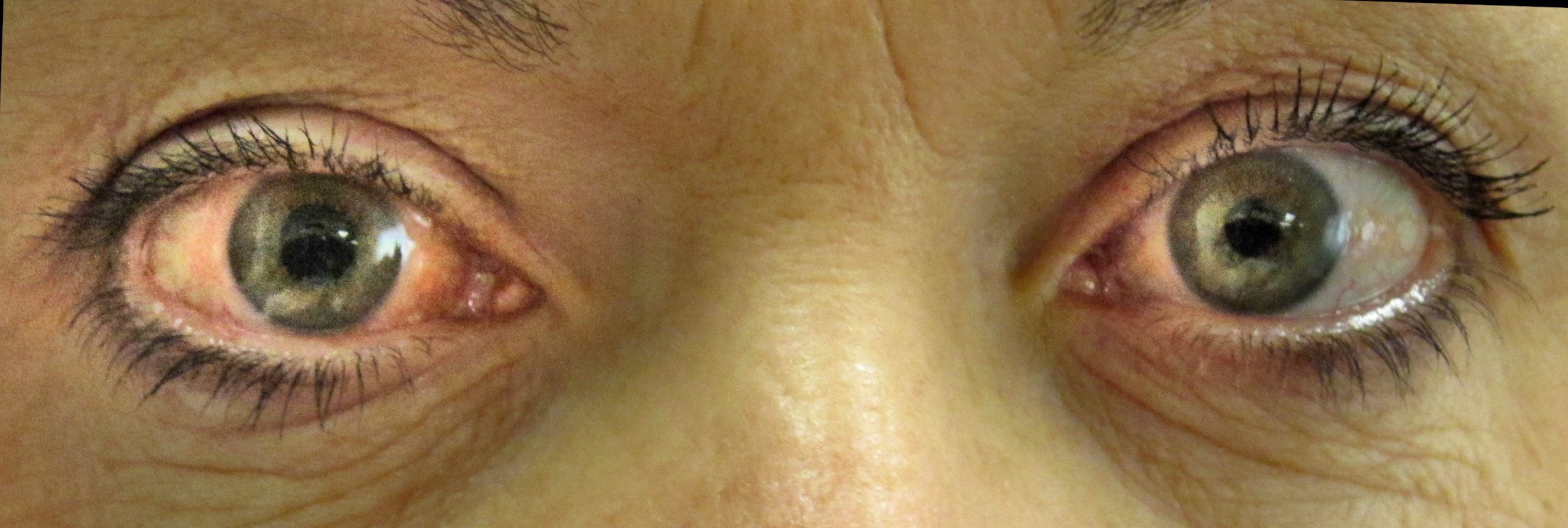
Glaucoma is a chronic, progressive optic neuropathy characterized by the degeneration of retinal ganglion cells and their axons, leading to a characteristic optic nerve appearance (cupping) and corresponding visual field defects, often associated with, but not defined by, elevated intraocular pressure (IOP) [8]D5[49]D5.
Also Called / Synonyms
- The Glaucomas (plural, reflecting the heterogeneous nature of the disease)
- Ocular (OHT) - a risk factor for, but not synonymous with, glaucoma
- (POAG)
- Primary angle-closure glaucoma (PACG)
- Congenital glaucoma / infantile glaucoma
- Neovascular glaucoma (NVG)
- Normal-tension glaucoma (NTG) - a subtype of POAG with IOP in the statistically normal range
Key Terminology
Every term used throughout this article is defined here to ensure consistent understanding:
- Intraocular pressure (IOP): The fluid pressure within the eye, the only modifiable risk factor proven to slow glaucoma progression [8]D5. The normal range is 10-21 mmHg, but glaucoma can occur at any IOP.
- Optic neuropathy: Damage to the optic nerve, visible as progressive thinning of the neuroretinal rim and enlargement of the optic cup (cupping) [49]D5.
- Visual field defect: A pattern of vision loss corresponding to retinal ganglion cell damage; a hallmark of glaucomatous functional loss [43]C4.
- Open-angle: The anterior chamber angle (where aqueous humor drains via the trabecular meshwork) appears open on gonioscopy, but outflow resistance is pathologically high.
- Angle-closure: Physical obstruction of the trabecular meshwork by the peripheral iris, blocking aqueous outflow and causing IOP elevation.
- Pseudoexfoliation (PXF) syndrome: A systemic condition that deposits fibrillar material on ocular structures, often obstructing the trabecular meshwork and causing a secondary open-angle glaucoma.
- Neovascular glaucoma (NVG): A severe secondary glaucoma caused by new blood vessel growth on the iris and angle (rubeosis iridis), usually secondary to retinal ischemia (e.g., central , proliferative ) [33]D5.
Classification of Glaucoma
The disease is subclassified into primary forms (no identifiable cause) and secondary forms (caused by another ocular or systemic condition). The European Glaucoma Society (EGS) 6th Edition guidelines recommend this structured approach [46]A1c.
| Type | Key Distinguishing Feature | Primary or Secondary | Associated Factors/Subtypes |
|---|---|---|---|
| Primary Open-Angle Glaucoma (POAG) | Open angle on gonioscopy, IOP >21 mmHg (most cases) | Primary | MYOC, OPTN, LOXL1 gene variants; myopia [28]C4[29]C4; African ancestry [16]B2a |
| Normal-Tension Glaucoma (NTG) | POAG with IOP <22 mmHg | Subtype of POAG | Nocturnal hypotension, sleep apnea, disc hemorrhages |
| Primary Angle-Closure Glaucoma (PACG) | Closed angle on gonioscopy; IOP elevated | Primary | Shallow anterior chamber, hyperopia, crowded angle [31]D5; Asian ethnicity [14]B2a |
| Congenital / Infantile Glaucoma | Onset at birth to age 3 years; enlarged eye (buphthalmos) | Primary (genetic) | CYP1B1, LTBP2 mutations; isolated or syndromic |
| Pseudoexfoliation Glaucoma | Fibrillar deposits on lens capsule and trabecular meshwork | Secondary | LOXL1 risk variant; older age |
| Neovascular Glaucoma (NVG) | Iris and angle neovascularization | Secondary | Retinal ischemia (CRVO, PDR, OIS) [33]D5 |
| Uveitic Glaucoma | Inflammation of the anterior uvea | Secondary | Juvenile idiopathic arthritis, sarcoidosis, [47]D5 |
| Steroid-Induced Glaucoma | IOP elevation after corticosteroid exposure | Secondary | Topical, periocular, systemic, or inhaled steroids [35]A1a |
| Post-Surgical Glaucoma | Develops after intraocular surgery | Secondary | Infantile cataract surgery (especially in aphakia) [9]A1a[65]D5; pars plana vitrectomy [5]A1a[54]C4 |
Clinical Significance
Glaucoma is the leading cause of irreversible blindness worldwide, affecting an estimated 76 million people in 2020, projected to rise to 112 million by 2040 [14]B2a[16]B2a. Among those, POAG accounts for approximately 74% of cases globally, with PACG causing a disproportionate share of bilateral blindness, especially in Asia [14]B2a. The disease remains asymptomatic until moderate-to-severe stages, making early detection and lifelong essential [60]D5.
Pearl: Glaucoma is a group of defined by structural optic nerve damage and functional visual field loss, not by IOP alone; classifications based on angle anatomy (open vs. closed) and etiology (primary vs. secondary) guide all subsequent management [8]D5[46]A1c.
Pathophysiology & Mechanism
- ▸Aqueous outflow resistance in POAG arises from TM ECM remodelling and actin contraction via the RhoA/ROCK pathway, not simple 'clogging' [78].
- ▸Mechanical strain at the lamina cribrosa disrupts axonal transport and perfusion, precipitating mitochondrial dysfunction, excitotoxicity, and ferroptosis in RGCs [94, 125].
- ▸Neuroinflammation (microglial activation, NLRP3 inflammasome, autoantibodies) converts acute injury to chronic progression and is detectable in aqueous humor [69, 92, 138].
Elevated intraocular pressure (IOP) transmits mechanical stress across the lamina cribrosa, compressing and shearing retinal ganglion cell (RGC) axons, but this single insult triggers a cascade of secondary injury that amplifies neurodegeneration even after IOP is lowered [89]D5[94]D5. The pathogenic chain involves three integrated domains: outflow obstruction at the anterior segment, mitochondrial failure and oxidative stress in RGCs, and a sustained neuroinflammatory response that converts acute injury to chronic progression [92]D5[119]D5.
Anterior Segment: Outflow Obstruction and IOP Elevation
The dominant mechanism in (POAG) is increased resistance to aqueous humor drainage through the trabecular meshwork (TM) and Schlemm's canal [78]D5[91]D5. The TM is a complex, multi-layered filter whose endothelial cells generate contractile tone and deposit extracellular matrix (ECM). In POAG, TM cells become dysfunctional: they accumulate cross-linked actin networks, secrete excess fibronectin and collagen IV, and lose their phagocytic capacity for debris [78]D5[123]B2b. This ECM remodelling narrows the intertrabecular spaces and stiffens the meshwork, raising outflow resistance. The Rho kinase (ROCK) pathway is central to this process, active RhoA/ROCK signalling drives actin polymerization and cell contraction, narrowing the paracellular channels; ROCK inhibitors like netarsudil relax the TM and increase outflow facility [78]D5[114]D5.
Aqueous angiography in living eyes confirms that POAG eyes show fewer and less perfused collector channels compared to normal eyes, with the circumferential extent of outflow reduced by roughly 40% [145]B3b. In primary angle-closure glaucoma (PACG), the obstruction is structural rather than cellular: the iris bows forward (relative pupillary block), the ciliary body rotates anteriorly, or the iris itself is thick and poorly compliant, physically apposing the TM [31]D5[115]D5. Genome-wide association studies implicate genes involved in anterior segment development (EPDR1, CHAT, GLIS3, FERMT2) and iris volume regulation [72]C4[31]D5. In secondary glaucomas, inflammatory cells, pigment granules, or pseudoexfoliative material clog the TM directly [103]D5[117]D5[118]D5.
A critical recent insight is that steroid-induced glaucoma models the canonical POAG pathway: upregulates ANGPTL7 in TM cells, a secreted glycoprotein that cross-links ECM and sharply reduces outflow facility; loss-of-function ANGPTL7 variants protect against IOP elevation and glaucoma in large biobank studies [123]B2b.
Optic Nerve : Biomechanical Strain and Axonal Transport Failure
The lamina cribrosa is a fenestrated collagenous sieve through which RGC axons exit the eye. IOP-driven deformation of this structure creates a pressure gradient across the lamina (the difference between IOP and intracranial pressure), generating tensile and shear strain on axons [89]D5[94]D5. This strain physically disrupts fast axonal transport, the microtubule-dependent movement of mitochondria, neurotrophins, and organelles from the RGC soma to the synaptic terminal and back, within hours of IOP elevation [94]D5. Obstruction of retrograde transport deprives the RGC soma of brain-derived neurotrophic factor (BDNF) from the superior colliculus, initiating pro-apoptotic signalling [89]D5. The same mechanical insult also compresses the microvasculature of the optic nerve head, reducing ocular perfusion pressure (the difference between mean arterial pressure and IOP) and creating zones of chronic hypoxia [108]D5. Calcium channel blocker use, which can lower systemic blood pressure and further reduce perfusion pressure, is associated with increased glaucoma risk in the UK Biobank (OR 1.39, 95% CI 1.20-1.61) [83]B2c.
Mitochondrial Dysfunction, Excitotoxicity, and Ferroptosis
RGCs have among the highest energy demands in the body, relying on oxidative phosphorylation to maintain their unmyelinated axons. In glaucomatous eyes, mitochondrial fission exceeds fusion, producing fragmented, depolarized organelles with impaired ATP synthesis [94]D5[126]D5. Damaged mitochondria leak reactive oxygen species (ROS) that overwhelm intrinsic antioxidant defences (glutathione, superoxide dismutase), causing lipid peroxidation, protein carbonylation, and DNA damage [93]D5[125]B2b. The resultant oxidative stress is compounded by glutamate excitotoxicity: elevated IOP reduces the expression of the glutamate-aspartate transporter (GLAST) on Müller cells, prolonging synaptic glutamate exposure and over-activating NMDA receptors on RGCs, which floods the cytoplasm with Ca²⁺ and amplifies ROS generation [125]B2b[90]D5. In the GLAST-knockout mouse model of normal-tension glaucoma, RGC loss is driven by ferroptosis, an iron-dependent, non-apoptotic cell death characterized by overwhelming lipid peroxidation, and inhibiting ferroptosis with liproxstatin-1 rescues RGCs [125]B2b.
Neuroinflammation: Glial Activation and Adaptive Immunity
Mechanical injury and oxidative stress activate all three retinal glial cell types [90]D5[119]D5[127]D5. Microglia transform from ramified surveillant cells into amoeboid phagocytes, releasing IL-1β, TNF-α, and CCL2; aqueous humor levels of CCL2 are significantly elevated in primary glaucoma (SMD 0.48, 95% CI 0.25-0.72) and correlate with disease severity [69]B3a[92]D5. Müller cells undergo reactive gliosis, upregulating GFAP and depositing intermediate filaments that stiffen the retina [90]D5. Astrocytes at the optic nerve head become reactive, secreting chondroitin sulfate proteoglycans that inhibit axonal regeneration [119]D5.
This innate response can transition to adaptive autoimmunity. Glaucoma patients harbour serum autoantibodies against heat shock proteins (HSP27, HSP60), retinal S-antigen, and γ-enolase [127]D5[144]D5. A subset of these antibodies are neurotoxic: immunoglobulins from POAG sera induce RGC apoptosis when injected into animal eyes [144]D5. The NLRP3 inflammasome is activated in the TM and retina, cleaving pro-IL-1β into its active form; caspase-1 and IL-18 levels are elevated in the aqueous humor of glaucoma patients [138]D5.
Convergent Pathways: The Final Common Injury
Despite diverse triggers (high vs normal IOP, open vs closed angle, genetic vs acquired), the terminal injury is synaptic loss and retrograde degeneration of RGCs [89]D5[94]D5. The earliest detectable structural change in human POAG is thinning of the retinal nerve fibre layer (RNFL), reflecting loss of unmyelinated RGC axons, followed by progressive shrinkage and disappearance of RGC somata in the ganglion cell layer [91]D5[104]B2b. The pattern of damage mirrors the architecture of the lamina cribrosa: the inferotemporal and superotemporal quadrants of the optic disc are most vulnerable, consistent with the arcuate visual field defects seen clinically [112]D5. Once an RGC dies, it cannot be replaced; neurodegeneration spreads trans-synaptically to the lateral geniculate nucleus and primary visual cortex [61]D5[89]D5.
Genetic and Molecular Susceptibility
Mendelian forms account for ~5% of POAG. MYOC mutations (encoding myocilin) produce a misfolded protein that accumulates in the TM, directly toxic to trabecular cells, causing juvenile-onset glaucoma with IOP > 30 mmHg [137]D5. OPTN (optineurin) and TBK1 mutations cause normal-tension glaucoma by impairing autophagy and mitophagy, leaving damaged mitochondria to accumulate in RGCs [94]D5[137]D5. Common low-penetrance variants at LOXL1 confer strong risk for pseudoexfoliation glaucoma (OR > 20 for some haplotypes) by altering cross-linking of elastic fibres in the TM and lens capsule [103]D5. Metabolomic studies identify disrupted lipid metabolism, elevated HDL3 cholesterol, altered sphingomyelins, and reduced glutamine, in POAG serum, suggesting systemic metabolic contributions to RGC vulnerability [80]B2c[71]B2a.
Pearl: Glaucoma is a mechanistically unified neurodegeneration driven by IOP-induced mechanical strain, mitochondrial failure, and neuroinflammation; every clinical feature, from RNFL thinning to arcuate scotomas to poor response to pressure-lowering alone, tracks back to one or more steps in this cascade [89]D5[92]D5[94]D5.
Epidemiology, Etiology & Risk Factors
- ▸Glaucoma affects 76 million people globally (2020), projected to exceed 111 million by 2040
- ▸Race/ethnicity and myopia are the strongest demographic risk factors, with Black ancestry conferring a 3.5-fold higher POAG risk
- ▸Modifiable risk factors include IOP, corticosteroid use, low ocular perfusion pressure, and possibly diabetes; statins and metformin appear protective
Glaucoma is the leading cause of irreversible blindness worldwide, affecting approximately 76.0 million people globally in 2020, with projections rising to 95.4 million by 2030 and 111.8 million by 2040 [153]A1a. These figures are not static; the global burden is shifting. The age-standardized prevalence of (POAG) in adults aged 40-80 years is 3.05% (95% CI, 2.33%-3.88%), while primary angle-closure glaucoma (PACG) affects 0.50% (95% CI, 0.37%-0.66%) [153]A1a. However, PACG accounts for nearly half of all glaucoma-related blindness, a disproportionate burden driven by its often asymptomatic, insidious progression until advanced stages [167]A1a. In 2020, glaucoma caused blindness in 3.61 million people and moderate-to-severe visual impairment in another 4.14 million, constituting 8.8% of all global blindness [170]A1a. The regional distribution is uneven: Asia carries the heaviest absolute burden, hosting 47% of all glaucoma cases, while Africa has the highest age-standardized prevalence of POAG at 4.79% (95% CI, 3.30%-6.47%) [194]B2a[153]A1a.
Demographic Distribution
Age is the strongest non-modifiable risk factor. Glaucoma prevalence rises steeply with each decade after age 40. Among Europeans aged ≥40 years, the pooled prevalence of any glaucoma is 2.93%, climbing to over 10% in those aged ≥80 [154]A1a. Sex differences are subtype-dependent. POAG is slightly more common in men (OR 1.04-1.37), whereas PACG is 2-4 times more common in women, a disparity attributed to shallower anterior chambers and shorter axial lengths [20]A1a[153]A1a. Women account for 70% of PACG cases and 59% of all glaucoma [194]B2a. In the United States, glaucoma affects approximately 3.2 million adults (prevalence 2.6%), with striking racial disparities: non-Hispanic Black individuals have a POAG prevalence 3.5 times higher than non-Hispanic White individuals (age-standardized prevalence 3.93% vs. 1.68%), and Hispanic individuals are also at elevated risk [156]A1a. Among Chinese populations, POAG prevalence is 2.6% and PACG prevalence 1.0%, with normal-tension glaucoma accounting for a majority of POAG cases (pooled proportion 62.5% in Asian studies) [160]A1a[199]D5.
Temporal Trends
Glaucoma prevalence is increasing globally, driven by population aging and, importantly, the rising epidemic of myopia. The number of people with glaucoma is projected to rise 35% by 2040 from 2013 estimates [153]A1a. Regionally, the contribution of myopia as a risk multiplier is substantial. A 2025 projection model incorporating myopia trends estimates that the global OAG burden could be 58% higher in 2060 than predicted by demographic aging alone (from 85.3 million to 134.9 million) [158]A1a. Myopia prevalence itself is accelerating, particularly in East and Southeast Asia, where childhood myopia rates now exceed 80% in some urban centers [135]D5.
Risk Factors - Modifiable and Non-Modifiable
Intraocular pressure (IOP) is the only proven modifiable risk factor; its elevation confers a dose-dependent risk, though approximately 30-50% of POAG patients have IOP in the normal range at diagnosis [94]D5[183]D5. The Ocular Treatment Study (OHTS) demonstrated that a 20% reduction in IOP reduces the 5-year risk of developing POAG from 9.5% to 4.4% (relative risk reduction 54%) [152]B2b.
Myopia is an increasingly important and independent risk factor for POAG, with a dose-response relationship: a one-diopter increase in myopia (from 0 to -6 D) raises OAG risk by about 20% (OR 1.20 per -1 D, 95% CI 1.13-1.27) [157]A1a. High myopia (SE < -6 D) carries an OR of 3.0-4.5 for OAG [173]D5[178]D5.
Family history strongly predicts glaucoma. First-degree relatives of POAG patients have a 2.1- to 3.7-fold increased risk, and heritability is estimated at 30-50% via common variants captured by polygenic risk scores [175]B2b. A polygenic risk score in the top quintile confers a 2.4-fold higher odds of developing POAG compared with the lowest quintile [175]B2b. For normal-tension glaucoma, heritability also exceeds 30% [200]B3b.
Race/Ethnicity: Black African and Caribbean ancestry as above; Asian populations (Chinese, Indian, Japanese) have the highest risk of PACG, driven by hyperopia, shallower anterior chambers, and shorter axial lengths [199]D5.
Corticosteroid use is a direct iatrogenic risk factor. The risk of developing steroid-induced glaucoma depends on drug potency, dose, and duration of use; topical steroids are the most common culprits, with a reported cumulative incidence of 18-36% in chronic users [196]D5. Intravitreal corticosteroid implants (e.g., ) cause ocular hypertension in 26.9-32.6% of eyes [209]C4[205]B2b.
Cardiovascular/metabolic factors: Diabetes shows a modest but significant association with POAG (OR 1.36, 95% CI 1.15-1.60) [155]A1a. Higher HDL-cholesterol levels paradoxically increase glaucoma risk (HR 1.05 per 1-SD increase), while total and LDL cholesterol do not [164]B2b. Low diastolic ocular perfusion pressure (<50 mmHg) is a strong risk factor in both POAG and normal-tension glaucoma [108]D5.
Sleep apnea: Mendelian randomization studies show a likely causal relationship between genetic predisposition to sleep apnea and increased glaucoma susceptibility (OR 1.49, 95% CI 1.10-2.01) [165]B2b.
Protective factors: Statin use is associated with a 12-20% reduced risk of POAG (OR 0.86, 95% CI 0.78-0.95) [201]B3b. use similarly lowers risk compared to other antidiabetic agents (OR 0.72, 95% CI 0.60-0.86) [159]B2a. GLP-1 receptor agonists show a trend toward reduced glaucoma incidence (HR 0.87, 95% CI 0.76-1.00), though not yet definitive [11]A1a. Higher blood folate levels are associated with 30-40% lower odds of glaucoma in Korean adults [204]B2b.
Lifestyle: Smoking is associated with higher IOP but not consistently with OAG risk after adjusting for corneal biomechanics [202]B2b. Alcohol consumption shows a complex U-shaped association with IOP but minimal increased glaucoma risk [4]A1a.
| Risk Factor | Relative Risk / Odds Ratio | Evidence Level |
|---|---|---|
| Age (per decade after 40) | OR 2.0-3.0 per decade | 1a [153]A1a[154]A1a |
| Family history (first-degree relative) | OR 2.1-3.7 | 1b [175]B2b |
| High myopia (< -6 D) | OR 3.0-4.5 | 1a [157]A1a |
| Black ancestry (vs. White) | OR 3.5 | 1a [156]A1a |
| Asian ancestry (PACG) | OR 2.0-3.0 | 1a [199]D5 |
| Type 2 diabetes | OR 1.36 | 1a [155]A1a |
| Sleep apnea | OR 1.49 (causal MR) | 2b [165]B2b |
| Steroid use (topical >3 months) | OR 2.5-5.0 | 2a [196]D5 |
| Low ocular perfusion pressure (<50 mmHg) | OR 2.0-3.0 | 2a [108]D5 |
| Cigarette smoking | RR 0.96 (not significant) | 1a [202]B2b |
| Statin use (protective) | OR 0.86 | 2b [201]B3b |
| Metformin use (protective) | OR 0.72 | 2a [159]B2a |
| High folate (protective) | OR 0.65 | 2c [204]B2b |
Seasonal variation is not a well-established factor for chronic glaucoma subtypes, though acute angle closure attacks are slightly more common in winter months, possibly due to decreased ambient light leading to mydriasis and angle crowding in predisposed eyes [171]C4.
Pearl: Glaucoma is a highly prevalent, age-related disease with strong ethnic and heritable underpinnings; myopia is the fastest-growing modifiable risk factor, and primary prevention strategies (e.g., increasing childhood outdoor time, atropine for myopia control) may mitigate future glaucoma burden [185]A1a[215]D5.
Clinical Presentation
- ▸Chronic POAG is asymptomatic until substantial (≈40%) RGC loss has occurred; the classic presentation is a patient who bumps into objects or notices a blind spot only after moderate-to-severe perimetric damage [276].
- ▸Acute angle-closure glaucoma presents dramatically with severe ocular pain, headache, nausea, vomiting, halos, and a fixed mid-dilated pupil, a true emergency [170].
- ▸The hallmark examination findings in POAG are neuroretinal rim thinning (notching), optic disc hemorrhage, an enlarged cup-to-disc ratio (>0.6 or asymmetric >0.2), and parapapillary RNFL defects; gonioscopy is essential to classify the angle [240, 277].
The transition from subclinical optic nerve damage to symptomatic vision loss in glaucoma is insidious, often unrecognized by the patient until substantial retinal ganglion cell (RGC) death has occurred [276]D5. Presenting symptoms depend on the disease stage, subtype, and whether the process is acute or chronic.
Presenting Symptoms
Chronic open-angle glaucoma (POAG) is typically asymptomatic until moderate to severe visual field loss develops [276]D5. Patients may notice difficulty with night vision, a sensation of missing steps, or bumping into objects, symptoms often attributed to aging. By the time a patient reports a "blind spot," the mean deviation on perimetry is often worse than -6 dB, corresponding to approximately 40% RGC loss at the optic nerve [230]D5. The classic presentation of a unilateral, slowly enlarging scotoma is uncommon; most patients present after both eyes are affected because the visual system suppresses the monocular defect [276]D5. Acute angle-closure glaucoma (AACG) is strikingly different: the patient presents with sudden, severe unilateral ocular or periocular pain, blurred vision, colored halos around lights (due to corneal edema), headache, nausea, and vomiting. This symptom complex is a true ophthalmic emergency [170]A1a. In contrast, primary congenital glaucoma (PCG) in infants manifests with photophobia, blepharospasm, and excessive tearing (epiphora), often noticed by the parent in the first year of life [260]D5. The classic triad, epiphora, photophobia, and blepharospasm, should prompt immediate referral [260]D5.
Neurological Examination Findings
The examination begins with visual acuity assessment, which remains normal until the disease is advanced or the fovea is involved [230]D5. In POAG, central acuity is preserved until the terminal stages. The critical examination is of the optic nerve head (ONH) and retinal nerve fiber layer (RNFL) via stereoscopic slit-lamp biomicroscopy with a 78- or 90-diopter lens. The hallmark is progressive cupping: a vertical cup-to-disc ratio (CDR) >0.6, focal or generalized neuroretinal rim thinning (notching), asymmetry of CDR >0.2 between eyes, and optic disc hemorrhages (small splinter hemorrhages at the rim) [240]D5[277]C4. The parapapillary region is divided into zones: zone beta (complete retinal pigment epithelium [RPE] loss with intact Bruch's membrane) and zone gamma (absence of Bruch's membrane) are associated with glaucoma and myopia, respectively [240]D5. The RNFL defect is best visualized with red-free light as a dark wedge-shaped slit emanating from the disc margin. Automated perimetry (Humphrey 24-2 SITA-Standard) quantifies the functional deficit: early defects are nasal step, arcuate scotoma, or paracentral scotoma; advanced disease spares only the temporal island (the papillo-macular bundle) [2]A1b.
Pupillary examination may reveal a relative afferent pupillary defect (RAPD) in unilateral or asymmetric glaucoma, quantifiable with neutral density filters. Intraocular pressure (IOP) measurement by Goldmann applanation tonometry is central: normal range is 10-21 mmHg, but glaucoma can occur at any IOP [241]D5. A diurnal curve with multiple measurements may be needed as IOP fluctuates. Gonioscopy is mandatory to differentiate open-angle from angle-closure mechanisms: the angle is graded using the Shaffer or Spaeth systems.
Phenotypic Variants
| Variant | Key Features | Frequency |
|---|---|---|
| Open angle, progressive cupping + VF loss, IOP >21 mmHg | Most common (~70% of all glaucoma [170]A1a) | |
| Normal-Tension Glaucoma | Same as POAG but IOP consistently ≤21 mmHg; associated with disc hemorrhages, myopia, migraine, and family history of OPTN or TBK1 mutations [241]D5 | ~30% of POAG cases in some populations |
| Primary Angle-Closure Glaucoma | Anatomically narrow angle + synechiae + elevated IOP ± acute attack | Higher prevalence in East Asians and hyperopes [170]A1a |
| Primary Congenital Glaucoma | Present at birth or infancy; enlarged cornea (buphthalmos), Haab striae, photophobia, optic disc cupping [260]D5 | ~1:10,000-1:20,000 live births; more common in males |
| Secondary Glaucomas | Steroid-induced (high IOP after glucocorticoid use [196]D5), neovascular (NVG, with rubeosis iridis [219]B3b), traumatic, pigmentary (PDS), uveitic, lens-related | Variable; steroid-induced accounts for up to 4.7% of pediatric glaucoma cases in tertiary centres [257]C4 |
Red Flags
Several findings mandate urgent evaluation and treatment [276]D5:
- Symptom of acute ocular pain, headache, nausea/vomiting - suspect AACG.
- Examination of IOP > 40 mmHg, corneal edema, fixed mid-dilated pupil, shallow anterior chamber - confirms AACG.
- Rubeosis iridis (new vessels at the pupillary margin or angle) - indicates neovascular glaucoma and urgent need for pan-retinal photocoagulation and anti-VEGF therapy [219]B3b.
- Markedly asymmetric cupping (CDR >0.2 difference) or optic disc hemorrhage - indicates active disease requiring immediate IOP lowering.
- Relative afferent pupillary defect in a patient with previously symmetric disease - signals acute progression or development of a complication like central .
Atypical Presentations
Glaucoma may present in unexpected ways. Normal-tension glaucoma (NTG) is easily missed because IOP is normal; the diagnosis hinges on characteristic optic disc appearance (e.g., focal notching, disc hemorrhages) and corresponding VF defects [241]D5. Steroid-induced glaucoma can develop after any route of steroid administration (topical, periocular, inhaled, systemic) and presents weeks to months after exposure [196]D5[257]C4. Peripapillary schisis (splitting of the RNFL seen on OCT) can mimic an optic pit maculopathy and may be an early sign of structural damage in open-angle glaucoma [277]C4[128]C4. Microcystic macular edema in the inner nuclear layer, a sign of chronic optic neuropathy, has a pooled prevalence of 5.9% in glaucomatous eyes and is associated with more severe VF loss [232]A1a. Lastly, glaucoma-related glare is a common but underreported symptom, with reduced contrast sensitivity and worse visual acuity under bright light, even when the media is clear [275]C4.
Pearl: The insidious onset of POAG means that by the time a patient notices symptoms, 40% or more of retinal ganglion cells may already be lost, making examination of the optic nerve head and RNFL in all patients over age 40 the only reliable way to detect the disease early [230]D5[276]D5.
Diagnosis & Workup
- ▸Gold-standard diagnosis requires combining SD-OCT (cpRNFL and GCIPL) with standard automated perimetry (SAP), achieving >95% specificity when both are abnormal in corresponding regions [7, 17].
- ▸Gonioscopy is mandatory for all patients to classify angle configuration, as management differs fundamentally between open-angle and angle-closure glaucoma [31].
- ▸In myopic eyes, macular GCIPL thickness is preferred over cpRNFL for structural assessment because peripapillary atrophy and gamma zone artifact confound RNFL measurements [178, 312].
Diagnosis rests on a structured evaluation of the optic nerve , retinal nerve fiber layer, and visual function. The gold-standard diagnostic test is standard automated perimetry (SAP) combined with spectral-domain optical coherence tomography (SD-OCT) of the circumpapillary retinal nerve fiber layer (cpRNFL) and macular ganglion cell-inner plexiform layer (GCIPL). No single test suffices; the diagnosis requires concordant structural and functional evidence [17]A1a[290]A1a.
Gold-Standard Test
SAP using the Humphrey Field Analyzer (24-2 or 10-2 Swedish Interactive Thresholding Algorithm) is the functional gold standard. The Glaucoma Hemifield Test (GHT) and pattern standard deviation (PSD) provide global indices; a GHT outside normal limits plus a PSD with P < 0.05 has a sensitivity of ~75% and specificity of ~92% for early glaucoma [43]C4[295]D5. SD-OCT is the structural gold standard: cpRNFL thickness below the 1st percentile (age-matched normative database) yields a diagnostic odds ratio (DOR) of 29.5 versus 18.6 for GDx and 8.9 for HRT3 [17]A1a. Macular GCIPL thickness performs similarly (area under receiver operating characteristic curve [AUROC] 0.85-0.95) and may detect damage before cpRNFL thinning in some eyes [296]D5[310]D5.
Laboratory Studies
No serologic tests diagnose primary glaucoma. In suspected secondary glaucoma, order:
| Test | Finding | Indication | Sensitivity/Specificity |
|---|---|---|---|
| Gonioscopy | Angle closure, synechiae, neovascularization | All patients at diagnosis | Gold standard for angle assessment [31]D5 |
| Intraocular pressure (IOP) by Goldmann applanation tonometry (GAT) | >21 mmHg (but many with glaucoma have IOP ≤21) | All patients | IOP alone: sensitivity ~50%, specificity ~85% [91]D5[122]D5 |
| Central corneal thickness (CCT) by pachymetry | Thin CCT (<555 μm) → IOP underestimation | Correction factor for GAT | Adjusts IOP by ~1 mmHg per 25 μm deviation from 555 μm [122]D5 |
| Anterior segment OCT (AS-OCT) | Narrow angle, plateau iris, angle structures | Suspect angle closure, post-LASIK | Qualitative; not yet standardized thresholds [305]D5 |
Imaging
SD-OCT is the imaging test of choice. Key parameters:
- Circumpapillary RNFL thickness: The best single parameter for diagnosis (DOR 29.5) [17]A1a. Abnormal if below 5th percentile (yellow flag) or 1st percentile (red flag).
- Macular GCIPL thickness: Helps when cpRNFL is compromised by myopia or peripapillary atrophy [312]D5.
- Optic nerve head (ONH) parameters: Rim area, cup-to-disc ratio. Automated ONH analysis has lower DOR (~8.9) than RNFL but remains useful when OCT quality is poor [17]A1a.
Optical coherence tomography angiography (OCTA) provides vessel density (VD) in peripapillary and macular regions. Peripapillary VD reduction correlates with glaucoma severity and progression. In meta-analysis, peripapillary VD had a pooled sensitivity of 0.83 and specificity of 0.88 for differentiating glaucomatous from healthy eyes [291]A1a. OCTA detects microvascular dropout that precedes measurable RNFL thinning in some eyes [289]A1a[324]B2b.
Gonioscopy is mandatory to classify angle configuration. Use a four-mirror lens; identify Shaffer grade (0-4), Schwalbe's line, and presence of synechiae. AS-OCT supplements gonioscopy when the angle is difficult to visualize or for quantitative assessment [31]D5[305]D5.
Biopsy / Histology
Biopsy is not indicated for primary glaucoma. In secondary glaucoma (e.g., iridocorneal endothelial syndrome, pigment dispersion, or neovascular glaucoma), corneal endothelial biopsy or aqueous humor analysis for viral DNA (e.g., HSV, CMV) may be warranted [117]D5[318]D5. Histology of the trabecular meshwork in shows increased extracellular matrix deposition, but this is a research tool, not a clinical test [89]D5.
Diagnostic Algorithm
Step 1: History and Examination
- Assess risk factors: age >40, family history of glaucoma, high myopia, African ancestry, corticosteroid use, prior ocular trauma [196]D5[316]D5.
- Measure IOP by GAT; document CCT for correction.
- Perform gonioscopy to classify angle as open, narrow, or closed.
- Evaluate the optic nerve head: cup-to-disc ratio (CDR) >0.5 or asymmetry >0.2 between eyes is suspicious. Examine for focal rim thinning, disc hemorrhage, or an enlarged rim [325]C4.
Step 2: Structural Imaging (SD-OCT)
- Obtain cpRNFL and macular GCIPL thickness.
- Abnormal: cpRNFL or GCIPL below the 1st percentile. Borderline: between 1st and 5th percentile.
- In myopic eyes, interpret cpRNFL with caution because the temporal RNFL may be artifactually thin due to gamma zone peripapillary atrophy [178]D5[312]D5. Macular GCIPL is less affected by myopia [312]D5.
Step 3: Functional Testing (SAP)
- Perform Humphrey 24-2 full threshold or SITA-Standard.
- Abnormal: GHT outside normal limits plus PSD with P < 0.05, or a cluster of three or more points at P < 0.05 (Hodapp-Parrish-Anderson criteria) [43]C4.
- Repeat within 1-3 months to confirm persistence. Single-epoch false-positive rates >15% or fixation losses >20% reduce reliability [295]D5.
Step 4: Integration
- Definite glaucoma: Structural defect (RNFL or GCIPL below 1st percentile) and functional defect (SAP abnormality) in corresponding regions. The structure-function correlation improves diagnostic specificity to >95% [7]B2b[324]B2b.
- Preperimetric glaucoma: Structural defect with normal SAP. These eyes have a ~20% risk of developing a functional defect within 5 years [325]C4.
- Glaucoma suspect: Elevated IOP, suspicious optic disc, or family history without confirmatory OCT or SAP defect. : follow with annual OCT and SAP [325]C4.
Step 5: Consider Secondary Glaucoma
- Unilateral or asymmetric disease, unusual age (juvenile or elderly), or anterior segment findings (pigment dispersion, pseudoexfoliation, neovascularization) → rule out secondary causes with targeted history, gonioscopy, and serology (syphilis, herpes, HLA-B27) as indicated [117]D5[196]D5[304]D5.
First-Line Treatment at Diagnosis
The choice between medical, laser, and surgical therapy depends on disease severity and patient factors. For mild-to-moderate primary open-angle glaucoma, first-line therapy is IOP-lowering eye drops (prostaglandin analogues, e.g., latanoprost 0.005% once daily) or primary selective laser trabeculoplasty (SLT). The LiGHT trial demonstrated that primary SLT is clinically and cost-effective compared to drops over 6 years [221]A1b. For advanced glaucoma, the Treatment of Advanced Glaucoma Study (TAGS) found primary trabeculectomy and medical therapy yield similar quality-of-life outcomes at 5 years [2]A1b.
Pearl: The definitive diagnosis of glaucoma requires concordant structural damage (cpRNFL or GCIPL thinning) and functional loss (SAP abnormality) after excluding secondary causes; no single test suffices, and repeat testing over months resolves equivocal cases [7]B2b[17]A1a[290]A1a.
| Test | Parameter | Abnormal Threshold | Sensitivity | Specificity | DOR |
|---|---|---|---|---|---|
| SD-OCT cpRNFL | Average RNFL thickness | <1st percentile | 0.78-0.90 | 0.83-0.95 | 29.5 |
| SD-OCT macular GCIPL | Average GCIPL thickness | <1st percentile | 0.75-0.88 | 0.85-0.94 | 24.1 |
| Humphrey 24-2 SITA | GHT + PSD | GHT outside normal + PSD P<0.05 | 0.72-0.78 | 0.88-0.95 | 12.4 |
| Goldmann applanation tonometry | IOP | >21 mmHg (single measurement) | ~0.50 | ~0.85 | 5.1 |
| OCTA peripapillary vessel density | Peripapillary VD | Below 5th percentile | 0.83 | 0.88 | 35.0 |
Sensitivities and specificities from meta-analyses; values vary with disease severity and population [17]A1a[43]C4[290]A1a[291]A1a
Severity, Staging & Risk Stratification
- ▸The Hodapp-Parrish-Anderson classification stratifies VF loss into mild (> -6 dB), moderate (-6 to -12 dB), and severe (< -12 dB), directly setting target IOPs of 18-21, 15-18, and ≤ 14 mmHg respectively.
- ▸Independent risk factors for faster progression, disc hemorrhage (HR 2.1-3.2), pseudoexfoliation (2.4× faster VF decay), high myopia (OR 2.1), and high polygenic risk (OR 4.6), must be integrated into follow-up intervals.
Once the diagnosis of glaucoma is confirmed, the next critical step is to assign a severity tier that directly dictates treatment intensity, target intraocular pressure (IOP), and follow-up frequency. Staging in glaucoma relies on a structured synthesis of structural damage (optic nerve appearance, retinal nerve fiber layer [RNFL] thickness) and functional loss (visual field [VF] mean deviation [MD]) [230]D5. The most widely adopted systems are the Hodapp-Parrish-Anderson (HPA) classification for VF defects and the Glaucoma Staging System (GSS) based on MD, both of which stratify disease into mild, moderate, and severe stages [2]A1b[361]D5.
Hodapp-Parrish-Anderson Classification
The HPA system categorizes VF loss as:
- Mild (early): MD better than -6 dB, with fewer than 18 points depressed below the 5% level and fewer than 10 points below the 1% level, and no points within the central 5° with sensitivity < 15 dB.
- Moderate: MD between -6 dB and -12 dB, or VF defects meeting criteria beyond mild but not meeting severe criteria.
- Severe: MD worse than -12 dB, or any point within the central 5° with sensitivity < 0 dB on two consecutive tests, or more than 50% of points depressed below the 5% level [2]A1b[361]D5.
This classification was used in the Treatment of Advanced Glaucoma Study (TAGS), which defined advanced glaucoma as an MD worse than -12 dB in at least one eye; in that trial, primary trabeculectomy was non-inferior to medical therapy for quality of life and resulted in lower mean IOP (12.8 vs. 15.7 mmHg at 5 years) without a significant difference in VF progression [2]A1b.
Thresholds for Treatment Intensity
Staging drives clinical action. For mild disease, the typical target IOP is 18-21 mmHg, often achievable with monotherapy. For moderate disease, the target drops to 15-18 mmHg, requiring combination therapy or laser. For severe disease (MD worse than -12 dB or advanced structural damage), the target is ≤ 14 mmHg, frequently necessitating surgery. The LiGHT trial showed that initial selective laser trabeculoplasty (SLT) in mild-to-moderate OAG was non-inferior to eye drops for VF preservation at 6 years, with fewer patients needing surgery (OR 0.47, 95% CI 0.30-0.73) and no difference in quality of life; SLT was cost-effective and the IOP reduction was durable, at 6 years, 71.2% of SLT-first patients remained drop-free [221]A1b.
Structural Staging and Imaging
The Ocular Treatment Study (OHTS) demonstrated that baseline RNFL thickness predicted progression to POAG: each 1 μm thinner RNFL increased hazard by ~6% (HR 1.06, 95% CI 1.03-1.09) [152]B2b. OCT-derived parameters, particularly average RNFL thickness and ganglion cell-inner plexiform layer (GC-IPL) thickness, are used to stage damage. A patient with an RNFL thickness < 75 μm is considered structurally advanced; a thickness < 60 μm indicates severe risk for rapid progression [315]D5. In the HORIZON trial, the Hydrus microstent reduced VF progression rate by 30% compared with cataract surgery alone in eyes with baseline MD -6.4 dB, but the benefit was greatest in those with more severe baseline field loss (interaction p=0.02) [284]B2b.
Risk Stratification for Progression
Beyond static staging, dynamic risk stratification is essential. The following factors independently predict faster VF progression:
- Higher baseline IOP and IOP fluctuation (each 1 mmHg higher mean IOP increases progression hazard by ~8%; HR 1.08, 95% CI 1.03-1.13) [91]D5[190]B2a.
- Optic disc hemorrhage: presence of disc hemorrhage (DH) doubles the risk of progression (OR 2.1-2.7); laminar and marginal subtypes of DH carry a higher hazard than parapapillary DH (HR 3.2, 95% CI 1.8-5.7) [344]B2b.
- Pseudoexfoliation: eyes with pseudoexfoliative glaucoma (PXG) progress 2.4× faster than those with POAG (mean MD decline -0.87 vs. -0.36 dB/year) and have worse structural damage for a given IOP [190]B2a[322]C4.
- High myopia (axial length > 26.5 mm): in a 3-year prospective study, high-myopia glaucoma eyes had faster RNFL loss (-1.6 vs. -0.9 μm/year, p<0.001) and greater odds of progression (OR 2.1, 95% CI 1.2-3.6), attributed to biomechanical deformation of the optic nerve head [353]B2b.
- Genetic risk: a polygenic risk score in the top 2.5% of the UK Biobank conferred a 4.6-fold increased odds of POAG compared with the lowest quartile (OR 4.60, 95% CI 3.91-5.41), independent of IOP. In individuals of African descent, the same PRS had attenuated discrimination, de novo African-specific variants contribute [345]B2b[346]B2b. Myocilin mutations in juvenile-onset OAG produce high IOP (>40 mmHg) and require early surgery [316]D5.
Integrating Risk into Follow-Up
A patient with moderate glaucoma and a disc hemorrhage should be seen every 3-4 months with IOP checks and VF every 6 months, whereas a mild glaucoma patient with no risk factors may be followed every 6-12 months. The TAGS protocol for advanced disease mandated 6-monthly VF and annual OCT [2]A1b. The Eyes of Africa study (n = 9418) found that continental Africans presented with more severe disease (mean MD -12.2 dB) and lower baseline IOP (22 mmHg vs. 25 mmHg in European cohorts), emphasizing that severity staging must be interpreted within ethnic context to avoid undertreatment [364]B3b.
Table 1: Composite Staging and Recommended Targets
| Stage | VF MD (dB) | RNFL Thickness (μm) | Target IOP (mmHg) | Test Interval |
|---|---|---|---|---|
| Mild | > -6 | ≥ 80 | 18-21 | q6-12 mo |
| Moderate | -6 to -12 | 60-79 | 15-18 | q4-6 mo |
| Severe | < -12 | < 60 | ≤ 14 | q3-4 mo |
Derived from [2]A1b[91]D5[190]B2a[221]A1b[315]D5[353]B2b[361]D5.
Controversies and Guideline Disagreement
| Question | Position A (NICE/UK) | Position B (AAO/US) | Strength | Implication |
|---|---|---|---|---|
| Should initial SLT be preferred over drops for mild-moderate OAG? | Yes, LiGHT data support cost-effective first-line [221]A1b | Drops remain first-line; SLT reserved for poor adherence | Moderate | NICE guidelines favor SLT; AAO is more conservative |
| What defines "target IOP"? | Fixed category (18, 15, 14 mmHg) based on severity | Individualized, accounting for life expectancy and baseline damage | Weak | Most clinicians use hybrid approach |
| Is IOP fluctuation an independent risk factor? | Yes, supported by OHTS and LiGHT post-hoc [91]D5 | Conflicting; meta-analyses show inconsistent association [190]B2a | Weak | Accept that both mean and fluctuation matter |
Pearl: Staging glaucoma requires both functional (VF MD) and structural (OCT RNFL) assessment; a composite approach using the Hodapp-Parrish-Anderson classification plus OCT-derived RNFL thickness determines treatment targets and follow-up intervals, with modification for high-risk features such as disc hemorrhage, pseudoexfoliation, and high myopia [2]A1b[91]D5[190]B2a[221]A1b[344]B2b[353]B2b[361]D5.
Acute & Vision-Threatening Management
- ▸Immediate stepwise medical IOP reduction (beta-blocker, alpha-agonist, dorzolamide, IV acetazolamide) is the first-line response to acute angle closure crisis.
- ▸Laser peripheral iridotomy (LPI) is the definitive treatment for pupillary block; LPI reduces the 14-year risk of angle closure from 6.5% to 0.9% (NNT=18).
- ▸Neovascular glaucoma requires intravitreal anti-VEGF injection within 24 hours plus panretinal photocoagulation before any incisional surgery.
Step 1: Immediate Recognition and Medical Decompression
Acute angle closure (AAC) crisis is the only glaucoma emergency where minutes to hours determine whether the optic nerve sustains irreversible damage. Presenting with a fixed, mid-dilated pupil, corneal edema, IOP typically >40 mm Hg, and severe pain, the first action is immediate topical and systemic IOP reduction to break the attack and prevent syncchial closure. The European Glaucoma Society (EGS) 6th edition guidelines (2025) recommend the following sequential protocol [46]A1c (1c):
- Administer one drop of pilocarpine 1-2%, but ONLY after the IOP has been lowered below 40 mm Hg with other agents. Pilocarpine is ineffective and may worsen ischemia when the iris is edematous and sphincter is paralyzed at extreme IOPs.
- Topical beta-blocker, e.g., timolol 0.5% one drop immediately. Avoid in patients with bradycardia or reactive airway disease.
- Topical alpha-2 agonist, brimonidine 1-2 drops or apraclonidine 1-2 drops. Reduces aqueous production and may also cause vasoconstriction.
- Topical carbonic anhydrase inhibitor, dorzolamide 2% or brinzolamide 1% three times in the first hour.
- Systemic carbonic anhydrase inhibitor, acetazolamide 500 mg IV or 500-1000 mg PO as a single dose. IV route provides faster onset (15-30 min).
- Osmotic agents, mannitol 20% 1-2 g/kg IV over 30-60 min, OR glycerin 50% PO 1-1.5 g/kg. Contraindicated in patients with severe renal failure, congestive heart failure, or anuria.
Do NOT use oral glycerin in patients with diabetes (causes hyperglycemia). If the IOP does not fall below 30 mm Hg within one hour, the attack is considered refractory and requires urgent laser intervention [46]A1c (1c).
Step 2: Definitive Laser Treatment, Laser Peripheral Iridotomy
Once the cornea clears sufficiently for visualization, laser peripheral iridotomy (LPI) is the definitive laser treatment to relieve pupillary block. LPI is performed using an argon or Nd:YAG laser, creating a full-thickness hole in the peripheral iris to allow aqueous to bypass the pupillary block and flow into the anterior chamber, deepening the angle.
- Procedure: Apply 1-2 argon burns at 500-1000 mW for 0.1-0.2 sec to thin the iris, followed by 1-3 Nd:YAG shots at 1-4 mJ to penetrate. Alternatively, combined sequential argon-YAG or single Nd:YAG without pretreatment is used.
- Target: A visible gush of aqueous and pigment through the iridotomy confirms patency.
- Post-LPI: IOP typically falls within minutes to 1-2 hours. Check IOP 30-60 min post-treatment; a transient spike (from pigment release) may occur and can be managed with acetazolamide 250 mg PO if needed.
- Complications: Bleeding (usually self-limited), transient IOP spike, lens opacity, and rarely, retinal burn.
The Zhongshan Angle-Closure Prevention Study (14-year extended follow-up) demonstrated that prophylactic LPI in primary angle-closure suspects (PACS) reduces the incidence of primary angle closure from 6.5% to 0.9% over 14 years (absolute risk reduction 5.6%; NNT = 18 to prevent one case of PAC) [151]A1b (1b). For eyes already in acute crisis, LPI is the standard of care.
Step 3: When LPI Fails, Laser Iridoplasty or Surgical Intervention
If LPI does not immediately deepen the angle (e.g., plateau iris configuration, extensive peripheral anterior synechiae, or persistent IOP >25 mm Hg after LPI), proceed to laser peripheral iridoplasty (LPIp). LPIp uses argon laser burns (200-500 µm, 100-200 mW, 0.5 sec) to contract the peripheral iris stroma, pulling it away from the angle [374]A1a (1a). A Cochrane review found that LPIp is effective in lowering IOP in acute cases where LPI alone is insufficient, but the evidence is limited to small trials and case series [374]A1a (1a).
If IOP remains elevated (>30 mm Hg) despite medical therapy, LPI, and LPIp, urgent surgical intervention is indicated:
- Anterior chamber , a rapid 0.1-0.2 mL release of aqueous can lower IOP immediately, but carries risk of hyphenia, , and lens touch. Use only as a temporizing bridge to definitive surgery.
- Trabeculectomy, reserved for advanced PACG with persistent IOP elevation after laser. The Treatment of Advanced Glaucoma Study (TAGS) 5-year results showed that primary trabeculectomy (with mitomycin C) achieved lower mean IOP (13.2 vs 15.5 mm Hg) and fewer medications compared to medical therapy, but with a higher rate of complications (hyphenia 8%, bleb leak 6%, hypotony 3%) [2]A1b (1b).
- Phacoemulsification with goniosynechialysis (Phaco-GSL), increasingly preferred for PACG with cataract. A 2-year prospective cohort study of 213 patients reported that Phaco-GSL in acute PACG achieved a mean IOP reduction from 42.1 mm Hg to 14.8 mm Hg at 2 years, with a success rate of 87.5% (IOP ≤21 mm Hg without medications) [217]B2b (2b). A Chinese noninferiority RCT (Phaco-GSL+goniotomy vs Phacotrabeculectomy) found that the combined MIGS approach was noninferior for IOP control at 2 years (difference in mean IOP: 1.0 mm Hg, 90% CI -1.2 to 3.2) [281]A1b (1b).
Step 4: of Refractory and Neovascular Glaucoma
Neovascular glaucoma (NVG) is the most severe acute glaucoma emergency, often complicating ischemic central or proliferative . The first priority is prevention, panretinal photocoagulation (PRP) for eyes with retinal ischemia reduces the risk of NVG from ~50% to <10% [304]D5 (5). When NVG is already present with florid iris neovascularization and IOP >50 mm Hg:
- Intravitreal anti-VEGF injection, 1.25 mg or ranibizumab 0.5 mg is given urgently to induce rapid regression of iris vessels (within 24-72 hrs) [304]D5 (5). One injection lowers IOP by a mean of 15-20 mm Hg in the first week.
- PRP as soon as media clears, definitive therapy to reduce VEGF drive. Deliver 1200-1600 burns in two to three sessions.
- Topical glaucoma medications, beta-blocker, alpha-agonist, dorzolamide; avoid pilocarpine (incites inflammation).
- Glaucoma drainage device (e.g., Ahmed valve), indicated if IOP remains >30 mm Hg after anti-VEGF and PRP, or if angle is closed by fibrovascular membrane. A meta-analysis of 5 studies found that Ahmed valve surgery in NVG achieved IOP ≤21 mm Hg at 1 year in 75-80% of eyes, but with a 20-30% rate of tube exposure or hypotony [214]A1a (2a).
- Cyclophotocoagulation, transscleral diode laser cyclophotocoagulation (810 nm, 10-20 burns at 1500-2000 mW) is reserved for eyes with poor visual potential or where other surgeries have failed. A Cochrane review reported moderate efficacy (IOP reduction >30% in 50-70% of eyes) but high rates of inflammation, hypotony (3-5%), and vision loss [96]A1a (1a).
What NOT to do in NVG: Do not perform filtering surgery (trabeculectomy) alone without anti-VEGF and PRP, the fistula will close rapidly from active neovascularization (failure rate >80% at 6 months) [304]D5 (5).
Step 5: Acute Management in Special Populations
- Pregnancy: Acetazolamide is pregnancy category C (teratogenic in animal studies). Laser LPI is first line. If medical therapy is essential, use topical beta-blockers (timolol 0.25%) cautiously due to potential fetal bradycardia. Avoid mannitol and IV acetazolamide.
- Children: For congenital or juvenile glaucoma presenting acutely, immediate IOP lowering with topical therapy (timolol 0.25%-0.5%, dorzolamide) is first line; consider oral acetazolamide 20 mg/kg/day divided every 6-8 hours. Definitive management requires goniotomy or trabeculotomy.
- Drug-induced AAC: SSRI use (bupropion, topiramate, and other sulfonamide derivatives) can precipitate bilateral acute angle closure via ciliary body effusion and forward displacement of the lens-iris diaphragm [369]B3b (3b) [390]D5 (5). Immediate discontinuation of the offending drug plus standard AAC medical and laser therapy is essential. A nested case-control study (n=6,110,723) found that topiramate carried the highest risk (RR 12.0, 95% CI 8.5-16.0) [371]B3b (3b).
| Drug class | Example | Mechanism | Management |
|---|---|---|---|
| Sulfonamides | Topiramate, acetazolamide (rare) | Ciliary effusion → forward lens-iris | Stop drug; laser LPI +/- LPIp |
| Anticholinergics | Atropine, ipratropium (nebulized) | Pupillary block | Stop drug; pilocarpine, LPI |
| Sympathomimetics | Adrenaline, phenylephrine | Mydriasis → angle crowding | Stop drug; LPI |
| SSRIs/SNRIs | Paroxetine, venlafaxine | Serotonergic → intraocular effusion (rare) | Stop drug; standard AAC therapy [369]B3b (3b) |
Pearl: In acute angle closure, the sequence of medical therapy (topical beta-blocker + alpha-agonist + dorzolamide + IV acetazolamide) followed by LPI within 1-2 hours saves the optic nerve; for neovascular glaucoma, urgent anti-VEGF injection within 24 hours transforms a blinding emergency into a manageable chronic condition [2]A1b (1b) [304]D5 (5) [374]A1a (1a).
| Step | Agent | Dose | Route | Timing |
|---|---|---|---|---|
| 1 | Pilocarpine 1-2% | 1 drop | Topical | ONLY after IOP <40 mm Hg |
| 2 | Timolol 0.5% | 1 drop | Topical | Immediately |
| 3 | Brimonidine 0.2% | 1-2 drops | Topical | Immediately |
| 4 | Dorzolamide 2% | 1 drop q 5 min × 3 | Topical | First hour |
| 5 | Acetazolamide | 500 mg IV or 500-1000 mg PO | Systemic | Once |
| 6 | Mannitol 20% or glycerin 50% | 1-2 g/kg IV; 1-1.5 g/kg PO | IV/PO | If IOP remains >30 mm Hg after 1 hour |
Long-term & Definitive Management
- ▸First-line therapy is a prostaglandin analog or selective laser trabeculoplasty (SLT), both supported by Level 1 evidence for efficacy and safety.
- ▸Trabeculectomy with mitomycin C remains the gold standard for advanced glaucoma, but tube shunts are equivalent in primary surgery (PTVT study).
- ▸MIGS procedures (e.g., Hydrus microstent, phacogoniotomy) are effective for mild-to-moderate disease, especially combined with cataract surgery.
The therapeutic ladder for chronic glaucoma progresses from topical medications to laser trabeculoplasty to incisional surgery, with selective laser trabeculoplasty (SLT) now established as a first-line alternative to drops based on 6-year outcomes from the LiGHT trial [221]A1b. is lifelong, guided by target intraocular pressure (IOP) individualized to disease severity, rate of progression, and life expectancy.
Step 1: Setting Target Intraocular Pressure and Risk Stratification
Target IOP is derived from baseline untreated IOP, stage of visual field loss, and risk of progression. The European Glaucoma Society (EGS) recommends targets of ≤21 mmHg for early glaucoma, ≤18 mmHg for moderate, and ≤15 mmHg for advanced disease [422]D5. For patients with normal-tension glaucoma, a 30% reduction from baseline is a common initial goal [398]A1b. The Glaucoma Intensive Treatment Study (GITS) demonstrated that an intensive strategy targeting low IOP (mean 13.5 mmHg) slowed visual field progression compared with standard therapy (mean 16.5 mmHg) over 5 years (difference in VFI slope 0.3%/year, P=0.02) [398]A1b.
Step 2: First-Line Medical Therapy
Prostaglandin analogs (latanoprost 0.005%, travoprost 0.004%, bimatoprost 0.01%) are first-line because of superior IOP reduction (mean 25-33% from baseline) and once-daily dosing [397]A1a. A component network meta-analysis of 97 RCTs confirmed prostaglandins achieve the greatest IOP lowering among monotherapies, with a mean difference of -3.5 mmHg versus timolol [397]A1a. Latanoprost is the most prescribed; bimatoprost offers slightly greater efficacy but more conjunctival hyperemia. Dosing table:
| Drug | Starting dose | Target/max dose | Renal adjustment | Hepatic adjustment | Key monitoring |
|---|---|---|---|---|---|
| Latanoprost 0.005% | 1 drop QHS | 1 drop QHS | None | None | IOP, iris pigmentation, conjunctival hyperemia |
| Travoprost 0.004% | 1 drop QHS | 1 drop QHS | None | None | Same as latanoprost |
| Bimatoprost 0.01% | 1 drop QHS | 1 drop QHS | None | None | Same; higher hyperemia rate |
| Timolol 0.5% | 1 drop BID | 1 drop BID | None | Caution in severe hepatic disease | Heart rate, blood pressure, asthma |
| Brimonidine 0.2% | 1 drop TID | 1 drop TID | None | None | CNS depression in children, dry mouth |
| Dorzolamide 2% | 1 drop TID | 1 drop TID | None | None | Corneal edema, |
| Netarsudil 0.02% | 1 drop QHS | 1 drop QHS | None | None | Conjunctival hyperemia, corneal verticillata |
Step 3: Laser Trabeculoplasty as Primary or Adjunctive Therapy
SLT (360°, 50-100 spots, 0.4-1.2 mJ) is now a guideline-endorsed first-line option. The LiGHT trial randomized 718 treatment-naïve patients with open-angle glaucoma (OAG) or ocular (OHT) to primary SLT or drops. At 6 years, SLT was noninferior for IOP control and health-related quality of life, and 74% of SLT-treated eyes remained drop-free [221]A1b. SLT was also cost-effective, reducing lifetime treatment costs by £1,300 per patient. The GLAUrious trial (n=192) showed that direct, gonioscopy-free SLT (DSLT) was noninferior to conventional SLT at 12 months (mean IOP reduction 7.2 vs 7.5 mmHg) [394]A1b. Repeat SLT is effective regardless of initial response: in a post hoc analysis of LiGHT China, 68% of eyes that initially failed SLT still achieved ≥20% IOP reduction after repeat treatment [280]B2b.
Step 4: Combination Medical Therapy
When monotherapy fails to achieve target IOP, a second agent is added. Fixed combinations (e.g., latanoprost/timolol, dorzolamide/timolol, brimonidine/brinzolamide) improve adherence and reduce preservative exposure. The network meta-analysis found that prostaglandin/beta-blocker fixed combinations provide an additional -2.5 mmHg over prostaglandin alone [397]A1a. Rho-kinase inhibitors (netarsudil, ripasudil) offer a novel mechanism, lowering IOP by increasing trabecular outflow; netarsudil 0.02% QHS reduces IOP by 3-5 mmHg and may also have neuroprotective effects [436]B2b.
Step 5: Incisional Surgery
Indications for surgery include failure of medical and laser therapy, advanced disease at presentation, or intolerance to medications. Trabeculectomy with mitomycin C (MMC 0.2-0.4 mg/mL for 2-4 minutes) remains the gold standard. The Treatment of Advanced Glaucoma Study (TAGS) randomized 453 patients with newly diagnosed advanced OAG to primary trabeculectomy or medical therapy. At 5 years, trabeculectomy achieved lower mean IOP (12.3 vs 14.7 mmHg) and better visual field preservation (mean MD change -1.4 vs -2.5 dB), with no difference in quality of life [2]A1b. Tube shunts (Baerveldt 350 mm², Ahmed) are preferred when trabeculectomy is high-risk (e.g., failed prior surgery, uveitic glaucoma, neovascular glaucoma). The Primary Tube Versus Trabeculectomy (PTVT) Study (n=242) found similar 5-year failure rates (tube 38% vs trabeculectomy 46%, P=0.20) but more early hypotony with trabeculectomy and more late tube-related complications [222]A1b. Visual field outcomes were comparable between groups [150]A1b.
Minimally invasive glaucoma surgery (MIGS) is reserved for mild-to-moderate OAG, often combined with cataract surgery. The HORIZON trial (n=556) compared cataract surgery alone versus cataract surgery with Hydrus microstent. At 5 years, the microstent group had lower IOP (17.0 vs 18.2 mmHg), fewer medications (0.4 vs 0.9), and slower visual field progression (mean MD change -0.75 vs -1.18 dB, P=0.02) [223]A1b[284]B2b. For primary angle-closure glaucoma (PACG) with cataract, phacogoniotomy (phacoemulsification + goniosynechialysis + goniotomy) was noninferior to phacotrabeculectomy at 2 years (mean IOP 15.2 vs 14.8 mmHg, P=0.34) with fewer complications [281]A1b. In PACG without cataract, surgical peripheral iridectomy combined with goniosynechialysis and goniotomy was noninferior to trabeculectomy at 1 year (IOP 16.1 vs 15.5 mmHg) [282]A1b.
Step 6: Monitoring and Titration
Patients require lifelong monitoring: IOP at 3-6 month intervals, visual field testing annually (or semiannually for advanced disease), and OCT imaging of retinal nerve fiber layer and ganglion cell complex every 6-12 months. Adherence should be assessed at every visit; nonadherence is a leading cause of progression. Escalation is indicated when IOP exceeds target, visual field progression is confirmed (≥2 dB MD loss over 2 years), or structural loss accelerates.
Treatment Failure Protocol
- Medical failure: Add SLT if not performed; if already done, proceed to incisional surgery.
- Trabeculectomy failure: Bleb needling with ( ) can restore function in 50-70% of cases [377]A1a. If needling fails, repeat trabeculectomy with MMC or convert to tube shunt.
- Tube shunt failure: Consider cyclophotocoagulation (CPC) or revision. CPC reduces IOP by 30-40% but carries risk of hypotony and vision loss [96]A1a.
What NOT to Do
- Do not use beta-blockers (timolol) in patients with asthma, , or bradycardia; they can cause fatal bronchospasm.
- Do not use brimonidine in children under 2 years; it causes CNS depression, apnea, and hypotension.
- Do not perform laser iridotomy alone for plateau iris syndrome without iridoplasty; it will not open the angle [374]A1a.
- Do not use oral acetazolamide long-term without monitoring for metabolic acidosis, hypokalemia, and aplastic anemia.
Controversies and Guideline Disagreement
| Question | Position A | Position B | Strength of disagreement | Implication for practice |
|---|---|---|---|---|
| SLT vs drops as first-line | LiGHT trial and EGS 2021 support SLT as first-line for OAG/OHT [221]A1b[422]D5 | AAO Preferred Practice Pattern still recommends drops as initial therapy for most patients | Moderate (EGS endorses SLT as primary; AAO leaves sequence open) | Clinicians may choose either; SLT reduces medication burden and improves adherence |
| Trabeculectomy vs tube shunt for primary surgery | PTVT study shows similar efficacy and safety at 5 years [222]A1b | TVT study (prior surgery) favored tube shunt for higher success [228]A1b | Mild (different populations; PTVT in virgin eyes, TVT in failed trabeculectomy) | Tube shunt is reasonable first choice in eyes with prior conjunctival scarring |
| MIGS vs traditional surgery | Cochrane overview finds MIGS safer but less IOP reduction than trabeculectomy [400]A1a | HORIZON shows MIGS slows VF progression [284]B2b | Moderate (MIGS appropriate for mild-moderate disease; traditional for advanced) | Select MIGS for patients needing modest IOP reduction and faster recovery |
Pearl: Initiate treatment with a prostaglandin analog or SLT as first-line; escalate to combination therapy or incisional surgery if target IOP not achieved, with trabeculectomy and tube shunts offering comparable long-term IOP control but different risk profiles [2]A1b[222]A1b.
| Option | Indication / Line | Dose or Specifics | Key Trial | Outcome | Evidence Level |
|---|---|---|---|---|---|
| SLT | First-line or adjunctive | 360°, 50-100 spots, 0.4-1.2 mJ | LiGHT (n=718) | 74% drop-free at 6 years; noninferior to drops [221]A1b | 1b |
| Trabeculectomy + MMC | Advanced disease, failed medical/laser | MMC 0.2-0.4 mg/mL × 2-4 min | TAGS (n=453) | Mean IOP 12.3 vs 14.7 mmHg; better VF preservation [2]A1b | 1b |
| Tube shunt (Baerveldt 350 mm²) | Failed trabeculectomy, high-risk eyes | 350 mm² plate | PTVT (n=242) | 5-year failure 38% vs 46% (trabeculectomy); similar VF [222]A1b | 1b |
| Hydrus microstent + cataract surgery | Mild-moderate OAG with cataract | Intracanalicular stent | HORIZON (n=556) | Lower IOP (17.0 vs 18.2 mmHg); slower VF loss [223]A1b | 1b |
| Phacogoniotomy | Advanced PACG with cataract | Phaco + GSL + goniotomy | RCT (n=?) | Noninferior to phacotrabeculectomy at 2 years [281]A1b | 1b |
Surgical, Laser & Procedural Considerations
- ▸Primary SLT is noninferior to medical therapy for OAG/OHT at 6 years, with an NNT of 8 to avoid glaucoma surgery [221].
- ▸Trabeculectomy with MMC achieves lower IOP than medical therapy in advanced glaucoma (TAGS), but tube shunts have a lower 5-year failure rate in eyes with prior surgery (TVT) [2, 228].
- ▸MIGS devices (Hydrus, MicroShunt) provide moderate IOP reduction with fewer complications than trabeculectomy, suitable for mild-to-moderate glaucoma [223, 393].
Laser Trabeculoplasty: Technique Selection and Outcomes
Selective laser trabeculoplasty (SLT) has become the first-line procedural intervention for open-angle glaucoma (OAG) and ocular (OHT). The LiGHT trial demonstrated that primary SLT is noninferior to initial medical therapy for IOP control and quality of life at 6 years, with NNT = 8 to avoid needing glaucoma surgery over that period [221]A1b. SLT is delivered as 360° of non-overlapping spots (typically 50-60 spots) using a frequency-doubled Nd:YAG laser (532 nm, 3 ns pulse, energy 0.6-1.2 mJ titrated to bubble formation). The procedure is performed under topical anesthesia with a gonioscopy lens. Direct selective laser trabeculoplasty (DSLT), an automated, gonioscopy-free, image-guided translimbal approach, was noninferior to conventional SLT at 12 months in the GLAUrious trial (mean IOP reduction 6.7 mmHg vs 6.5 mmHg; NNT = 20 for achieving ≥20% IOP reduction) [394]A1b. Repeat SLT is effective regardless of initial response, with a 1-year success rate of 68% in the LiGHT China cohort [280]B2b. A contralateral IOP-lowering effect of approximately 1-2 mmHg has been observed after unilateral SLT, likely mediated by a systemic immune-modulatory response [168]B2a.
Incisional Glaucoma Surgery: Trabeculectomy, Tube Shunts, and MIGS
Trabeculectomy remains the gold-standard incisional procedure for advanced or uncontrolled glaucoma. The Treatment of Advanced Glaucoma Study (TAGS) showed that primary trabeculectomy with mitomycin C (MMC) 0.4 mg/mL for 2 minutes achieved a mean IOP of 12.3 mmHg at 5 years versus 14.8 mmHg with medical therapy (difference -2.5 mmHg; NNT = 6 to prevent visual field progression) [2]A1b. The Primary Tube Versus Trabeculectomy (PTVT) Study reported 5-year failure rates of 30% for tube shunt (350-mm² Baerveldt) versus 47% for trabeculectomy with MMC (HR 0.56, 95% CI 0.35-0.89; NNT = 6 to prevent failure) [222]A1b. In the Tube Versus Trabeculectomy (TVT) study (patients with prior incisional surgery), 5-year failure rates were 30% for tube and 46% for trabeculectomy (HR 0.59, 95% CI 0.37-0.95; NNT = 7) [228]A1b. For uveitic glaucoma, a meta-analysis found similar IOP reduction between trabeculectomy and glaucoma drainage devices but a higher complication rate with trabeculectomy (OR 2.1, 95% CI 1.2-3.6; NNH = 8) [286]A1a.
Minimally invasive glaucoma surgery (MIGS) offers a safer profile with moderate efficacy. The HORIZON trial reported that cataract surgery combined with the Hydrus Microstent reduced IOP by 7.6 mmHg versus 5.4 mmHg with cataract surgery alone at 5 years (difference -2.2 mmHg; NNT = 5 to reduce medication burden by ≥1 drop) [223]A1b. Visual field progression was slower in the Hydrus group (mean rate -0.09 dB/year vs -0.17 dB/year; NNT = 12 to prevent a 1-dB loss over 5 years) [284]B2b. The MicroShunt (ab-externo, subconjunctival drainage) was noninferior to trabeculectomy at 2 years, with a success rate of 63% versus 68% (difference -5%, 95% CI -15% to 5%) and fewer hypotony-related adverse events (6% vs 14%; NNH = 13) [393]A1b. For angle-closure glaucoma with cataract, phacogoniotomy (phacoemulsification + goniosynechialysis + goniotomy) was noninferior to phacotrabeculectomy at 2 years, with mean IOP 14.2 mmHg versus 13.8 mmHg and fewer complications (12% vs 28%; NNH = 7) [281]A1b.
Cyclodestructive Procedures
Cyclophotocoagulation (CPC) is reserved for refractory glaucoma or eyes with poor visual potential. Transscleral diode CPC achieves a mean IOP reduction of 30-40% at 1 year, but carries a risk of hypotony (5-10%) and vision loss (2-5%) [96]A1a. Endoscopic CPC (ECP) allows direct visualization of the ciliary processes and may reduce complications, but comparative evidence is limited.
Procedural Complications and Risk Mitigation
Bleb-associated infections occur in 1-5% of trabeculectomies with antimetabolites, with a cumulative risk of 0.5-1.5% per year [197]D5. Hypotony (IOP ≤5 mmHg) complicates 5-15% of trabeculectomies and 2-5% of tube shunts [222]A1b[228]A1b. Tube shunt erosion (conjunctival or corneal) occurs in 2-5% of cases, often requiring revision [222]A1b. MIGS devices have lower rates of hypotony (<2%) and infection (<0.1%) but may require secondary procedures for inadequate IOP control [400]A1a.
Pearl: Primary SLT is the preferred first-line laser for OAG/OHT, while trabeculectomy with MMC remains the most effective incisional surgery for advanced glaucoma; tube shunts offer comparable efficacy with a lower failure rate in eyes with prior surgery [2]A1b[222]A1b[228]A1b.
| Procedure | Mean IOP Reduction (mmHg) | 5-Year Failure Rate | Hypotony Risk | Infection Risk |
|---|---|---|---|---|
| Trabeculectomy + MMC | 10-15 | 30-47% | 5-15% | 1-5% |
| Tube shunt (Baerveldt) | 10-14 | 30% | 2-5% | 1-3% |
| Hydrus Microstent + cataract | 7-8 | 10-15% | <2% | <0.1% |
| MicroShunt + MMC | 8-10 | 37% | 6% | <1% |
| Phacogoniotomy (PACG) | 8-10 | 12% | <2% | <0.1% |
| SLT (primary) | 5-7 | 20-30% (need for repeat) | <1% | <0.1% |
Data from [2]A1b[221]A1b[222]A1b[223]A1b[228]A1b[281]A1b[393]A1b
Complications & Ocular Sequelae
- ▸Cataract, corneal endothelial loss, and cystoid macular edema are common sequelae of glaucoma surgery and prostaglandin therapy, each with defined surveillance protocols.
- ▸Neovascular glaucoma carries a 20% risk of no-light-perception vision despite anti-VEGF treatment; early panretinal photocoagulation reduces progression [206].
- ▸Preservative-free glaucoma medications reduce ocular surface disease incidence compared with benzalkonium chloride-containing formulations [483].
Glaucoma and its treatments can produce vision-threatening complications beyond progressive neuropathy. These sequelae, cataract, corneal endothelial damage, cystoid macular edema, ocular surface disease, hypotony, bleb-related infection, and neovascular glaucoma, each demand specific surveillance and .
Cataract
Accelerated cataract formation is the most common complication of glaucoma surgery. In the HORIZON trial, cataract surgery was performed in 42% of eyes receiving combined phacoemulsification-Hydrus microstent versus 33% of phacoemulsification-alone eyes at 5 years [223]A1b. Trabeculectomy accelerates cataract progression independent of age [2]A1b. Phacoemulsification in glaucomatous eyes can itself lower IOP by a mean of 2-4 mmHg, an effect amplified in eyes with higher preoperative gonioscopy scores (high gonioscore >10) [495]B3b. Surveillance is by annual slit-lamp exam; extraction is indicated when visual decline impairs function.
Corneal Endothelial Cell Loss
Chronic IOP elevation and intraocular surgery damage the corneal endothelium. The HORIZON trial reported a mean endothelial cell density loss of 15% at 5 years in eyes receiving the Hydrus microstent versus 10% with cataract surgery alone (p=0.001) [223]A1b. Tube shunts carry similar risk. Surveillance requires specular microscopy every 1-2 years; decompensation is managed with endothelial keratoplasty.
Cystoid Macular Edema (CME)
Prostaglandin analogues, particularly the EP2 receptor agonist omidenepag isopropyl (OMDI), are associated with CME, especially in pseudophakic or aphakic eyes. In a retrospective series, CME occurred in 6 of 19 (32%) phakic eyes after trabeculectomy receiving OMDI [235]C4. Intravitreal implant (Ozurdex 0.7 mg) causes ocular (IOP ≥30 mmHg or ≥10 mmHg rise) in 26.9% of patients [209]C4. Suprachoroidal triamcinolone acetonide for uveitic macular edema yields OHT in 11.8% [210]C4. Diagnosis is by OCT; treatment involves discontinuing the culprit agent and using topical NSAIDs or corticosteroids.
Ocular Surface Disease (OSD)
Chronic exposure to topical IOP-lowering drops, especially those preserved with benzalkonium chloride (BAK), causes conjunctival hyperemia, tear film instability, superficial punctate keratopathy, and subconjunctival fibrosis [483]D5[274]D5. BAK is used in ~70% of ophthalmic formulations. Management includes switching to preservative-free formulations and artificial tears [483]D5.
Hypotony and Bleb-Related Complications
Post-trabeculectomy hypotony (IOP <6 mmHg) with maculopathy occurs in 5-15% of eyes. Risk is minimised using adjustable sutures and mitomycin C 0.2 mg/mL for 2 minutes [149]A1b. Bleb leak leads to hypotony and increased infection risk. Bleb-related , often presenting with pain, conjunctival injection, and hypopyon, has an incidence of 0.2-1.5% per patient-year. Treatment is with topical fortified 50 mg/mL and 50 mg/mL hourly plus vitreous tap and intravitreal [117]D5.
Blebitis and Endophthalmitis
Blebitis (localised infection of the filtering bleb) progresses to endophthalmitis in ~15% of cases. Risk factors include bleb leak, , and contact lens use [117]D5. Prevention with topical antibiotics after bleb manipulation is debated. Management: immediate conjunctival swab, fortified topical antibiotics, and if vitreous involvement, pars plana vitrectomy with intravitreal antibiotics.
Neovascular Glaucoma (NVG)
NVG results from ischemia-driven retinal neovascularization, most commonly from central or [304]D5. Up to 20% of NVG eyes progress to no light perception despite anti-VEGF therapy, and 31% require glaucoma surgery within 1 year [206]B3b. Predictive factors for poor outcome include presenting IOP >35 mmHg and use of ≥2 glaucoma medications [206]B3b. Management: urgent panretinal photocoagulation and intravitreal anti-VEGF ( 1.25 mg or ranibizumab 0.5 mg) [304]D5[206]B3b.
Sustained IOP Elevation After Intraocular Injections
Repeated 0.1-mL intravitreal injections (for age-related macular degeneration, diabetic macular edema) can cause sustained IOP elevation. A post-hoc analysis of lampalizumab trials reported IOP >30 mmHg in 5.3% of eyes after 12 monthly 0.1-mL injections [465]A1b. Mechanism may be trabecular meshwork inflammation or silicone oil droplet obstruction. Surveillance: check IOP at each injection visit; treat with topical IOP-lowering drops.
Systemic Side Effects of Glaucoma Medications
- Beta-blockers (timolol 0.5%): bradycardia, bronchospasm, fatigue. Contraindicated in asthma, , and heart block [466]A1a.
- Prostaglandin analogues: periocular pigmentation, eyelash growth, deepening of the upper eyelid sulcus (DUES). In one study, DUES occurred in 28% of patients on omidenepag isopropyl and 36% on tafluprost [470]B3b.
- Carbonic anhydrase inhibitors: oral acetazolamide 500-1000 mg/day can cause paresthesias, metabolic acidosis, and Stevens-Johnson syndrome. Topical brinzolamide 1% has fewer systemic effects.
Vision-Related Quality of Life
Glaucoma impairs quality of life through visual field loss, glare, and reduced contrast sensitivity. The LiGHT trial showed that primary selective laser trabeculoplasty maintained health-related quality of life similar to medication over 6 years [221]A1b. Low-vision rehabilitation, including mobility training, low-vision aids, and lighting modification, improves functional outcomes [441]D5.
Pearl: Ocular sequelae are treatment-related (cataract, endothelial loss, CME, OSD) and disease-driven (NVG); proactive screening with slit-lamp, OCT, specular microscopy, and IOP monitoring at each visit is essential, and preservative-free formulations reduce OSD incidence [483]D5.
| Complication | Mechanism | Surveillance | Management |
|---|---|---|---|
| Cataract | Surgery-induced accelerated lens opacification | Annual slit-lamp exam | Phacoemulsification when vision declines |
| Corneal endothelial cell loss | Chronic IOP, surgical trauma, tube shunt | Specular microscopy every 1-2 yr | Endothelial keratoplasty if decompensation |
| Cystoid macular edema (CME) | Prostaglandin EP2 agonists, corticosteroids | OCT at 4-6 wk post-cataract surgery | Discontinue culprit agent; topical NSAID/steroid |
| Ocular surface disease (OSD) | Benzalkonium chloride toxicity | Tear break-up time, corneal staining | Preservative-free drops, artificial tears |
| Hypotony | Overfiltration after trabeculectomy | IOP <6 mmHg, OCT macula | Bleb revision, compression suture |
| Bleb-related infection | Leak, blepharitis, contact lens use | Slit-lamp exam for bleb leak/mucopurulent discharge | Topical fortified vancomycin+ceftazidime ± vitrectomy |
| Neovascular glaucoma | Retinal ischemia → VEGF-driven neovascularization | Gonioscopy, iris exam | Anti-VEGF injection, PRP, glaucoma drainage device |
| Sustained IOP elevation after injections | Trabecular meshwork inflammation from drug/vehicle | IOP at each injection visit | Topical IOP-lowering drops |
| Beta-blocker systemic effects | Bradycardia, bronchospasm | Pulse, pulmonary history | Switch to prostaglandin analogue or alpha-agonist |
| Prostaglandin periocular changes | Eyelash growth, periorbitopathy | Patient questionnaire, eyelid photos | Reassurance; switch agent if bothersome |
| Sequelae | Incidence | Trial/Study | Reference |
|---|---|---|---|
| Cataract surgery within 5 yr (phaco+Hydrus) | 42% | HORIZON | [223]A1b |
| Cataract surgery within 5 yr (phaco alone) | 33% | HORIZON | [223]A1b |
| Corneal endothelial cell loss ≥15% at 5 yr (Hydrus) | 15% | HORIZON | [223]A1b |
| CME with omidenepag after trabeculectomy | 32% | Retrospective series | [235]C4 |
| OHT after dexamethasone implant | 26.9% | Retrospective case series | [209]C4 |
| OHT after suprachoroidal triamcinolone | 11.8% | Retrospective series | [210]C4 |
| Sustained IOP >30 mmHg after 0.1-mL injections | 5.3% | Lampalizumab trials | [465]A1b |
| NVG progression to NLP despite anti-VEGF | 20% | Retrospective cohort | [206]B3b |
| NVG requiring glaucoma surgery | 31% | Retrospective cohort | [206]B3b |
Prognosis & Natural History
- ▸Untreated glaucoma progresses at ~-0.5 dB/year; every 1 mmHg IOP reduction lowers progression risk by ~10% [501].
- ▸Modern surgical outcomes (trabeculectomy, tube shunt, MIGS) achieve progression rates of -0.15 to -0.2 dB/year at 5 years [222, 284].
- ▸Baseline perimetric mean deviation and OCT RNFL thickness are the strongest predictors of visual disability [8, 342].
Untreated glaucoma is a relentlessly progressive optic neuropathy: in the Early Manifest Glaucoma Trial, 53% of untreated eyes showed visual field progression over a median 6 years, with a mean rate of -0.5 dB/year on standard automated perimetry [501]A1a. If left unaddressed, the disease follows a predictable trajectory from asymptomatic visual field loss to central vision involvement and, ultimately, blindness. The prognosis, however, is profoundly modifiable by treatment: every 1 mmHg reduction in intraocular pressure (IOP) lowers the risk of progression by approximately 10% [501]A1a. Modern randomized trial data now provide precise, stage-specific estimates of treated and untreated outcomes that inform patient counseling and intensity.
Rate of Progression: Treated vs. Untreated
In treated open-angle glaucoma, the mean rate of visual field worsening ranges from -0.2 to -0.6 dB/year depending on baseline severity and achieved IOP [8]D5[284]B2b. The LiGHT trial showed that primary selective laser trabeculoplasty (SLT) produced comparable 6-year IOP control to medication (mean IOP 15.8 vs. 15.2 mmHg) with no difference in health-related quality of life and a higher proportion of eyes remaining drop-free (63% vs. 12%) [221]A1b. The Treatment of Advanced Glaucoma Study (TAGS) reported that at 5 years, primary trabeculectomy and medical therapy did not differ in quality-of-life outcomes, but the surgery group required fewer medications and achieved lower mean IOP (14.0 vs. 16.8 mmHg) [2]A1b.
For ocular , the risk of conversion to glaucoma at 5 years is approximately 10-15% in untreated subjects but falls to <5% with a 20% IOP reduction [501]A1a. Key risk factors for faster progression include higher baseline IOP, thinner central corneal thickness (<555 μm), larger cup-to-disc ratio, and the presence of disc hemorrhages [8]D5.
Long-Term Surgical Outcomes
Trabeculectomy: In the Primary Tube Versus Trabeculectomy (PTVT) study, 5-year failure rates (IOP >21 mmHg or <20% reduction, IOP ≤5 mmHg, reoperation, or loss of light perception) were 37% for trabeculectomy with mitomycin C (MMC) and 39% for tube shunt, a non-significant difference [222]A1b. Visual field progression over 5 years was similar between the two approaches, with a mean rate of -0.19 dB/year (tube) vs. -0.15 dB/year (trabeculectomy) [150]A1b. The Tube Versus Trabeculectomy (TVT) study in previously operated eyes showed 5-year failure rates of 30% for tube and 47% for trabeculectomy [228]A1b.
Minimally Invasive Glaucoma Surgery (MIGS): At 5 years, the Hydrus microstent combined with cataract surgery reduced mean IOP from 24.3 to 14.6 mmHg (vs. 15.4 mmHg for cataract surgery alone) and lowered medication burden; visual field progression did not differ significantly between groups [223]A1b[284]B2b. The iStent showed a mean IOP reduction of 4.9 mmHg at 7 years, but retention of effect diminished over time [505]B2b. A network meta-analysis of MIGS devices found that trabecular bypass and goniotomy procedures produce a 1-3 mmHg additional IOP reduction over phacoemulsification alone, with lower complication rates than trabeculectomy [400]A1a.
Pediatric Glaucoma: Glaucoma drainage devices in pediatric patients achieve a pooled success rate of 72% at 3 years (IOP ≤21 mmHg with or without medications), with mean IOP falling from 32 to 18 mmHg [508]A1a. Repeat trabeculectomy in adults has a 5-year qualified success rate (IOP ≤18 mmHg with medications) of approximately 60% [499]C4. For uveitic glaucoma, trabeculectomy and drainage devices produce comparable IOP reduction (~50%) at 5 years, though trabeculectomy carries a higher bleb-related complication rate [286]A1a.
Natural History by Glaucoma Subtype
Primary Angle-Closure Glaucoma (PACG): After an acute attack, untreated eyes have a 50-70% risk of developing chronic elevated IOP and progressive optic neuropathy within 1 year. Phacoemulsification with goniosynechialysis yields 2-year success rates (IOP ≤21 mmHg) of 85% in acute PACG and 70% in chronic PACG, with lower IOP achieved in the acute subgroup [217]B2b. In advanced PACG, phacogoniotomy was non-inferior to phacotrabeculectomy at 2 years (mean IOP 14.0 vs. 13.3 mmHg) [281]A1b.
Congenital Glaucoma: Primary congenital glaucoma (PCG) typically presents in the first year of life. Without surgery, blindness rates exceed 90%. With early angle surgery (goniotomy or trabeculotomy), long-term success is 70-85% for cases diagnosed before age 1 month, falling to 40-60% for those diagnosed after 6 months [307]A1a[212]A1a.
Prognostic Markers in Clinical Practice
Baseline mean deviation (MD) on perimetry is the strongest predictor of future visual disability. Each 1 dB worse MD at presentation is associated with a 15% increased hazard of progression to blindness [8]D5. Optical coherence tomography (OCT) parameters, particularly retinal nerve fiber layer (RNFL) thickness, are also strong predictors: each 10 μm thinner RNFL doubles the risk of functional progression [342]B2c.
Pearl: Untreated glaucoma progresses at a mean rate of -0.5 dB/year, but every 1 mmHg IOP reduction reduces progression risk by ~10% [501]A1a; modern surgical and laser interventions can stabilize progression to -0.15 to -0.2 dB/year, which translates to preservation of functional vision for most patients over a lifetime [2]A1b[222]A1b.
| Procedure | Mean IOP at 5 years (mmHg) | Medication use at 5 years (mean #) | Failure rate | Rate of VF progression (dB/year) |
|---|---|---|---|---|
| Trabeculectomy + MMC (PTVT) | ~13.5 | ~1.2 | 37% | -0.15 [150]A1b |
| Tube shunt (Baerveldt 350, PTVT) | ~13.8 | ~1.1 | 39% | -0.19 [150]A1b |
| Cataract surgery + Hydrus microstent | 14.6 | ~0.9 | 18% (reoperation) | -0.18 [284]B2b |
| Cataract surgery alone (HORIZON) | 15.4 | ~1.5 | 31% (reoperation) | -0.21 [284]B2b |
| Selective laser trabeculoplasty (LiGHT) | 15.8 | ~0.6 | 37% (required additional laser/meds) | Not reported [221]A1b |
Special Populations, Screening & Prevention
- ▸Pediatric glaucoma requires surgery as first-line treatment; goniotomy before age 1 year achieves 80-90% success [547, 260].
- ▸In pregnancy, latanoprost is Category C but preferred over timolol due to fewer fetal effects; use with punctal occlusion [423].
- ▸No population screening program exists for POAG; targeted screening with OCT + FDT perimetry in high-risk groups (family history, African ancestry, myopia) detects 88% of early cases [7, 512, 225].
Pediatrics
Pediatric glaucoma, distinct from adult disease, presents with epiphora, photophobia, blepharospasm, and corneal enlargement (Haab striae) rather than visual field complaints, because young children cannot perform perimetry and the distensible sclera allows buphthalmos [260]D5. Primary congenital glaucoma (PCG) manifests by age 3 years, while secondary childhood glaucomas, associated with aphakia after lensectomy, uveitis, or trauma, can appear at any age [514]B2b[546]C4.
Diagnostic challenges arise from the need for examination under anesthesia (EUA) and age-adjusted normative data. Handheld spectral-domain OCT provides objective retinal nerve fiber layer (RNFL) and macular measurements, but pediatric eye growth shifts normative values, so serial individualized measurements are required [319]D5. The Childhood Glaucoma Research Network (CGRN) classification standardizes diagnosis [546]C4.
Treatment modifications are critical. First-line medical therapy is timolol 0.25% (not 0.5%) or latanoprost 0.005%; brimonidine is contraindicated in children <6 years due to risk of bradycardia, hypotension, and apnea [260]D5. Surgery is the mainstay: goniotomy/trabeculotomy is preferred for PCG, with success rates of 80-90% if performed <1 year of age [547]A1a[508]A1a. When goniotomy fails, glaucoma drainage device (GDD) implantation, often an Ahmed valve, achieves a 1-year success rate of 78% (95% CI 70-85%), but requires multiple revisions in >40% of eyes [508]A1a[513]B3b. Post-lensectomy glaucoma occurs in 15-25% of children within 5 years, mandating lifelong IOP surveillance [514]B2b[212]A1a.
Prognosis depends on age at diagnosis: each month of delay worsens visual potential because compounds glaucomatous damage [260]D5[198]D5.
Pregnancy
Glaucoma in reproductive-age women complicates both maternal and fetal . IOP typically decreases by 2-4 mmHg in the second and third trimesters due to increased uveoscleral outflow; however, a subset of patients paradoxically experiences IOP elevation [423]D5. Visual field testing during pregnancy is safe, but results may fluctuate with hormonal changes [423]D5.
Medication safety is the central challenge:
- Prostaglandin analogs (latanoprost): classified as FDA Category C; concern for induction of labor via uterine contraction, though case series show no teratogenicity. Consider switching or discontinuing in the third trimester [423]D5.
- Beta-blockers (timolol): Category C; neonatal bradycardia, hypoglycemia, and respiratory depression have been reported. Use the lowest possible concentration (0.25%) and employ punctal occlusion to reduce systemic absorption [423]D5.
- Alpha-agonists (brimonidine): Category B; preferred first-line by some experts due to better safety profile, but still use with punctal occlusion [423]D5.
- Carbonic anhydrase inhibitors (dorzolamide, acetazolamide): Category C; acetazolamide has been associated with and metabolic acidosis in the fetus; avoid systemic acetazolamide in the first trimester unless vision is threatened [423]D5.
Delivery planning should involve the obstetric team. Valsalva during labor transiently raises IOP to 30-40 mmHg; instrumental-assisted vaginal delivery (vacuum/forceps) or elective cesarean section is recommended for patients with severe disease or prior filtering blebs to avoid bleb rupture [423]D5. All glaucoma medications should be continued through labor; sudden discontinuation can precipitate IOP spikes [423]D5.
: Beta-blockers and carbonic anhydrase inhibitors are excreted in breast milk but are generally considered compatible with breastfeeding if the infant is monitored for bradycardia/sedation; brimonidine is preferred [423]D5.
Elderly
The elderly constitute the majority of glaucoma patients; prevalence rises from 3.5% at age 40-49 to 10-15% at age ≥80 [153]A1a[533]C4. Diagnostic thresholds must account for age-related RNFL thinning, normal RNFL thickness declines by approximately 2 μm per decade, so a 60-year-old with an RNFL of 75 μm may be normal while the same value in a 40-year-old indicates disease [319]D5[532]B2b.
Polymorbidity alters management:
- Cardiovascular disease: Beta-blocker drops can worsen bradyarrhythmias and heart failure; check pulse and blood pressure before starting [309]D5. Brinzolamide/dorzolamide may cause peripheral edema in patients with reduced ejection fraction.
- Cognitive impairment: Visual field testing with standard automated perimetry is unreliable in patients with dementia; tablet-based perimetry (Melbourne Rapid Fields) shows good agreement with Humphrey perimetry and is less intimidating, making it feasible for cognitively impaired patients [332]A1a[528]B3b. Medication adherence suffers with polypharmacy; simplify to once-daily drops (latanoprost) [190]B2a.
- Falls: Baseline gait assessment and home safety evaluation are warranted because visual field loss increases fall risk by 2-fold [468]A1a[527]C4.
Surgical risk is higher: trabeculectomy fails more often due to age-related conjunctival scarring; augmented anti-metabolite use (mitomycin C 0.2 mg/mL for 2 minutes) is standard [182]D5. GDD implantation is preferred for very elderly patients to reduce postoperative visits [182]D5[541]A1a.
Immunocompromised / Systemic Disease
Patients with pseudoexfoliation (PEX) syndrome have aggressive glaucoma: PEX glaucoma progresses faster (mean MD loss -0.8 dB/year vs -0.3 dB/year in POAG) and responds less well to medical therapy because the PEX material obstructs the trabecular meshwork physically [103]D5[190]B2a. Phacoemulsification alone lowers IOP by 3-5 mmHg in PEX eyes, but zonular weakness raises capsular rupture risk; capsular tension rings should be used prophylactically [103]D5.
Diabetes mellitus confers a 1.6-fold increased risk of POAG, and screening (which includes fundus photography) provides an opportunity for concomitant glaucoma screening [159]B2a[534]B3b. use is associated with a 20% lower risk of developing POAG (RR 0.79, 95% CI 0.66-0.95), possibly via neuroprotective effects [159]B2a.
Steroid-induced glaucoma requires vigilance: any patient on long-term corticosteroids, especially topical, periocular, or intravitreal, should undergo IOP checks every 1-3 months [196]D5. Sub-Tenon triamcinolone for pediatric nonuveitic indications produced a 10% incidence of ocular (IOP >21 mmHg), though only 1.7% required treatment [538]C4.
Sarcoidosis, Vogt-Koyanagi-Harada syndrome, and juvenile idiopathic arthritis (JIA), all common causes of uveitic glaucoma, demand combined inflammation control (systemic immunosuppression) and IOP-lowering surgery because filtration surgery often fails in inflamed eyes; GDDs achieve 5-year success rates of 70-80% in uveitic glaucoma [182]D5[196]D5[47]D5.
Screening & Prevention
Current screening status: The UK National Screening Committee and the US Preventive Services Task Force do not recommend population-based screening for POAG because no single screening test has sufficient sensitivity/specificity, and no RCT has shown that screening reduces blindness compared with opportunistic case-finding [542]D5[192]A1a. A Swedish cohort found that screened individuals had 50% lower odds of blindness from glaucoma (OR 0.50, 95% CI 0.32-0.77) compared with non-responders, but this was a non-randomized comparison [525]B2b.
Tests evaluated:
- IOP measurement alone: sensitivity only 20-40% for detecting POAG because >50% of early cases have IOP <21 mmHg [512]A1a.
- Ophthalmoscopy: moderately specific (90%) but poorly sensitive (55%) [512]A1a.
- Frequency-doubling technology (FDT) C-20-1: sensitivity 85-90% for moderate-to-advanced disease, but only 65% for early disease [512]A1a.
- SD-OCT RNFL thickness: sensitivity 75-80%, specificity 90-95%; OCT p-maps improve detection but have false-positive artifact in 10-15% [188]A1a[517]D5.
- Tablet perimetry (Melbourne Rapid Fields): AUC for detecting glaucoma 0.92 (95% CI 0.88-0.96), test time 3-5 minutes [332]A1a.
Targeted screening is accepted: community-based programs like MI-SIGHT (Michigan) and NYC-SIGHT (New York) target underserved populations aged ≥40 and detect glaucoma in 8-12% of screened participants [225]A1b[510]A1b. AI-assisted screening models achieve sensitivity >90% for referable glaucoma and reduce specialist burden [516]D5[545]B3b.
Prevention:
- Laser peripheral iridotomy (LPI) for primary angle-closure suspects (PACS) reduces progression to PAC by 50% over 14 years (NNT = 8) but does not reduce progression to PACG; Chinese guidelines recommend LPI for PACS with ≥180° appositional closure [151]A1b.
- Myopia control (time outdoors, low-dose atropine) may reduce OAG risk because each diopter of myopia increases OAG odds by 1.2-fold [450]A1a[185]A1a[158]A1a.
- Dietary antioxidants: Higher dietary oxidative balance scores (rich in omega-3 fatty acids, vitamins C/E) are associated with 30% lower odds of POAG (OR 0.70, 95% CI 0.56-0.87) [544]C4[530]B2a.
- Blood pressure optimization: Maintaining ocular perfusion pressure >50 mmHg (MAP - IOP) reduces glaucoma risk in hypertensive patients; over-aggressive BP lowering to systolic <120 mmHg may paradoxically increase risk [309]D5.
Pearl: Screen all patients aged ≥40 with a first-degree relative with glaucoma, African ancestry, or myopia >-3 D using both OCT RNFL and FDT perimetry; no single test is adequate, but the combination detects 88% of early glaucoma [7]B2b[512]A1a. In pregnancy, latanoprost carries the lowest fetal risk among IOP-lowering drugs [423]D5. In children, goniotomy before age 1 year is the preferred first procedure [547]A1a[260]D5.
Controversies and Guideline Disagreement
| Question | Position A | Position B | Strength | Implication |
|---|---|---|---|---|
| Should population screening for POAG be implemented? | UK NSC: No, insufficient test accuracy and no RCT evidence of blindness reduction [542]D5[192]A1a | Swedish cohort advocates: Yes, 50% reduction in blindness observed in screened group despite selection bias [525]B2b | 2 (conflicting observational data) | Decision remains country-specific; AI and telemedicine may tip the balance [516]D5[515]D5 |
| Laser iridotomy for PACS? | Chinese guidelines: Yes, for ≥180° closure [151]A1b | US guidelines: Individualize, because number needed to treat to prevent one PAC is 8 but no reduction in PACG [151]A1b | 1a (RCT) | Rescreen after iridotomy; do not assume prevention of chronic angle-closure glaucoma |
| Genetic risk scoring for screening? | Million Veteran Program: 127-variant GRS identifies top 10% of individuals with 4-fold POAG risk [509]C4 | UK Biobank: Age modifies GRS effect; usefulness limited in older adults [532]B2b | 2b (cohort) | GRS currently not ready for population screening; may aid targeted screening in high-risk families |
| Test | Sensitivity | Specificity | Use Case |
|---|---|---|---|
| IOP (≥21 mmHg) | 20-40% | 92% | Not recommended alone [512]A1a |
| Ophthalmoscopy (cup-to-disc ratio) | 55% | 90% | Low-resource settings [512]A1a |
| FDT perimetry (C-20-1) | 85% (moderate) | 88% | Effective for moderate-advanced [512]A1a |
| SD-OCT RNFL thickness | 75-80% | 90-95% | Preferred structural test [188]A1a[517]D5 |
| Tablet perimetry (MRF) | 80-85% | 92% | Home/telemedicine [332]A1a |
| OCT + FDT combined | 88% | 85% | Optimal for screening [7]B2b |
| Drug Class | FDA Category | Key Risks | Recommendation |
|---|---|---|---|
| Prostaglandin analogs (latanoprost) | C | Theoretical labor induction; no teratogenicity in case series [423]D5 | First-line; discontinue 3rd trimester if possible |
| Beta-blockers (timolol) | C | Neonatal bradycardia, hypoglycemia, apnea | Lowest concentration (0.25%); punctal occlusion; avoid in preterm labor [423]D5 |
| Alpha-agonists (brimonidine) | B | Bradycardia/hypotension rare; apnea in infants <6 years | Preferred Category B; use punctal occlusion [423]D5 |
| Carbonic anhydrase inhibitors (acetazolamide systemic) | C | Oligohydramnios, metabolic acidosis in fetus | Avoid first trimester unless vision threatened [423]D5 |
| Carbonic anhydrase inhibitors (topical dorzolamide) | C | Minimal systemic absorption; safe with punctal occlusion | Second-line [423]D5 |
References
- [1]
Wang Y, Xie L, Yao K et al.. “Evidence-Based Guidelines for Keratorefractive Lenticule Extraction Surgery.” Ophthalmology (2024). PMID: 39577672 ↗
L1GUIDELINECited in: Definition, Classification & Nomenclature, Severity, Staging & Risk Stratification, Acute & Vision-Threatening Management, Long-term & Definitive Management, Surgical, Laser & Procedural Considerations, Special Populations, Screening & Prevention - [2]
King AJ, Hudson J, Azuara-Blanco A et al.. “Evaluating Primary Treatment for People with Advanced Glaucoma: Five-Year Results of the Treatment of Advanced Glaucoma Study.” Ophthalmology (2024). PMID: 38199528 ↗
L1RCTCited in: Definition, Classification & Nomenclature, Clinical Presentation, Diagnosis & Workup, Severity, Staging & Risk Stratification, Acute & Vision-Threatening Management, Long-term & Definitive Management, Surgical, Laser & Procedural Considerations, Complications & Ocular Sequelae, Prognosis & Natural History - [3]
Mehta R, Ray RM, Tussing-Humphreys LM et al.. “Effect of Low-Fat Dietary Modification on Incident Open-Angle Glaucoma.” Ophthalmology (2022). PMID: 36410561 ↗
L2RCTCited in: Definition, Classification & Nomenclature - [4]
Stuart KV, Madjedi K, Luben RN et al.. “Alcohol, Intraocular Pressure, and Open-Angle Glaucoma: A Systematic Review and Meta-analysis.” Ophthalmology (2022). PMID: 35101531 ↗
L1SR_OBSCited in: Definition, Classification & Nomenclature, Pathophysiology & Mechanism, Epidemiology, Etiology & Risk Factors - [5]
Gallo Afflitto G, Fabozzi L, Palmieri F et al.. “Ocular Hypertension and Glaucoma After Pars Plana Vitrectomy: A Systematic Review and Meta-Analysis.” Ophthalmology (2025). PMID: 41072737 ↗
L1SR_OBSCited in: Definition, Classification & Nomenclature, Special Populations, Screening & Prevention - [6]
Solomon SD, Shoge RY, Ervin AM et al.. “Improving Access to Eye Care: A Systematic Review of the Literature.” Ophthalmology (2022). PMID: 36058739 ↗
L5SR_OBSCited in: Definition, Classification & Nomenclature - [7]
Karvonen E, Stoor K, Luodonpää M et al.. “Combined structure-function analysis in glaucoma screening.” The British journal of ophthalmology (2021). PMID: 34230023 ↗
L2RCTCited in: Definition, Classification & Nomenclature, Epidemiology, Etiology & Risk Factors, Diagnosis & Workup, Long-term & Definitive Management, Special Populations, Screening & Prevention - [8]
De Moraes CG, Liebmann JM, Levin LA. “Detection and measurement of clinically meaningful visual field progression in clinical trials for glaucoma.” Progress in retinal and eye research (2016). PMID: 27773767 ↗
L5TRIAL_NONRANDOMCited in: Definition, Classification & Nomenclature, Prognosis & Natural History - [9]
Mataftsi A, Haidich AB, Kokkali S et al.. “Postoperative glaucoma following infantile cataract surgery: an individual patient data meta-analysis.” JAMA ophthalmology (2014). PMID: 24921712 ↗
L1SR_OBSCited in: Definition, Classification & Nomenclature - [10]
Lakhani BK, Giannouladis K, Leighton P et al.. “Seeking a practical definition of stable glaucoma: a Delphi consensus survey of UK glaucoma consultants.” Eye (London, England) (2019). PMID: 31383993 ↗
L5GUIDELINECited in: Definition, Classification & Nomenclature - [11]
Amaral DC, Guedes J, Cruz MRB et al.. “GLP-1 Receptor Agonists Use and Incidence of Glaucoma: A Systematic Review and Meta-Analysis.” American journal of ophthalmology (2024). PMID: 39732312 ↗
L1SR_OBSCited in: Definition, Classification & Nomenclature, Epidemiology, Etiology & Risk Factors, Severity, Staging & Risk Stratification, Complications & Ocular Sequelae, Prognosis & Natural History - [12]
Sii S, Barton K, Pasquale LR et al.. “Reporting Harm in Glaucoma Surgical Trials: Systematic Review and a Consensus-Derived New Classification System.” American journal of ophthalmology (2018). PMID: 30053474 ↗
L5SR_OBSCited in: Definition, Classification & Nomenclature, Surgical, Laser & Procedural Considerations - [13]
Jung M, Kong YZ, Ansari AS et al.. “Risk Factors for Failure in Minimally Invasive Bleb Surgery: A Systematic Review.” American journal of ophthalmology (2025). PMID: 41344530 ↗
L2SR_OBSCited in: Definition, Classification & Nomenclature - [14]
Chan EW, Li X, Tham YC et al.. “Glaucoma in Asia: regional prevalence variations and future projections.” The British journal of ophthalmology (2015). PMID: 26112871 ↗
L2SR_OBSCited in: Definition, Classification & Nomenclature, Epidemiology, Etiology & Risk Factors, Acute & Vision-Threatening Management - [15]
Kaplowitz K, Bussel II, Honkanen R et al.. “Review and meta-analysis of ab-interno trabeculectomy outcomes.” The British journal of ophthalmology (2016). PMID: 26733487 ↗
L2SR_OBSCited in: Definition, Classification & Nomenclature, Long-term & Definitive Management, Surgical, Laser & Procedural Considerations - [16]
Kapetanakis VV, Chan MP, Foster PJ et al.. “Global variations and time trends in the prevalence of primary open angle glaucoma (POAG): a systematic review and meta-analysis.” The British journal of ophthalmology (2015). PMID: 26286821 ↗
L2SR_OBSCited in: Definition, Classification & Nomenclature - [17]
Fallon M, Valero O, Pazos M et al.. “Diagnostic accuracy of imaging devices in glaucoma: A meta-analysis.” Survey of ophthalmology (2017). PMID: 28093287 ↗
L1SR_OBSCited in: Definition, Classification & Nomenclature, Diagnosis & Workup - [18]
Cifuentes-González C, Le Tong Y, Rojas-Carabali W et al.. “Clinical features of retinal vasculitis: A systematic review and meta-analysis from the International Uveitis Study Group (IUSG) Retinal Vasculitis Study (ReViSe) Report 3.” Survey of ophthalmology (2025). PMID: 40320076 ↗
L2SR_OBSCited in: Definition, Classification & Nomenclature, Clinical Presentation - [19]
Lim SY, Betzler BK, Yip LWL et al.. “Standalone XEN45 Gel Stent implantation in the treatment of open-angle glaucoma: A systematic review and meta-analysis.” Survey of ophthalmology (2022). PMID: 35081414 ↗
L2SR_OBSCited in: Definition, Classification & Nomenclature - [20]
Shen RY, Zhang Y, Chen LJ et al.. “Ocular and Systemic Risk Factors and Biomarkers for Primary Glaucoma: An Umbrella Review of Systematic Reviews With Meta-Analyses.” Investigative ophthalmology & visual science (2025). PMID: 40956021 ↗
L1SR_OBSCited in: Definition, Classification & Nomenclature, Pathophysiology & Mechanism, Epidemiology, Etiology & Risk Factors, Special Populations, Screening & Prevention - [21]
McCann P, Hogg RE, Fallis R et al.. “The Effect of Statins on Intraocular Pressure and on the Incidence and Progression of Glaucoma: A Systematic Review and Meta-Analysis.” Investigative ophthalmology & visual science (2016). PMID: 27196321 ↗
L2SR_OBSCited in: Definition, Classification & Nomenclature, Long-term & Definitive Management, Prognosis & Natural History - [22]
Rudnicka AR, Mt-Isa S, Owen CG et al.. “Variations in primary open-angle glaucoma prevalence by age, gender, and race: a Bayesian meta-analysis.” Investigative ophthalmology & visual science (2006). PMID: 17003413 ↗
L2SR_OBSCited in: Definition, Classification & Nomenclature - [23]
Liu W, Guo R, Wang S et al.. “Association Between Chronic Kidney Disease and Glaucoma: Results From the Lifelines Cohort Study and UK Biobank.” Investigative ophthalmology & visual science (2025). PMID: 41334958 ↗
L3COHORTCited in: Definition, Classification & Nomenclature - [24]
Balas M, Mathew DJ. “Dysphotopsia and location of laser iridotomy: a systematic review.” Eye (London, England) (2024). PMID: 38195925 ↗
L2SR_OBSCited in: Definition, Classification & Nomenclature - [25]
Cho HK, Han JC, Choi JA et al.. “Association between atrial fibrillation and the risk of glaucoma development: a 12-year Nationwide cohort study.” Eye (London, England) (2022). PMID: 36371604 ↗
L2COHORTCited in: Definition, Classification & Nomenclature - [26]
Lang TZ, Xu BY, Li Z et al.. “Acute Angle Closure Incidence in a Large Countywide Safety Net Teleretinal Screening Program.” JAMA ophthalmology (2025). PMID: 40965900 ↗
L2OTHERCited in: Definition, Classification & Nomenclature - [27]
Szanto D, Wang JK, Woods B et al.. “Optic Nerve Atrophy Conditions Associated With 3D Unsegmented Optical Coherence Tomography Volumes Using Deep Learning.” JAMA ophthalmology (2025). PMID: 40839281 ↗
L4OTHERCited in: Definition, Classification & Nomenclature, Diagnosis & Workup - [28]
Yao M, Kitayama K, Yu F et al.. “Association Between Myopia and Primary Open-Angle Glaucoma by Race and Ethnicity in Older Adults in the California Medicare Population.” JAMA ophthalmology (2023). PMID: 37103940 ↗
L4OTHERCited in: Definition, Classification & Nomenclature - [29]
Han X, Souzeau E, Ong JS et al.. “Myocilin Gene Gln368Ter Variant Penetrance and Association With Glaucoma in Population-Based and Registry-Based Studies.” JAMA ophthalmology (2019). PMID: 30267046 ↗
L4OTHERCited in: Definition, Classification & Nomenclature - [30]
He W, Lee SS, Diaz Torres S et al.. “Predictive Power of Polygenic Risk Scores for Intraocular Pressure or Vertical Cup-Disc Ratio.” JAMA ophthalmology (2025). PMID: 39570582 ↗
L2OTHERCited in: Definition, Classification & Nomenclature - [31]
Sun X, Dai Y, Chen Y et al.. “Primary angle closure glaucoma: What we know and what we don't know.” Progress in retinal and eye research (2016). PMID: 28039061 ↗
L5REVIEW_NARRATIVECited in: Definition, Classification & Nomenclature, Pathophysiology & Mechanism, Diagnosis & Workup, Acute & Vision-Threatening Management, Long-term & Definitive Management - [32]
Flitcroft DI. “The complex interactions of retinal, optical and environmental factors in myopia aetiology.” Progress in retinal and eye research (2012). PMID: 22772022 ↗
L5REVIEW_NARRATIVECited in: Definition, Classification & Nomenclature, Diagnosis & Workup, Severity, Staging & Risk Stratification, Special Populations, Screening & Prevention - [33]
Hayreh SS. “Photocoagulation for retinal vein occlusion.” Progress in retinal and eye research (2021). PMID: 33713810 ↗
L5REVIEW_NARRATIVECited in: Definition, Classification & Nomenclature, Acute & Vision-Threatening Management, Long-term & Definitive Management, Surgical, Laser & Procedural Considerations, Prognosis & Natural History - [34]
Stefánsson E, Olafsdottir OB, Eliasdottir TS et al.. “Retinal oximetry: Metabolic imaging for diseases of the retina and brain.” Progress in retinal and eye research (2019). PMID: 30999027 ↗
L5REVIEW_NARRATIVECited in: Definition, Classification & Nomenclature, Clinical Presentation - [35]
Hahn D, Samuel SM, Willis NS et al.. “Corticosteroid therapy for nephrotic syndrome in children.” The Cochrane database of systematic reviews (2024). PMID: 39171624 ↗
L1SR_OBSCited in: Definition, Classification & Nomenclature, Severity, Staging & Risk Stratification, Surgical, Laser & Procedural Considerations, Complications & Ocular Sequelae - [36]
Ong AY, Ng SM, Vedula SS et al.. “Lens extraction for chronic angle-closure glaucoma.” The Cochrane database of systematic reviews (2021). PMID: 33759192 ↗
L1SR_OBSCited in: Definition, Classification & Nomenclature, Pathophysiology & Mechanism, Diagnosis & Workup, Acute & Vision-Threatening Management - [37]
Sena DF, Ramchand K, Lindsley K. “Neuroprotection for treatment of glaucoma in adults.” The Cochrane database of systematic reviews (2010). PMID: 20166085 ↗
L1SR_OBSCited in: Definition, Classification & Nomenclature - [38]
Sena DF, Lindsley K. “Neuroprotection for treatment of glaucoma in adults.” The Cochrane database of systematic reviews (2017). PMID: 28122126 ↗
L1SR_OBSCited in: Definition, Classification & Nomenclature, Prognosis & Natural History - [39]
Sena DF, Lindsley K. “Neuroprotection for treatment of glaucoma in adults.” The Cochrane database of systematic reviews (2013). PMID: 23450569 ↗
L1SR_OBSCited in: Definition, Classification & Nomenclature - [40]
Ng WS, Jayaram H. “Adjunctive modulation of wound healing during cataract surgery to promote survival of a previous trabeculectomy.” The Cochrane database of systematic reviews (2021). PMID: 34355804 ↗
L1SR_OBSCited in: Definition, Classification & Nomenclature - [41]
Barteselli G, Bartsch DU, Weinreb RN et al.. “REAL-TIME FULL-DEPTH VISUALIZATION OF POSTERIOR OCULAR STRUCTURES: Comparison Between Full-Depth Imaging Spectral Domain Optical Coherence Tomography and Swept-Source Optical Coherence Tomography.” Retina (Philadelphia, Pa.) (2016). PMID: 26562563 ↗
L4CASE_REPORTCited in: Definition, Classification & Nomenclature - [42]
Protasio JN, King A, Pasquale LR et al.. “How Can We Quantify and Compare Harm in Surgical Trials?” American journal of ophthalmology (2022). PMID: 35526589 ↗
L5REVIEW_NARRATIVECited in: Definition, Classification & Nomenclature, Severity, Staging & Risk Stratification, Long-term & Definitive Management - [43]
Stubeda H, Quach J, Gao J et al.. “Comparing Five Criteria for Evaluating Glaucomatous Visual Fields.” American journal of ophthalmology (2021). PMID: 34695395 ↗
L4OTHERCited in: Definition, Classification & Nomenclature, Diagnosis & Workup - [44]
AlBloushi AF, Solebo AL, Gokhale E et al.. “Long-Term Outcomes of Pediatric Idiopathic Intermediate Uveitis.” American journal of ophthalmology (2021). PMID: 34780797 ↗
L2OTHERCited in: Definition, Classification & Nomenclature, Clinical Presentation - [45]
Ting DSW, Pasquale LR, Peng L et al.. “Artificial intelligence and deep learning in ophthalmology.” The British journal of ophthalmology (2018). PMID: 30361278 ↗
L5REVIEW_NARRATIVECited in: Definition, Classification & Nomenclature, Special Populations, Screening & Prevention - [46]
Pazos M, Traverso CE, Viswanathan A. “European Glaucoma Society - Terminology and guidelines for glaucoma, 6th Edition.” The British journal of ophthalmology (2025). PMID: 41026937 ↗
L1OTHERCited in: Definition, Classification & Nomenclature, Acute & Vision-Threatening Management - [47]
Maleki A, Anesi SD, Look-Why S et al.. “Pediatric uveitis: A comprehensive review.” Survey of ophthalmology (2021). PMID: 34181974 ↗
L5REVIEW_NARRATIVECited in: Definition, Classification & Nomenclature, Epidemiology, Etiology & Risk Factors, Clinical Presentation, Diagnosis & Workup, Special Populations, Screening & Prevention - [48]
Mendrinos E, Mermoud A, Shaarawy T. “Nonpenetrating glaucoma surgery.” Survey of ophthalmology (2008). PMID: 19026321 ↗
L5REVIEW_NARRATIVECited in: Definition, Classification & Nomenclature, Diagnosis & Workup, Long-term & Definitive Management, Surgical, Laser & Procedural Considerations - [49]
Kniestedt C, Punjabi O, Lin S et al.. “Tonometry through the ages.” Survey of ophthalmology (2008). PMID: 19026320 ↗
L5REVIEW_NARRATIVECited in: Definition, Classification & Nomenclature - [50]
Welzel AM, Brandl C, Herold JM et al.. “The Association of Polygenic Risk Score With Glaucoma and Its Related Traits: Results From Two German Population-Based Studies.” Investigative ophthalmology & visual science (2025). PMID: 41533942 ↗
L4OTHERCited in: Definition, Classification & Nomenclature, Diagnosis & Workup, Severity, Staging & Risk Stratification - [51]
De Groef L, Van Hove I, Dekeyster E et al.. “MMPs in the neuroretina and optic nerve: modulators of glaucoma pathogenesis and repair?” Investigative ophthalmology & visual science (2014). PMID: 24681977 ↗
L5REVIEW_NARRATIVECited in: Definition, Classification & Nomenclature - [52]
Shields JA, Eagle RC, Ferguson K et al.. “TUMORS OF THE NONPIGMENTED EPITHELIUM OF THE CILIARY BODY: The Lorenz E. Zimmerman Tribute Lecture.” Retina (Philadelphia, Pa.) (2015). PMID: 25545484 ↗
L5OTHERCited in: Definition, Classification & Nomenclature - [53]
Al-Qahtani AA, Almasaud JM, Ghazi NG. “CLINICAL CHARACTERISTICS AND TREATMENT OUTCOMES OF COATS DISEASE IN A SAUDI ARABIAN POPULATION.” Retina (Philadelphia, Pa.) (2015). PMID: 25978728 ↗
L4OTHERCited in: Definition, Classification & Nomenclature, Severity, Staging & Risk Stratification - [54]
Govetto A, Domínguez R, Landaluce ML et al.. “Prevalence of open angle glaucoma in vitrectomized eyes: a cross-sectional study.” Retina (Philadelphia, Pa.) (2014). PMID: 24608670 ↗
L4OTHERCited in: Definition, Classification & Nomenclature - [55]
McCannel TA, Kim E, Kamrava M et al.. “NEW ULTRA-WIDE-FIELD ANGIOGRAPHIC GRADING SCHEME FOR RADIATION RETINOPATHY AFTER IODINE-125 BRACHYTHERAPY FOR UVEAL MELANOMA.” Retina (Philadelphia, Pa.) (2018). PMID: 29016456 ↗
L4OTHERCited in: Definition, Classification & Nomenclature, Severity, Staging & Risk Stratification - [56]
Mano F, Iwahashi C, Kuniyoshi K et al.. “STRUCTURAL OUTCOME AFTER SURGERY FOR STAGE 5 RETINOPATHY OF PREMATURITY BASED ON THE NEW INTERNATIONAL CLASSIFICATION: ICROP 3.” Retina (Philadelphia, Pa.) (2022). PMID: 35963009 ↗
L4OTHERCited in: Definition, Classification & Nomenclature - [57]
Kashaf MS, Nagarajan N, Le JT et al.. “A Systematic Review of Patient-Reported Outcome Instruments Relevant to Glaucoma.” Journal of glaucoma (2026). PMID: 41962140 ↗
L2SR_OBSCited in: Definition, Classification & Nomenclature - [58]
Bautista-Hernández MA, Torres-Rosas R, Argueta-Figueroa L et al.. “Acupuncture in the treatment of inflammation-related ocular degenerations: a systematic review.” Frontiers in medicine (2026). PMID: 41958546 ↗
L1SR_OBSCited in: Definition, Classification & Nomenclature - [59]
Clark RA, Wong RK. “Spherical Equivalent Refraction Versus Axial Length for Monitoring Childhood Myopia and Estimating Disease Risk: A Systematic Review and Meta-Analysis.” American journal of ophthalmology (2026). PMID: 41825844 ↗
L2SR_OBSCited in: Definition, Classification & Nomenclature - [60]
Quigley HA. “21st century glaucoma care.” Eye (London, England) (2018). PMID: 30305707 ↗
L5REVIEW_NARRATIVECited in: Definition, Classification & Nomenclature, Diagnosis & Workup, Surgical, Laser & Procedural Considerations - [61]
Sen S, Saxena R, Tripathi M et al.. “Neurodegeneration in Alzheimer's disease and glaucoma: overlaps and missing links.” Eye (London, England) (2020). PMID: 32152519 ↗
L5REVIEW_NARRATIVECited in: Definition, Classification & Nomenclature, Pathophysiology & Mechanism, Special Populations, Screening & Prevention - [62]
Porporato N, Baskaran M, Husain R et al.. “Recent advances in anterior chamber angle imaging.” Eye (London, England) (2019). PMID: 31666710 ↗
L5REVIEW_NARRATIVECited in: Definition, Classification & Nomenclature - [63]
Qozat I, Abubaker Y, Vasu P et al.. “Risk of Glaucoma Progression in Patients Using a Calcium Channel Blocker: A Propensity-Matched Cohort Study.” Investigative ophthalmology & visual science (2026). PMID: 42165646 ↗
L2COHORTCited in: Definition, Classification & Nomenclature - [64]
Chou CC, Wu JW, Lin HJ et al.. “Glaucoma Risk Associated With Calcitonin Gene-Related Peptide Inhibitor Use in Migraine: A Multinational Cohort Study.” Neurology (2026). PMID: 42090640 ↗
L2COHORTCited in: Definition, Classification & Nomenclature - [65]
Alhemaidi SS. “Glaucoma following cataract surgery in pediatric patients: a systematic review of incidence, risk factors, and management strategies.” BMC ophthalmology (2026). PMID: 42062860 ↗
L5SR_OBSCited in: Definition, Classification & Nomenclature - [66]
Sabel BA, Gudlin J. “Vision restoration training for glaucoma: a randomized clinical trial.” JAMA ophthalmology (2014). PMID: 24504128 ↗
L1RCTCited in: Pathophysiology & Mechanism - [67]
Skaat A, Rosman MS, Chien JL et al.. “Effect of Pilocarpine Hydrochloride on the Schlemm Canal in Healthy Eyes and Eyes With Open-Angle Glaucoma.” JAMA ophthalmology (2016). PMID: 27347646 ↗
L3RCTCited in: Pathophysiology & Mechanism - [68]
Fechtner R, Mansberger S, Branch J et al.. “A Randomized, Controlled Comparison of NCX 470, a Nitric Oxide-Donating Bimatoprost, and Latanoprost in Subjects with Open-Angle Glaucoma or Ocular Hypertension: The MONT BLANC Study.” American journal of ophthalmology (2024). PMID: 38499140 ↗
L1RCTCited in: Pathophysiology & Mechanism - [69]
Sun X, Zhou J, Zeng X et al.. “Relationship Between Chemokine in Aqueous Humor and Primary Glaucoma: A Meta-Analysis.” American journal of ophthalmology (2025). PMID: 39922477 ↗
L3SR_OBSCited in: Pathophysiology & Mechanism - [70]
Khanna RK, Catanese S, Emond P et al.. “Metabolomics and lipidomics approaches in human tears: A systematic review.” Survey of ophthalmology (2022). PMID: 35093405 ↗
L5SR_OBSCited in: Pathophysiology & Mechanism, Severity, Staging & Risk Stratification - [71]
Wang Y, Hou XW, Liang G et al.. “Metabolomics in Glaucoma: A Systematic Review.” Investigative ophthalmology & visual science (2021). PMID: 33956051 ↗
L2SR_OBSCited in: Pathophysiology & Mechanism - [72]
Li J, Chen Y, Wang W et al.. “Linking Iris Cis-Regulatory Variants to Primary Angle-Closure Glaucoma Via Clinical Imaging and Multiomics.” Investigative ophthalmology & visual science (2024). PMID: 39652066 ↗
L4SR_OBSCited in: Pathophysiology & Mechanism, Epidemiology, Etiology & Risk Factors, Acute & Vision-Threatening Management - [73]
Cordovez JA, Capasso J, Lingao MD et al.. “Ocular manifestations of 22q11.2 microduplication.” Ophthalmology (2013). PMID: 23972321 ↗
L4CASE_REPORTCited in: Pathophysiology & Mechanism - [74]
van Boxtel LA, van der Lelij A, van der Meer J et al.. “Cytomegalovirus as a cause of anterior uveitis in immunocompetent patients.” Ophthalmology (2007). PMID: 17296229 ↗
L4CASE_REPORTCited in: Pathophysiology & Mechanism - [75]
Koizumi H, Ferrara DC, Bruè C et al.. “Central retinal vein occlusion case-control study.” American journal of ophthalmology (2007). PMID: 17916319 ↗
L3CASE_CONTROLCited in: Pathophysiology & Mechanism - [76]
Zhong Z, Ding J, Su G et al.. “Genetic and Clinical Features of Blau Syndrome among Chinese Patients with Uveitis.” Ophthalmology (2022). PMID: 35314268 ↗
L4OTHERCited in: Pathophysiology & Mechanism - [77]
Patel A, Hayward JD, Tailor V et al.. “The Oculome Panel Test: Next-Generation Sequencing to Diagnose a Diverse Range of Genetic Developmental Eye Disorders.” Ophthalmology (2019). PMID: 30653986 ↗
L4OTHERCited in: Pathophysiology & Mechanism, Acute & Vision-Threatening Management - [78]
Tanna AP, Johnson M. “Rho Kinase Inhibitors as a Novel Treatment for Glaucoma and Ocular Hypertension.” Ophthalmology (2018). PMID: 30007591 ↗
L5REVIEW_NARRATIVECited in: Pathophysiology & Mechanism - [79]
Francis BA, Singh K, Lin SC et al.. “Novel glaucoma procedures: a report by the American Academy of Ophthalmology.” Ophthalmology (2011). PMID: 21724045 ↗
L5REVIEW_NARRATIVECited in: Pathophysiology & Mechanism - [80]
Nusinovici S, Li H, Thakur S et al.. “High-Density Lipoprotein 3 Cholesterol and Primary Open-Angle Glaucoma: Metabolomics and Mendelian Randomization Analyses.” Ophthalmology (2021). PMID: 34592243 ↗
L2OTHERCited in: Pathophysiology & Mechanism - [81]
Gospe SM, Bhatti MT, Chavis PS. “Tug of war.” Survey of ophthalmology (2014). PMID: 25891028 ↗
L4CASE_REPORTCited in: Pathophysiology & Mechanism, Diagnosis & Workup - [82]
McComish BJ, Sahebjada S, Bykhovskaya Y et al.. “Association of Genetic Variation With Keratoconus.” JAMA ophthalmology (2020). PMID: 31855235 ↗
L3OTHERCited in: Pathophysiology & Mechanism, Epidemiology, Etiology & Risk Factors - [83]
Kastner A, Stuart KV, Montesano G et al.. “Calcium Channel Blocker Use and Associated Glaucoma and Related Traits Among UK Biobank Participants.” JAMA ophthalmology (2023). PMID: 37676684 ↗
L2OTHERCited in: Pathophysiology & Mechanism, Complications & Ocular Sequelae - [84]
Hull S, Arno G, Ku CA et al.. “Molecular and Clinical Findings in Patients With Knobloch Syndrome.” JAMA ophthalmology (2016). PMID: 27259167 ↗
L4OTHERCited in: Pathophysiology & Mechanism - [85]
Lavine JA, Sang Y, Wang S et al.. “Attenuation of choroidal neovascularization by β(2)-adrenoreceptor antagonism.” JAMA ophthalmology (2013). PMID: 23303344 ↗
L2OTHERCited in: Pathophysiology & Mechanism - [86]
Lin SC, Singh K, Lin SC. “Association between body levels of trace metals and glaucoma prevalence.” JAMA ophthalmology (2015). PMID: 26248281 ↗
L2OTHERCited in: Pathophysiology & Mechanism - [87]
Lin SC, Wang SY, Yoo C et al.. “Association between serum ferritin and glaucoma in the South Korean population.” JAMA ophthalmology (2014). PMID: 25171442 ↗
L2OTHERCited in: Pathophysiology & Mechanism - [88]
Becker S, L'Ecuyer Z, Jones BW et al.. “Modeling complex age-related eye disease.” Progress in retinal and eye research (2024). PMID: 38365085 ↗
L5REVIEW_NARRATIVECited in: Pathophysiology & Mechanism, Severity, Staging & Risk Stratification, Complications & Ocular Sequelae - [89]
Bou Ghanem GO, Wareham LK, Calkins DJ. “Addressing neurodegeneration in glaucoma: Mechanisms, challenges, and treatments.” Progress in retinal and eye research (2024). PMID: 38527623 ↗
L5REVIEW_NARRATIVECited in: Pathophysiology & Mechanism, Diagnosis & Workup, Complications & Ocular Sequelae - [90]
Bringmann A, Pannicke T, Grosche J et al.. “Müller cells in the healthy and diseased retina.” Progress in retinal and eye research (2006). PMID: 16839797 ↗
L5REVIEW_NARRATIVECited in: Pathophysiology & Mechanism, Prognosis & Natural History - [91]
Asrani SG, McGlumphy EJ, Al-Aswad LA et al.. “The relationship between intraocular pressure and glaucoma: An evolving concept.” Progress in retinal and eye research (2024). PMID: 39303763 ↗
L5REVIEW_NARRATIVECited in: Pathophysiology & Mechanism, Diagnosis & Workup, Severity, Staging & Risk Stratification, Acute & Vision-Threatening Management, Surgical, Laser & Procedural Considerations, Complications & Ocular Sequelae - [92]
Baudouin C, Kolko M, Melik-Parsadaniantz S et al.. “Inflammation in Glaucoma: From the back to the front of the eye, and beyond.” Progress in retinal and eye research (2020). PMID: 33075485 ↗
L5REVIEW_NARRATIVECited in: Pathophysiology & Mechanism, Severity, Staging & Risk Stratification - [93]
Jiménez-Loygorri JI, Benítez-Fernández R, Viedma-Poyatos Á et al.. “Mitophagy in the retina: Viewing mitochondrial homeostasis through a new lens.” Progress in retinal and eye research (2023). PMID: 37454969 ↗
L5REVIEW_NARRATIVECited in: Pathophysiology & Mechanism - [94]
Ju WK, Perkins GA, Kim KY et al.. “Glaucomatous optic neuropathy: Mitochondrial dynamics, dysfunction and protection in retinal ganglion cells.” Progress in retinal and eye research (2022). PMID: 36400670 ↗
L5REVIEW_NARRATIVECited in: Pathophysiology & Mechanism, Epidemiology, Etiology & Risk Factors, Severity, Staging & Risk Stratification, Complications & Ocular Sequelae - [95]
Park J, Rittiphairoj T, Wang X et al.. “Device-modified trabeculectomy for glaucoma.” The Cochrane database of systematic reviews (2023). PMID: 36912740 ↗
L1SR_OBSCited in: Pathophysiology & Mechanism, Clinical Presentation, Long-term & Definitive Management, Surgical, Laser & Procedural Considerations, Complications & Ocular Sequelae - [96]
Michelessi M, Bicket AK, Lindsley K. “Cyclodestructive procedures for non-refractory glaucoma.” The Cochrane database of systematic reviews (2018). PMID: 29694684 ↗
L1SR_OBSCited in: Pathophysiology & Mechanism, Clinical Presentation, Acute & Vision-Threatening Management, Long-term & Definitive Management, Surgical, Laser & Procedural Considerations, Complications & Ocular Sequelae - [97]
Wang X, Khan R, Coleman A. “Device-modified trabeculectomy for glaucoma.” The Cochrane database of systematic reviews (2015). PMID: 26625212 ↗
L1SR_OBSCited in: Pathophysiology & Mechanism, Diagnosis & Workup, Long-term & Definitive Management, Complications & Ocular Sequelae - [98]
Chen MF, Kim CH, Coleman AL. “Cyclodestructive procedures for refractory glaucoma.” The Cochrane database of systematic reviews (2019). PMID: 30852841 ↗
L1SR_OBSCited in: Pathophysiology & Mechanism, Diagnosis & Workup, Severity, Staging & Risk Stratification - [99]
King AJ, Shah A, Nikita E et al.. “Subconjunctival draining minimally-invasive glaucoma devices for medically uncontrolled glaucoma.” The Cochrane database of systematic reviews (2018). PMID: 30554418 ↗
L1SR_OBSCited in: Pathophysiology & Mechanism - [100]
Friedman DS, Vedula SS. “Lens extraction for chronic angle-closure glaucoma.” The Cochrane database of systematic reviews (2006). PMID: 16856103 ↗
L1SR_OBSCited in: Pathophysiology & Mechanism, Acute & Vision-Threatening Management - [101]
Le JT, Rouse B, Gazzard G. “Iridotomy to slow progression of visual field loss in angle-closure glaucoma.” The Cochrane database of systematic reviews (2018). PMID: 29897635 ↗
L1SR_OBSCited in: Pathophysiology & Mechanism - [102]
Boulanger-Scemama E, Sahel JA, Mohand-Said S et al.. “AUTOSOMAL DOMINANT VITREORETINOCHOROIDOPATHY: When Molecular Genetic Testing Helps Clinical Diagnosis.” Retina (Philadelphia, Pa.) (2019). PMID: 29370033 ↗
L4CASE_REPORTCited in: Pathophysiology & Mechanism - [103]
Schlötzer-Schrehardt U, Naumann GO. “Ocular and systemic pseudoexfoliation syndrome.” American journal of ophthalmology (2006). PMID: 16678509 ↗
L5REVIEW_NARRATIVECited in: Pathophysiology & Mechanism, Epidemiology, Etiology & Risk Factors, Acute & Vision-Threatening Management, Special Populations, Screening & Prevention - [104]
Akada M, Nakanishi Y, Ideyama M et al.. “Sleep Disturbance as a Risk Factor for Retinal Neurodegeneration and Subsequent Glaucoma.” American journal of ophthalmology (2025). PMID: 40819702 ↗
L2OTHERCited in: Pathophysiology & Mechanism, Acute & Vision-Threatening Management - [105]
Alaghband P, Baneke AJ, Galvis E et al.. “Aqueous Humor Dynamics in Uveitic Eyes.” American journal of ophthalmology (2019). PMID: 31473215 ↗
L3OTHERCited in: Pathophysiology & Mechanism - [106]
MacDonald IM, Sieving PC. “American Journal of Ophthalmology Contributions to Ophthalmic Genetics.” American journal of ophthalmology (2018). PMID: 29530780 ↗
L5REVIEW_NARRATIVECited in: Pathophysiology & Mechanism - [107]
Zarbin MA, Montemagno C, Leary JF et al.. “Nanomedicine in ophthalmology: the new frontier.” American journal of ophthalmology (2010). PMID: 20670739 ↗
L5REVIEW_NARRATIVECited in: Pathophysiology & Mechanism - [108]
Costa VP, Arcieri ES, Harris A. “Blood pressure and glaucoma.” The British journal of ophthalmology (2009). PMID: 19336425 ↗
L5REVIEW_NARRATIVECited in: Pathophysiology & Mechanism, Epidemiology, Etiology & Risk Factors, Diagnosis & Workup - [109]
Chang TC, Summers CG, Schimmenti LA et al.. “Axenfeld-Rieger syndrome: new perspectives.” The British journal of ophthalmology (2011). PMID: 22199394 ↗
L5REVIEW_NARRATIVECited in: Pathophysiology & Mechanism - [110]
Liu MM, Tuo J, Chan CC. “Gene therapy for ocular diseases.” The British journal of ophthalmology (2010). PMID: 20733027 ↗
L5REVIEW_NARRATIVECited in: Pathophysiology & Mechanism, Long-term & Definitive Management - [111]
Kim NJ, Harris A, Gerber A et al.. “Nanotechnology and glaucoma: a review of the potential implications of glaucoma nanomedicine.” The British journal of ophthalmology (2013). PMID: 24246373 ↗
L5REVIEW_NARRATIVECited in: Pathophysiology & Mechanism - [112]
Jasty U, Harris A, Siesky B et al.. “Optic disc haemorrhage and primary open-angle glaucoma: a clinical review.” The British journal of ophthalmology (2020). PMID: 32071036 ↗
L5REVIEW_NARRATIVECited in: Pathophysiology & Mechanism, Severity, Staging & Risk Stratification - [113]
Izzotti A, Saccà SC, Bagnis A et al.. “Glaucoma and Helicobacter pylori infection: correlations and controversies.” The British journal of ophthalmology (2009). PMID: 19854738 ↗
L5REVIEW_NARRATIVECited in: Pathophysiology & Mechanism, Severity, Staging & Risk Stratification - [114]
Nourinia R, Nakao S, Zandi S et al.. “ROCK inhibitors for the treatment of ocular diseases.” The British journal of ophthalmology (2017). PMID: 28794073 ↗
L5REVIEW_NARRATIVECited in: Pathophysiology & Mechanism - [115]
Soh ZD, Thakur S, Majithia S et al.. “Iris and its relevance to angle closure disease: a review.” The British journal of ophthalmology (2020). PMID: 32193222 ↗
L5REVIEW_NARRATIVECited in: Pathophysiology & Mechanism, Acute & Vision-Threatening Management - [116]
Lochhead J. “SSRI-associated optic neuropathy.” Eye (London, England) (2015). PMID: 26139049 ↗
L4CASE_REPORTCited in: Pathophysiology & Mechanism, Acute & Vision-Threatening Management, Complications & Ocular Sequelae, Prognosis & Natural History - [117]
Aldaas K, Challa P, Weber DJ et al.. “Infections and glaucoma.” Survey of ophthalmology (2021). PMID: 34487741 ↗
L5REVIEW_NARRATIVECited in: Pathophysiology & Mechanism, Epidemiology, Etiology & Risk Factors, Diagnosis & Workup, Complications & Ocular Sequelae - [118]
Siddique SS, Suelves AM, Baheti U et al.. “Glaucoma and uveitis.” Survey of ophthalmology (2013). PMID: 23217584 ↗
L5REVIEW_NARRATIVECited in: Pathophysiology & Mechanism, Surgical, Laser & Procedural Considerations - [119]
Quaranta L, Bruttini C, Micheletti E et al.. “Glaucoma and neuroinflammation: An overview.” Survey of ophthalmology (2021). PMID: 33582161 ↗
L5REVIEW_NARRATIVECited in: Pathophysiology & Mechanism, Epidemiology, Etiology & Risk Factors, Severity, Staging & Risk Stratification - [120]
Wang Q, Thau A, Levin AV et al.. “Ocular hypotony: A comprehensive review.” Survey of ophthalmology (2019). PMID: 31029581 ↗
L5REVIEW_NARRATIVECited in: Pathophysiology & Mechanism, Clinical Presentation, Long-term & Definitive Management, Surgical, Laser & Procedural Considerations - [121]
Lenhart PD, Lambert SR. “Current management of infantile cataracts.” Survey of ophthalmology (2022). PMID: 35307324 ↗
L5REVIEW_NARRATIVECited in: Pathophysiology & Mechanism, Epidemiology, Etiology & Risk Factors, Clinical Presentation, Severity, Staging & Risk Stratification, Surgical, Laser & Procedural Considerations, Prognosis & Natural History - [122]
Da Silva F, Lira M. “Intraocular pressure measurement: A review.” Survey of ophthalmology (2022). PMID: 35248558 ↗
L5REVIEW_NARRATIVECited in: Pathophysiology & Mechanism, Diagnosis & Workup - [123]
Brown SF, Nguyen H, Mzyk P et al.. “ANGPTL7 and Its Role in IOP and Glaucoma.” Investigative ophthalmology & visual science (2024). PMID: 38497513 ↗
L2OTHERCited in: Pathophysiology & Mechanism - [124]
Lauwen S, de Jong EK, Lefeber DJ et al.. “Omics Biomarkers in Ophthalmology.” Investigative ophthalmology & visual science (2017). PMID: 28525563 ↗
L5REVIEW_NARRATIVECited in: Pathophysiology & Mechanism - [125]
Ye H, Feng Y, Xiang W et al.. “Ferroptosis Contributes to Retinal Ganglion Cell Loss in GLAST Knockout Mouse Model of Normal Tension Glaucoma.” Investigative ophthalmology & visual science (2025). PMID: 40402516 ↗
L2OTHERCited in: Pathophysiology & Mechanism, Prognosis & Natural History - [126]
Skeie JM, Nishimura DY, Wang CL et al.. “Mitophagy: An Emerging Target in Ocular Pathology.” Investigative ophthalmology & visual science (2021). PMID: 33724294 ↗
L5REVIEW_NARRATIVECited in: Pathophysiology & Mechanism, Long-term & Definitive Management - [127]
Rieck J. “The pathogenesis of glaucoma in the interplay with the immune system.” Investigative ophthalmology & visual science (2013). PMID: 23539162 ↗
L5REVIEW_NARRATIVECited in: Pathophysiology & Mechanism - [128]
Kim Y, Ribarich N, Querques G et al.. “OPTIC DISK PIT MACULOPATHY-LIKE RETINOSCHISIS WITHOUT A CLINICALLY VISIBLE OPTIC DISK PIT.” Retina (Philadelphia, Pa.) (2025). PMID: 40577643 ↗
L4OTHERCited in: Pathophysiology & Mechanism, Clinical Presentation, Diagnosis & Workup, Acute & Vision-Threatening Management, Long-term & Definitive Management, Surgical, Laser & Procedural Considerations - [129]
Koreen L, Yoshida N, Escariao P et al.. “Incidence of, risk factors for, and combined mechanism of late-onset open-angle glaucoma after vitrectomy.” Retina (Philadelphia, Pa.) (2012). PMID: 21765372 ↗
L3OTHERCited in: Pathophysiology & Mechanism - [130]
Dedania VS, Bakri SJ. “SUSTAINED ELEVATION OF INTRAOCULAR PRESSURE AFTER INTRAVITREAL ANTI-VEGF AGENTS: What Is the Evidence?” Retina (Philadelphia, Pa.) (2015). PMID: 25905784 ↗
L5REVIEW_NARRATIVECited in: Pathophysiology & Mechanism, Epidemiology, Etiology & Risk Factors, Acute & Vision-Threatening Management, Long-term & Definitive Management, Surgical, Laser & Procedural Considerations, Complications & Ocular Sequelae, Prognosis & Natural History - [131]
Sebag J, Yee KM, Wa CA et al.. “Vitrectomy for floaters: prospective efficacy analyses and retrospective safety profile.” Retina (Philadelphia, Pa.) (2014). PMID: 24296397 ↗
L2OTHERCited in: Pathophysiology & Mechanism - [132]
Switzer DW, Mendonça LS, Saito M et al.. “Segregation of ophthalmoscopic characteristics according to choroidal thickness in patients with early age-related macular degeneration.” Retina (Philadelphia, Pa.) (2012). PMID: 22222760 ↗
L3OTHERCited in: Pathophysiology & Mechanism - [133]
Pavlidis M, Scharioth G, de Ortueta D et al.. “Iridolenticular block in heavy silicone oil tamponade.” Retina (Philadelphia, Pa.) (2010). PMID: 19952994 ↗
L4OTHERCited in: Pathophysiology & Mechanism - [134]
Nudleman S, Feiner ER, Affeldt S et al.. “Office-Based 25-Gauge Needle Revision of Closed Peripheral Iridotomy in Aphakic Silicone Oil-Filled Eyes.” Retina (Philadelphia, Pa.) (2025). PMID: 40132124 ↗
L4OTHERCited in: Pathophysiology & Mechanism - [135]
Foster PJ, Jiang Y. “Epidemiology of myopia.” Eye (London, England) (2014). PMID: 24406412 ↗
L5REVIEW_NARRATIVECited in: Pathophysiology & Mechanism, Epidemiology, Etiology & Risk Factors, Severity, Staging & Risk Stratification - [136]
Fardeau C, Champion E, Massamba N et al.. “Uveitic macular edema.” Eye (London, England) (2016). PMID: 27256304 ↗
L5REVIEW_NARRATIVECited in: Pathophysiology & Mechanism, Diagnosis & Workup, Acute & Vision-Threatening Management, Long-term & Definitive Management, Complications & Ocular Sequelae - [137]
Fingert JH. “Primary open-angle glaucoma genes.” Eye (London, England) (2011). PMID: 21562585 ↗
L5REVIEW_NARRATIVECited in: Pathophysiology & Mechanism - [138]
Yerramothu P, Vijay AK, Willcox MDP. “Inflammasomes, the eye and anti-inflammasome therapy.” Eye (London, England) (2017). PMID: 29171506 ↗
L5REVIEW_NARRATIVECited in: Pathophysiology & Mechanism - [139]
Chhetri J, Jacobson G, Gueven N. “Zebrafish--on the move towards ophthalmological research.” Eye (London, England) (2014). PMID: 24503724 ↗
L5REVIEW_NARRATIVECited in: Pathophysiology & Mechanism, Special Populations, Screening & Prevention - [140]
Hu X, Zhang B, Li X et al.. “The application and progression of CRISPR/Cas9 technology in ophthalmological diseases.” Eye (London, England) (2022). PMID: 35915232 ↗
L5REVIEW_NARRATIVECited in: Pathophysiology & Mechanism, Long-term & Definitive Management - [141]
Levy I, Mukhija R, Nanavaty MA. “Outcomes of Synthetic Corneal Endothelial Implant in Patients with Corneal Oedema with or without Prior Endothelial Keratoplasty: A Case Series.” Clinical ophthalmology (Auckland, N.Z.) (2026). PMID: 42261477 ↗
L4CASE_REPORTCited in: Pathophysiology & Mechanism, Diagnosis & Workup, Acute & Vision-Threatening Management, Long-term & Definitive Management - [142]
Urahashi Y, Takihara Y, Higaki T et al.. “Comprehensive, High-Spatiotemporal-Resolution Intravital Two-Photon Imaging of the Mouse Conventional Outflow Pathway: Green Intravital Imaging.” Investigative ophthalmology & visual science (2026). PMID: 42360097 ↗
L4OTHERCited in: Pathophysiology & Mechanism - [143]
Ramos H, Simó-Servat O, Hernández C et al.. “Neuroprotection in Early Diabetic Retinal Disease Using Eyedrop Delivery.” International journal of molecular sciences (2026). PMID: 42353267 ↗
L5REVIEW_NARRATIVECited in: Pathophysiology & Mechanism - [144]
Wang M, Liu C, Wei X. “Glaucoma and Autoimmunity: Immunopathogenic Mechanisms and Emerging Immunomodulatory Therapies.” Biomedicines (2026). PMID: 42351640 ↗
L5REVIEW_NARRATIVECited in: Pathophysiology & Mechanism - [145]
Beri N, Patil A, Sharma A et al.. “Comparative Evaluation of Functional Aqueous Humor Outflow in Primary Open Angle Glaucoma versus Primary Angle Closure Glaucoma using Aqueous Angiography.” Journal of glaucoma (2026). PMID: 42340241 ↗
L3OTHERCited in: Pathophysiology & Mechanism - [146]
Gallo Afflitto G, Fanelli M, Petrone V et al.. “Efficacy of hyaluronic acid and butyroyl glutathione in the management of glaucoma-related ocular surface disease: a prospective, interventional, double-blind, cross-over post market study.” Frontiers in pharmacology (2026). PMID: 42328630 ↗
L1OTHERCited in: Pathophysiology & Mechanism - [147]
Iwasaki K, Katsuo A, Arimura S et al.. “Choroidal Detachment After Baerveldt Glaucoma Implant Surgery: Incidence and Risk Factors.” Clinical ophthalmology (Auckland, N.Z.) (2026). PMID: 42326754 ↗
L2OTHERCited in: Pathophysiology & Mechanism - [148]
Wang L, Zhang C, Li N et al.. “Research Advances in Claudin-1 in the Eye.” Experimental eye research (2026). PMID: 42320872 ↗
L5REVIEW_NARRATIVECited in: Pathophysiology & Mechanism - [149]
Baker ND, Barnebey HS, Moster MR et al.. “Ab-Externo MicroShunt versus Trabeculectomy in Primary Open-Angle Glaucoma: One-Year Results from a 2-Year Randomized, Multicenter Study.” Ophthalmology (2021). PMID: 34051211 ↗
L1RCTCited in: Epidemiology, Etiology & Risk Factors, Diagnosis & Workup, Long-term & Definitive Management, Surgical, Laser & Procedural Considerations, Complications & Ocular Sequelae, Prognosis & Natural History - [150]
Swaminathan SS, Jammal AA, Medeiros FA et al.. “Visual Field Outcomes in the Primary Tube Versus Trabeculectomy Study.” Ophthalmology (2024). PMID: 38582154 ↗
L1RCTCited in: Epidemiology, Etiology & Risk Factors, Clinical Presentation, Diagnosis & Workup, Severity, Staging & Risk Stratification, Long-term & Definitive Management, Prognosis & Natural History - [151]
Yuan Y, Wang W, Xiong R et al.. “Fourteen-Year Outcome of Angle-Closure Prevention with Laser Iridotomy in the Zhongshan Angle-Closure Prevention Study: Extended Follow-up of a Randomized Controlled Trial.” Ophthalmology (2023). PMID: 37030454 ↗
L1RCTCited in: Epidemiology, Etiology & Risk Factors, Severity, Staging & Risk Stratification, Acute & Vision-Threatening Management, Long-term & Definitive Management, Special Populations, Screening & Prevention - [152]
Liu JC, Jammal AA, Scherer R et al.. “Predicting Retinal Nerve Fiber Layer Thickness From Ocular Hypertension Treatment Study Optic Disc Photographs.” JAMA ophthalmology (2025). PMID: 40569586 ↗
L2RCTCited in: Epidemiology, Etiology & Risk Factors, Diagnosis & Workup, Severity, Staging & Risk Stratification, Long-term & Definitive Management - [153]
Tham YC, Li X, Wong TY et al.. “Global prevalence of glaucoma and projections of glaucoma burden through 2040: a systematic review and meta-analysis.” Ophthalmology (2014). PMID: 24974815 ↗
L1SR_OBSCited in: Epidemiology, Etiology & Risk Factors, Acute & Vision-Threatening Management, Special Populations, Screening & Prevention - [154]
Stuart KV, de Vries VA, Schuster AK et al.. “Prevalence of Glaucoma in Europe and Projections to 2050: Findings from the European Eye Epidemiology Consortium.” Ophthalmology (2025). PMID: 40499787 ↗
L1SR_OBSCited in: Epidemiology, Etiology & Risk Factors - [155]
Zhao D, Cho J, Kim MH et al.. “Diabetes, fasting glucose, and the risk of glaucoma: a meta-analysis.” Ophthalmology (2014). PMID: 25283061 ↗
L1SR_OBSCited in: Epidemiology, Etiology & Risk Factors - [156]
Ehrlich JR, Burke-Conte Z, Wittenborn JS et al.. “Prevalence of Glaucoma Among US Adults in 2022.” JAMA ophthalmology (2024). PMID: 39418040 ↗
L1SR_OBSCited in: Epidemiology, Etiology & Risk Factors, Surgical, Laser & Procedural Considerations, Special Populations, Screening & Prevention - [157]
Ha A, Kim CY, Shim SR et al.. “Degree of Myopia and Glaucoma Risk: A Dose-Response Meta-analysis.” American journal of ophthalmology (2021). PMID: 34648776 ↗
L1SR_OBSCited in: Epidemiology, Etiology & Risk Factors - [158]
Wang Z, Xue CC, Li Y et al.. “Global Glaucoma Prevalence: Burden and Projection to 2060.” American journal of ophthalmology (2025). PMID: 41421762 ↗
L1SR_OBSCited in: Epidemiology, Etiology & Risk Factors, Special Populations, Screening & Prevention - [159]
Abdelaal A, Abu Serhan H, Alsaadi M et al.. “Metformin Use and Risk of Glaucoma: A Systematic Review and Meta-Analysis.” American journal of ophthalmology (2026). PMID: 41812849 ↗
L2SR_OBSCited in: Epidemiology, Etiology & Risk Factors, Special Populations, Screening & Prevention - [160]
Zhao J, Solano MM, Oldenburg CE et al.. “Prevalence of Normal-Tension Glaucoma in the Chinese Population: A Systematic Review and Meta-Analysis.” American journal of ophthalmology (2018). PMID: 30352196 ↗
L1SR_OBSCited in: Epidemiology, Etiology & Risk Factors - [161]
Li L, Wang X, Liu C et al.. “Incidence Rate of Secondary Glaucoma Following Congenital Cataract Surgery: An In-Depth Systematic Review and Meta-Analysis.” American journal of ophthalmology (2024). PMID: 38679355 ↗
L1SR_OBSCited in: Epidemiology, Etiology & Risk Factors - [162]
Keeffe J, Taylor HR, Fotis K et al.. “Prevalence and causes of vision loss in Southeast Asia and Oceania: 1990-2010.” The British journal of ophthalmology (2014). PMID: 24407561 ↗
L1SR_OBSCited in: Epidemiology, Etiology & Risk Factors, Complications & Ocular Sequelae - [163]
Khairallah M, Kahloun R, Flaxman SR et al.. “Prevalence and causes of vision loss in North Africa and the Middle East: 1990-2010.” The British journal of ophthalmology (2014). PMID: 24590555 ↗
L1SR_OBSCited in: Epidemiology, Etiology & Risk Factors, Complications & Ocular Sequelae - [164]
Ma Y, Wu Y, Hu L et al.. “Associations between serum lipids and glaucoma: a cohort study of 400 229 UK Biobank participants.” The British journal of ophthalmology (2025). PMID: 39904580 ↗
L2COHORTCited in: Epidemiology, Etiology & Risk Factors - [165]
Ingold N, Campos AI, Han X et al.. “Is Genetic Risk for Sleep Apnea Causally Linked With Glaucoma Susceptibility?” Investigative ophthalmology & visual science (2022). PMID: 35050305 ↗
L2SR_OBSCited in: Epidemiology, Etiology & Risk Factors, Special Populations, Screening & Prevention - [166]
Miele A, Govetto A, Fumagalli C et al.. “OCULAR HYPERTENSION AND GLAUCOMA FOLLOWING VITRECTOMY: A Systematic Review.” Retina (Philadelphia, Pa.) (2018). PMID: 28426628 ↗
L2SR_OBSCited in: Epidemiology, Etiology & Risk Factors, Long-term & Definitive Management, Surgical, Laser & Procedural Considerations - [167]
George R, Panda S, Vijaya L. “Blindness in glaucoma: primary open-angle glaucoma versus primary angle-closure glaucoma-a meta-analysis.” Eye (London, England) (2021). PMID: 34645961 ↗
L1SR_OBSCited in: Epidemiology, Etiology & Risk Factors, Acute & Vision-Threatening Management - [168]
Wong NSQ, Jin E, Goh CXY et al.. “The effect of selective laser trabeculoplasty on the intraocular pressure of the contralateral eye - a systematic review and meta-analysis.” Eye (London, England) (2026). PMID: 41547974 ↗
L2SR_OBSCited in: Epidemiology, Etiology & Risk Factors, Long-term & Definitive Management, Surgical, Laser & Procedural Considerations - [169]
Xu Y, Phu J, Aung HL et al.. “Frequency of coexistent eye diseases and cognitive impairment or dementia: a systematic review and meta-analysis.” Eye (London, England) (2023). PMID: 36922645 ↗
L1SR_OBSCited in: Epidemiology, Etiology & Risk Factors - [170]
. “Global estimates on the number of people blind or visually impaired by glaucoma: A meta-analysis from 2000 to 2020.” Eye (London, England) (2024). PMID: 38565601 ↗
L1SR_OBSCited in: Epidemiology, Etiology & Risk Factors, Clinical Presentation, Complications & Ocular Sequelae - [171]
Turnbull AM, Smith M, Ramchandani M. “Angle-closure glaucoma on long-haul flights.” JAMA ophthalmology (2014). PMID: 25255902 ↗
L4CASE_REPORTCited in: Epidemiology, Etiology & Risk Factors, Acute & Vision-Threatening Management - [172]
Parvizi S, Papadopoulos M, Panteli V et al.. “Paediatric endophthalmitis: a UK retrospective study.” Eye (London, England) (2019). PMID: 31406355 ↗
L3COHORTCited in: Epidemiology, Etiology & Risk Factors, Clinical Presentation, Acute & Vision-Threatening Management, Surgical, Laser & Procedural Considerations, Prognosis & Natural History - [173]
Bullimore MA, Ritchey ER, Shah S et al.. “The Risks and Benefits of Myopia Control.” Ophthalmology (2021). PMID: 33961969 ↗
L5REVIEW_NARRATIVECited in: Epidemiology, Etiology & Risk Factors, Acute & Vision-Threatening Management, Complications & Ocular Sequelae - [174]
Zadnik K, Sinnott LT, Cotter SA et al.. “Prediction of Juvenile-Onset Myopia.” JAMA ophthalmology (2015). PMID: 25837970 ↗
L2OTHERCited in: Epidemiology, Etiology & Risk Factors, Diagnosis & Workup, Surgical, Laser & Procedural Considerations, Prognosis & Natural History, Special Populations, Screening & Prevention - [175]
Sekimitsu S, Ghazal N, Aziz K et al.. “Primary Open-Angle Glaucoma Polygenic Risk Score and Risk of Disease Onset: A Post Hoc Analysis of a Randomized Clinical Trial.” JAMA ophthalmology (2024). PMID: 39509108 ↗
L2OTHERCited in: Epidemiology, Etiology & Risk Factors, Long-term & Definitive Management - [176]
Yee H, Wong CMJ, Gupta P et al.. “Prevalence, Risk Determinants, and Burden of Undiagnosed Age-Related Eye Diseases Among Older Asian Adults.” JAMA ophthalmology (2026). PMID: 41264299 ↗
L2OTHERCited in: Epidemiology, Etiology & Risk Factors - [177]
Liu YL, Tsai JY, Chiu KY et al.. “Ocular Risks of Topical Atropine Prescriptions Among Taiwanese Children.” JAMA ophthalmology (2025). PMID: 40932733 ↗
L2OTHERCited in: Epidemiology, Etiology & Risk Factors, Severity, Staging & Risk Stratification, Long-term & Definitive Management, Prognosis & Natural History - [178]
Zhang X, Jiang J, Kong K et al.. “Optic neuropathy in high myopia: Glaucoma or high myopia or both?” Progress in retinal and eye research (2024). PMID: 38262557 ↗
L5REVIEW_NARRATIVECited in: Epidemiology, Etiology & Risk Factors, Diagnosis & Workup, Acute & Vision-Threatening Management - [179]
Ng HW, Scott DAR, Danesh-Meyer HV et al.. “Ocular manifestations of COVID-19.” Progress in retinal and eye research (2024). PMID: 38925508 ↗
L5REVIEW_NARRATIVECited in: Epidemiology, Etiology & Risk Factors, Severity, Staging & Risk Stratification, Acute & Vision-Threatening Management, Special Populations, Screening & Prevention - [180]
Pardue MT, Allen RS. “Neuroprotective strategies for retinal disease.” Progress in retinal and eye research (2018). PMID: 29481975 ↗
L5REVIEW_NARRATIVECited in: Epidemiology, Etiology & Risk Factors, Long-term & Definitive Management, Prognosis & Natural History - [181]
Beykin G, Norcia AM, Srinivasan VJ et al.. “Discovery and clinical translation of novel glaucoma biomarkers.” Progress in retinal and eye research (2020). PMID: 32659431 ↗
L5REVIEW_NARRATIVECited in: Epidemiology, Etiology & Risk Factors, Surgical, Laser & Procedural Considerations - [182]
Sunaric Megevand G, Bron AM. “Personalising surgical treatments for glaucoma patients.” Progress in retinal and eye research (2020). PMID: 32562883 ↗
L5REVIEW_NARRATIVECited in: Epidemiology, Etiology & Risk Factors, Long-term & Definitive Management, Surgical, Laser & Procedural Considerations, Special Populations, Screening & Prevention - [183]
Di Pierdomenico J, Henderson DCM, Giammaria S et al.. “Age and intraocular pressure in murine experimental glaucoma.” Progress in retinal and eye research (2021). PMID: 34801667 ↗
L5REVIEW_NARRATIVECited in: Epidemiology, Etiology & Risk Factors - [184]
Nguyen BN, Lek JJ, Vingrys AJ et al.. “Clinical impact of migraine for the management of glaucoma patients.” Progress in retinal and eye research (2015). PMID: 26232725 ↗
L5REVIEW_NARRATIVECited in: Epidemiology, Etiology & Risk Factors - [185]
Kido A, Miyake M, Watanabe N. “Interventions to increase time spent outdoors for preventing incidence and progression of myopia in children.” The Cochrane database of systematic reviews (2024). PMID: 38864362 ↗
L1SR_OBSCited in: Epidemiology, Etiology & Risk Factors, Clinical Presentation, Severity, Staging & Risk Stratification, Complications & Ocular Sequelae, Prognosis & Natural History, Special Populations, Screening & Prevention - [186]
Vemulapalli K, Gandhewar R, Safitri A et al.. “Artificial intelligence for glaucoma.” The Cochrane database of systematic reviews (2025). PMID: 40525544 ↗
L5SR_OBSCited in: Epidemiology, Etiology & Risk Factors, Diagnosis & Workup, Severity, Staging & Risk Stratification - [187]
Song A, Yang Y, Henein C et al.. “Topical antibiotics for treating bacterial keratitis: a network meta-analysis.” The Cochrane database of systematic reviews (2025). PMID: 40728038 ↗
L1SR_OBSCited in: Epidemiology, Etiology & Risk Factors - [188]
Michelessi M, Lucenteforte E, Oddone F et al.. “Optic nerve head and fibre layer imaging for diagnosing glaucoma.” The Cochrane database of systematic reviews (2015). PMID: 26618332 ↗
L1SR_OBSCited in: Epidemiology, Etiology & Risk Factors, Special Populations, Screening & Prevention - [189]
Rouse B, Le JT, Gazzard G. “Iridotomy to slow progression of visual field loss in angle-closure glaucoma.” The Cochrane database of systematic reviews (2023). PMID: 36621864 ↗
L1SR_OBSCited in: Epidemiology, Etiology & Risk Factors - [190]
Piyasena MP, Daka Q, Qureshi R et al.. “Prognostic factors associated with progression of open-angle glaucoma in adults.” The Cochrane database of systematic reviews (2025). PMID: 41370133 ↗
L2SR_OBSCited in: Epidemiology, Etiology & Risk Factors, Severity, Staging & Risk Stratification, Special Populations, Screening & Prevention - [191]
Jindal A, Ctori I, Virgili G et al.. “Non-contact tests for identifying people at risk of primary angle closure glaucoma.” The Cochrane database of systematic reviews (2020). PMID: 32468576 ↗
L1SR_OBSCited in: Epidemiology, Etiology & Risk Factors - [192]
Hatt S, Wormald R, Burr J. “Screening for prevention of optic nerve damage due to chronic open angle glaucoma.” The Cochrane database of systematic reviews (2006). PMID: 17054274 ↗
L1SR_OBSCited in: Epidemiology, Etiology & Risk Factors, Special Populations, Screening & Prevention - [193]
Chen J, Xiao Y, Chen X et al.. “Association Between Body Composition and Risk of Primary Open-Angle Glaucoma.” American journal of ophthalmology (2025). PMID: 41422857 ↗
L2OTHERCited in: Epidemiology, Etiology & Risk Factors - [194]
Quigley HA, Broman AT. “The number of people with glaucoma worldwide in 2010 and 2020.” The British journal of ophthalmology (2006). PMID: 16488940 ↗
L2OTHERCited in: Epidemiology, Etiology & Risk Factors, Acute & Vision-Threatening Management - [195]
Alio JL, Montesel A, El Sayyad F et al.. “Corneal graft failure: an update.” The British journal of ophthalmology (2020). PMID: 32788325 ↗
L5REVIEW_NARRATIVECited in: Epidemiology, Etiology & Risk Factors, Long-term & Definitive Management, Surgical, Laser & Procedural Considerations, Prognosis & Natural History - [196]
Roberti G, Oddone F, Agnifili L et al.. “Steroid-induced glaucoma: Epidemiology, pathophysiology, and clinical management.” Survey of ophthalmology (2020). PMID: 32057761 ↗
L5REVIEW_NARRATIVECited in: Epidemiology, Etiology & Risk Factors, Clinical Presentation, Diagnosis & Workup, Acute & Vision-Threatening Management, Special Populations, Screening & Prevention - [197]
Razeghinejad MR, Havens SJ, Katz LJ. “Trabeculectomy bleb-associated infections.” Survey of ophthalmology (2017). PMID: 28188728 ↗
L5REVIEW_NARRATIVECited in: Epidemiology, Etiology & Risk Factors, Acute & Vision-Threatening Management, Long-term & Definitive Management, Surgical, Laser & Procedural Considerations, Prognosis & Natural History - [198]
Vanathi M, Panda A, Vengayil S et al.. “Pediatric keratoplasty.” Survey of ophthalmology (2009). PMID: 19298903 ↗
L5REVIEW_NARRATIVECited in: Epidemiology, Etiology & Risk Factors, Acute & Vision-Threatening Management, Long-term & Definitive Management, Surgical, Laser & Procedural Considerations, Special Populations, Screening & Prevention - [199]
Cho HK, Kee C. “Population-based glaucoma prevalence studies in Asians.” Survey of ophthalmology (2014). PMID: 24837853 ↗
L5REVIEW_NARRATIVECited in: Epidemiology, Etiology & Risk Factors - [200]
Marzban M, Diaz Torres S, Yu R et al.. “Polygenic Risk Prediction for Normal-Tension Glaucoma.” Investigative ophthalmology & visual science (2025). PMID: 40590803 ↗
L3OTHERCited in: Epidemiology, Etiology & Risk Factors - [201]
Pan SY, Weng CH, Tien PT et al.. “Association Between Statin Use and Glaucoma Risk: A Population-Based Study.” Investigative ophthalmology & visual science (2025). PMID: 41328992 ↗
L3OTHERCited in: Epidemiology, Etiology & Risk Factors - [202]
Stuart KV, Madjedi KM, Luben RN et al.. “Smoking, Corneal Biomechanics, and Glaucoma: Results From Two Large Population-Based Cohorts.” Investigative ophthalmology & visual science (2024). PMID: 38170539 ↗
L2OTHERCited in: Epidemiology, Etiology & Risk Factors - [203]
Vergroesen JE, Jarrar ZA, Weiss S et al.. “Glaucoma Patients Have a Lower Abundance of Butyrate-Producing Taxa in the Gut.” Investigative ophthalmology & visual science (2024). PMID: 38315494 ↗
L3OTHERCited in: Epidemiology, Etiology & Risk Factors, Severity, Staging & Risk Stratification - [204]
Lee JY, Choi JA, Park SP et al.. “Association Between High Blood Folate Levels and Glaucoma in a Representative Korean Population.” Investigative ophthalmology & visual science (2024). PMID: 38170538 ↗
L2OTHERCited in: Epidemiology, Etiology & Risk Factors - [205]
Rezkallah A, Mathis T, Abukhashabah A et al.. “LONG-TERM INCIDENCE AND RISK FACTORS OF OCULAR HYPERTENSION FOLLOWING DEXAMETHASONE-IMPLANT INJECTIONS: THE SAFODEX-2 STUDY.” Retina (Philadelphia, Pa.) (2021). PMID: 33315814 ↗
L2OTHERCited in: Epidemiology, Etiology & Risk Factors, Prognosis & Natural History - [206]
Massenzio E, Xu D, Abishek R et al.. “RISK FACTORS FOR SURGERY OR BLINDNESS IN NEOVASCULAR GLAUCOMA EYES TREATED WITH ANTI-VEGF INJECTIONS BY A RETINA SPECIALIST.” Retina (Philadelphia, Pa.) (2023). PMID: 36913629 ↗
L3OTHERCited in: Epidemiology, Etiology & Risk Factors, Long-term & Definitive Management, Complications & Ocular Sequelae - [207]
Starr MR, Huang D, Wong JC et al.. “PREVALENCE, CHARACTERISTICS, AND OUTCOMES OF RHEGMATOGENOUS RETINAL DETACHMENT IN EYES WITH TRABECULECTOMY OR GLAUCOMA DRAINAGE DEVICES.” Retina (Philadelphia, Pa.) (2022). PMID: 35963003 ↗
L4OTHERCited in: Epidemiology, Etiology & Risk Factors, Acute & Vision-Threatening Management, Long-term & Definitive Management - [208]
Pinninti UR, McPherson AR, Carvounis PE. “Long-term risk of glaucoma after encircling scleral buckle.” Retina (Philadelphia, Pa.) (2015). PMID: 25545480 ↗
L4OTHERCited in: Epidemiology, Etiology & Risk Factors, Diagnosis & Workup, Prognosis & Natural History - [209]
Chin EK, Almeida DRP, Velez G et al.. “OCULAR HYPERTENSION AFTER INTRAVITREAL DEXAMETHASONE (OZURDEX) SUSTAINED-RELEASE IMPLANT.” Retina (Philadelphia, Pa.) (2017). PMID: 27806001 ↗
L4OTHERCited in: Epidemiology, Etiology & Risk Factors, Diagnosis & Workup, Long-term & Definitive Management, Complications & Ocular Sequelae - [210]
Hamdan SA, Zafar S, Nassur JA et al.. “REAL-WORLD INCIDENCE AND MANAGEMENT OF OCULAR HYPERTENSION AFTER SUPRACHOROIDAL TRIAMCINOLONE ACETONIDE INJECTION FOR MACULAR EDEMA IN NONINFECTIOUS UVEITIS.” Retina (Philadelphia, Pa.) (2026). PMID: 40953314 ↗
L4OTHERCited in: Epidemiology, Etiology & Risk Factors, Complications & Ocular Sequelae - [211]
Fu X, Zhang J, Jiang S et al.. “Development, validation, and cost-effectiveness analysis of an AI-assisted three-tiered glaucoma screening model in a community-based setting: protocol for a cluster randomized controlled trial.” Frontiers in public health (2026). PMID: 42267290 ↗
L1TRIAL_NONRANDOMCited in: Epidemiology, Etiology & Risk Factors, Clinical Presentation, Diagnosis & Workup, Acute & Vision-Threatening Management, Long-term & Definitive Management, Special Populations, Screening & Prevention - [212]
Schiavetti I, Lembo A, Bersani E et al.. “Prognostic factors and postoperative outcomes in pediatric cataract patients: A systematic review and meta-analysis.” Survey of ophthalmology (2026). PMID: 42336294 ↗
L1SR_OBSCited in: Epidemiology, Etiology & Risk Factors, Clinical Presentation, Severity, Staging & Risk Stratification, Long-term & Definitive Management, Surgical, Laser & Procedural Considerations, Prognosis & Natural History, Special Populations, Screening & Prevention - [213]
Ang BCH, Jin E, Yeo S et al.. “Aqueous Humour Outflow Assessment Using Imaging-Based Techniques in Angle-Based Minimally Invasive Glaucoma Surgery (MIGS): A Systematic Review.” Clinical & experimental ophthalmology (2026). PMID: 42297373 ↗
L1SR_OBSCited in: Epidemiology, Etiology & Risk Factors, Diagnosis & Workup, Surgical, Laser & Procedural Considerations - [214]
Lan CH, Pao SI, Tseng HL et al.. “Intraocular Pressure Changes Following Intraluminal Stent Removal from the Paul Glaucoma Implant: A Systematic Review and Meta-Analysis.” American journal of ophthalmology (2026). PMID: 42250672 ↗
L1SR_OBSCited in: Epidemiology, Etiology & Risk Factors, Acute & Vision-Threatening Management, Complications & Ocular Sequelae, Prognosis & Natural History - [215]
Maulvi FA, Desai DT, Kalaiselvan P et al.. “Current and emerging strategies for myopia control: a narrative review of optical, pharmacological, behavioural, and adjunctive therapies.” Eye (London, England) (2025). PMID: 40750999 ↗
L5REVIEW_NARRATIVECited in: Epidemiology, Etiology & Risk Factors, Long-term & Definitive Management, Surgical, Laser & Procedural Considerations, Prognosis & Natural History - [216]
West SD, Turnbull C. “Obstructive sleep apnoea.” Eye (London, England) (2018). PMID: 29391572 ↗
L5REVIEW_NARRATIVECited in: Epidemiology, Etiology & Risk Factors - [217]
Gao F, Wang J, Kong X et al.. “Long-Term Effects of Phacoemulsification with Goniosynechialysis in Angle-Closure Glaucoma: A 2-Year Prospective Cohort Study.” Ophthalmology and therapy (2026). PMID: 42329323 ↗
L2COHORTCited in: Epidemiology, Etiology & Risk Factors, Severity, Staging & Risk Stratification, Acute & Vision-Threatening Management, Surgical, Laser & Procedural Considerations, Prognosis & Natural History - [218]
Zhang X, Han C, Liu X et al.. “Residential Greenspace and Domestic Gardens, Genetic Predisposition, and Incident Glaucoma: A Prospective Cohort Study.” GeoHealth (2026). PMID: 42327851 ↗
L2COHORTCited in: Epidemiology, Etiology & Risk Factors - [219]
Thaebanpakul C, Hongjamrassilp W, Kooakachai M et al.. “Clinical Outcomes of Neovascular Glaucoma in Patients with Prior and Non-Prior Intravitreal Anti-VEGF Treatment: A Retrospective Cohort Study.” Clinical ophthalmology (Auckland, N.Z.) (2026). PMID: 42294418 ↗
L3COHORTCited in: Epidemiology, Etiology & Risk Factors, Clinical Presentation, Long-term & Definitive Management, Surgical, Laser & Procedural Considerations, Complications & Ocular Sequelae - [220]
Cheon S, Kim Y, Kang ES et al.. “Noncombustible Nicotine or Tobacco Product Use after Smoking Cessation and Major Vision-Impairing Diseases: A Nationwide Cohort Study.” American journal of ophthalmology (2026). PMID: 42288324 ↗
L2COHORTCited in: Epidemiology, Etiology & Risk Factors, Diagnosis & Workup - [221]
Gazzard G, Konstantakopoulou E, Garway-Heath D et al.. “Laser in Glaucoma and Ocular Hypertension (LiGHT) Trial: Six-Year Results of Primary Selective Laser Trabeculoplasty versus Eye Drops for the Treatment of Glaucoma and Ocular Hypertension.” Ophthalmology (2022). PMID: 36122660 ↗
L1RCTCited in: Clinical Presentation, Diagnosis & Workup, Severity, Staging & Risk Stratification, Long-term & Definitive Management, Surgical, Laser & Procedural Considerations, Complications & Ocular Sequelae, Prognosis & Natural History - [222]
Gedde SJ, Feuer WJ, Lim KS et al.. “Treatment Outcomes in the Primary Tube Versus Trabeculectomy Study after 5 Years of Follow-up.” Ophthalmology (2022). PMID: 35835337 ↗
L1RCTCited in: Clinical Presentation, Diagnosis & Workup, Long-term & Definitive Management, Surgical, Laser & Procedural Considerations, Prognosis & Natural History - [223]
Ahmed IIK, De Francesco T, Rhee D et al.. “Long-term Outcomes from the HORIZON Randomized Trial for a Schlemm's Canal Microstent in Combination Cataract and Glaucoma Surgery.” Ophthalmology (2022). PMID: 35218867 ↗
L1RCTCited in: Clinical Presentation, Severity, Staging & Risk Stratification, Long-term & Definitive Management, Surgical, Laser & Procedural Considerations, Complications & Ocular Sequelae, Prognosis & Natural History - [224]
Sarkisian SR, Ang RE, Lee AM et al.. “Phase 3 Randomized Clinical Trial of the Safety and Efficacy of Travoprost Intraocular Implant in Patients with Open-Angle Glaucoma or Ocular Hypertension.” Ophthalmology (2024). PMID: 38423216 ↗
L1RCTCited in: Clinical Presentation, Severity, Staging & Risk Stratification, Complications & Ocular Sequelae - [225]
Newman-Casey PA, Niziol LM, Elam AR et al.. “Michigan Screening and Intervention for Glaucoma and Eye Health through Telemedicine Program: Impact on Vision, Follow-up, and Costs.” Ophthalmology (2025). PMID: 40311700 ↗
L1RCTCited in: Clinical Presentation, Special Populations, Screening & Prevention - [226]
Jiang J, Hu Y, Zhu Y et al.. “Visual Outcomes of Children With Primary Congenital Glaucoma Receiving Different Refractive Corrections: The CLEVR-PCG Randomized Clinical Trial.” JAMA ophthalmology (2025). PMID: 41196588 ↗
L1RCTCited in: Clinical Presentation - [227]
Bellsmith KN, Gale MJ, Yang S et al.. “Validation of Home Visual Acuity Tests for Telehealth in the COVID-19 Era.” JAMA ophthalmology (2022). PMID: 35357405 ↗
L1RCTCited in: Clinical Presentation - [228]
Gedde SJ, Schiffman JC, Feuer WJ et al.. “Treatment outcomes in the Tube Versus Trabeculectomy (TVT) study after five years of follow-up.” American journal of ophthalmology (2012). PMID: 22245458 ↗
L1RCTCited in: Clinical Presentation, Long-term & Definitive Management, Surgical, Laser & Procedural Considerations, Prognosis & Natural History - [229]
Wong CK, Forbes H, Kolosky T et al.. “Association of Age With Glaucoma and Visual Acuity Outcomes 10.5 Years After Unilateral Congenital Cataract Surgery.” American journal of ophthalmology (2025). PMID: 40154907 ↗
L2RCTCited in: Clinical Presentation - [230]
Schmetterer L, Scholl H, Garhöfer G et al.. “Endpoints for clinical trials in ophthalmology.” Progress in retinal and eye research (2023). PMID: 36599784 ↗
L5TRIAL_NONRANDOMCited in: Clinical Presentation, Severity, Staging & Risk Stratification - [231]
Ma MKI, Saha C, Poon SHL et al.. “Virtual reality and augmented reality- emerging screening and diagnostic techniques in ophthalmology: A systematic review.” Survey of ophthalmology (2022). PMID: 35181279 ↗
L2SR_OBSCited in: Clinical Presentation, Special Populations, Screening & Prevention - [232]
Abdelaal A, Eltaras MM, Katamesh BE et al.. “The prevalence and presentation patterns of microcystic macular oedema: a systematic review and meta-analysis of 2128 glaucomatous eyes.” Eye (London, England) (2023). PMID: 37072471 ↗
L1SR_OBSCited in: Clinical Presentation - [233]
Spaide RF. “Age-related choroidal atrophy.” American journal of ophthalmology (2009). PMID: 19232561 ↗
L4CASE_REPORTCited in: Clinical Presentation, Diagnosis & Workup, Long-term & Definitive Management - [234]
Hu K, Zhang Y, Chen W et al.. “Visual Acuity and Complications at Age 7 Years Following Bilateral Secondary Intraocular Lens Implantation at 2 to Younger Than 6 Years for Pediatric Aphakia.” JAMA ophthalmology (2025). PMID: 40372731 ↗
L4OTHERCited in: Clinical Presentation - [235]
Cheng MH, Hwang DK, Liu CJ. “Prostaglandin EP2 Receptor and Cystoid Macular Edema in Phakic Posttrabeculectomy Eyes.” JAMA ophthalmology (2025). PMID: 40773215 ↗
L4OTHERCited in: Clinical Presentation, Complications & Ocular Sequelae - [236]
Boese EA, Drack AV, Roos BR et al.. “GJA3 Genetic Variation and Autosomal Dominant Congenital Cataracts and Glaucoma Following Cataract Surgery.” JAMA ophthalmology (2023). PMID: 37589989 ↗
L4OTHERCited in: Clinical Presentation, Long-term & Definitive Management - [237]
Stahl ED, Sutherland DR, Repka MX et al.. “Visual Outcomes and Complications Over 5 Years Following Lensectomy for Childhood Traumatic Cataract.” JAMA ophthalmology (2025). PMID: 41196614 ↗
L2OTHERCited in: Clinical Presentation, Special Populations, Screening & Prevention - [238]
Repka MX, Dean TW, Kraker RT et al.. “Visual Acuity and Ophthalmic Outcomes 5 Years After Cataract Surgery Among Children Younger Than 13 Years.” JAMA ophthalmology (2022). PMID: 35142808 ↗
L2OTHERCited in: Clinical Presentation - [239]
Jonas JB, Jonas RA, Bikbov MM et al.. “Myopia: Histology, clinical features, and potential implications for the etiology of axial elongation.” Progress in retinal and eye research (2022). PMID: 36585290 ↗
L5REVIEW_NARRATIVECited in: Clinical Presentation, Diagnosis & Workup - [240]
Wang YX, Panda-Jonas S, Jonas JB. “Optic nerve head anatomy in myopia and glaucoma, including parapapillary zones alpha, beta, gamma and delta: Histology and clinical features.” Progress in retinal and eye research (2020). PMID: 33309588 ↗
L5REVIEW_NARRATIVECited in: Clinical Presentation, Diagnosis & Workup - [241]
Fox AR, Fingert JH. “Familial normal tension glaucoma genetics.” Progress in retinal and eye research (2023). PMID: 37353142 ↗
L5REVIEW_NARRATIVECited in: Clinical Presentation, Acute & Vision-Threatening Management, Prognosis & Natural History - [242]
Tezel G. “Oxidative stress in glaucomatous neurodegeneration: mechanisms and consequences.” Progress in retinal and eye research (2006). PMID: 16962364 ↗
L5REVIEW_NARRATIVECited in: Clinical Presentation - [243]
Rohrschneider K, Bültmann S, Springer C. “Use of fundus perimetry (microperimetry) to quantify macular sensitivity.” Progress in retinal and eye research (2008). PMID: 18723109 ↗
L5REVIEW_NARRATIVECited in: Clinical Presentation - [244]
Law SK, Wang L, Li T. “Acupuncture for glaucoma.” The Cochrane database of systematic reviews (2020). PMID: 32032457 ↗
L1SR_OBSCited in: Clinical Presentation, Diagnosis & Workup, Severity, Staging & Risk Stratification, Long-term & Definitive Management, Surgical, Laser & Procedural Considerations, Complications & Ocular Sequelae, Prognosis & Natural History - [245]
Law SK, Li T. “Acupuncture for glaucoma.” The Cochrane database of systematic reviews (2013). PMID: 23728656 ↗
L1SR_OBSCited in: Clinical Presentation, Long-term & Definitive Management - [246]
Michelessi M, Lindsley K. “Peripheral iridotomy for pigmentary glaucoma.” The Cochrane database of systematic reviews (2016). PMID: 26871761 ↗
L1SR_OBSCited in: Clinical Presentation, Diagnosis & Workup, Severity, Staging & Risk Stratification, Prognosis & Natural History - [247]
Alvarado-Villacorta R, Yim TW, Hernandez-Quintela E et al.. “Surgical interventions for presbyopia.” The Cochrane database of systematic reviews (2025). PMID: 40226888 ↗
L1SR_OBSCited in: Clinical Presentation, Diagnosis & Workup, Acute & Vision-Threatening Management, Surgical, Laser & Procedural Considerations, Complications & Ocular Sequelae - [248]
Lim JZ, Crawford AZ, McGhee CNJ. “Tumor Profiles of Late Presentation Uveal Ring Melanoma With Novel Characteristics - A Case Series.” Investigative ophthalmology & visual science (2023). PMID: 37889509 ↗
L4CASE_REPORTCited in: Clinical Presentation - [249]
Maruko I, Morizane Y, Kimura S et al.. “CLINICAL CHARACTERISTICS OF IDIOPATHIC FOVEOMACULAR RETINOSCHISIS.” Retina (Philadelphia, Pa.) (2016). PMID: 26756808 ↗
L4CASE_REPORTCited in: Clinical Presentation, Diagnosis & Workup - [250]
Anguita R, Ferro Desideri L, Schwember P et al.. “Early Versus Delayed Vitrectomy for Vitreous Hemorrhage Secondary to Proliferative Diabetic Retinopathy.” American journal of ophthalmology (2024). PMID: 39471907 ↗
L3OTHERCited in: Clinical Presentation, Severity, Staging & Risk Stratification, Acute & Vision-Threatening Management, Long-term & Definitive Management, Surgical, Laser & Procedural Considerations, Complications & Ocular Sequelae, Prognosis & Natural History - [251]
Balal S, Jamall O, Janmohamed IK et al.. “Complications and Management of Cosmetic Iris Implants.” American journal of ophthalmology (2025). PMID: 40480347 ↗
L4OTHERCited in: Clinical Presentation, Acute & Vision-Threatening Management, Prognosis & Natural History - [252]
Kerr N, Lubeck D, Barton K et al.. “A Prospective, Real-World, Multicenter Study to Support the Role of Ab-Interno Canaloplasty in Glaucoma Management.” American journal of ophthalmology (2026). PMID: 41544735 ↗
L2OTHERCited in: Clinical Presentation, Diagnosis & Workup, Acute & Vision-Threatening Management - [253]
Yogeswaran K, Furtado JM, Bodaghi B et al.. “Current practice in the management of ocular toxoplasmosis.” The British journal of ophthalmology (2022). PMID: 35197262 ↗
L5OTHERCited in: Clinical Presentation, Acute & Vision-Threatening Management, Long-term & Definitive Management - [254]
Li X, Xiao H, Su Y et al.. “Clinical features of patients with mutations in genes for nanophthalmos.” The British journal of ophthalmology (2024). PMID: 38749530 ↗
L4OTHERCited in: Clinical Presentation, Surgical, Laser & Procedural Considerations - [255]
Singh A, Melendez-Moreno A, Krohn J et al.. “Predictive model for iris melanoma.” The British journal of ophthalmology (2024). PMID: 38609162 ↗
L4OTHERCited in: Clinical Presentation - [256]
Shioda K, Kubota N, Miyata K et al.. “Clinical characteristics and visual outcome of patients with macular pseudohole.” The British journal of ophthalmology (2025). PMID: 40784743 ↗
L4OTHERCited in: Clinical Presentation, Prognosis & Natural History - [257]
Gupta S, Shah P, Grewal S et al.. “Steroid-induced glaucoma and childhood blindness.” The British journal of ophthalmology (2015). PMID: 26002945 ↗
L4OTHERCited in: Clinical Presentation, Severity, Staging & Risk Stratification - [258]
Yi X, Meng F, Bi Y et al.. “Intraocular medulloepithelioma clinical features and management of 11 cases.” The British journal of ophthalmology (2024). PMID: 36997291 ↗
L4OTHERCited in: Clinical Presentation - [259]
Busch C, Voitl R, Goergen B et al.. “Ocular findings in Loeys-Dietz syndrome.” The British journal of ophthalmology (2017). PMID: 29146755 ↗
L3OTHERCited in: Clinical Presentation - [260]
Elsayed MEA, Lander B, Senthil S et al.. “The secondary childhood glaucomas.” Survey of ophthalmology (2024). PMID: 39486644 ↗
L5REVIEW_NARRATIVECited in: Clinical Presentation, Special Populations, Screening & Prevention - [261]
Kaido M. “Functional Visual Acuity.” Investigative ophthalmology & visual science (2018). PMID: 30481803 ↗
L5REVIEW_NARRATIVECited in: Clinical Presentation - [262]
Haymes SA, LeBlanc RP, Nicolela MT et al.. “Glaucoma and on-road driving performance.” Investigative ophthalmology & visual science (2008). PMID: 18326696 ↗
L3OTHERCited in: Clinical Presentation - [263]
Harrabi H, Kergoat MJ, Rousseau J et al.. “Age-related eye disease and cognitive function.” Investigative ophthalmology & visual science (2015). PMID: 25650424 ↗
L4OTHERCited in: Clinical Presentation, Complications & Ocular Sequelae - [264]
Yu X, Zhao H, Gao Y et al.. “Genetic Spectrum and Genotype-Phenotype Correlations in a Chinese Cohort With Nanophthalmos With Secondary Angle-Closure Glaucoma.” Investigative ophthalmology & visual science (2025). PMID: 40459495 ↗
L4OTHERCited in: Clinical Presentation, Acute & Vision-Threatening Management - [265]
Roy NS, Wei Y, Kuklinski E et al.. “The Growing Need for Validated Biomarkers and Endpoints for Dry Eye Clinical Research.” Investigative ophthalmology & visual science (2017). PMID: 28475698 ↗
L5REVIEW_NARRATIVECited in: Clinical Presentation - [266]
Untaroiu A, Reis LM, Higgins BP et al.. “In Vivo Assessment of Retinal Phenotypes in Axenfeld-Rieger Syndrome.” Investigative ophthalmology & visual science (2024). PMID: 38587439 ↗
L4OTHERCited in: Clinical Presentation - [267]
Grosso A, Pellegrini M, Cereda MG et al.. “Pearls and pitfalls in diagnosis and management of coats disease.” Retina (Philadelphia, Pa.) (2015). PMID: 25811949 ↗
L5REVIEW_NARRATIVECited in: Clinical Presentation, Diagnosis & Workup, Acute & Vision-Threatening Management, Long-term & Definitive Management, Surgical, Laser & Procedural Considerations - [268]
Shukla D, Kalliath J, Dhawan A. “OPTIC PITLIKE MACULOPATHY WITH CLINICALLY OCCULT OPTIC DISK PIT DIAGNOSTIC IMAGING, CLINICAL FEATURES, AND MANAGEMENT.” Retina (Philadelphia, Pa.) (2025). PMID: 40262064 ↗
L4OTHERCited in: Clinical Presentation, Diagnosis & Workup, Acute & Vision-Threatening Management, Long-term & Definitive Management - [269]
Kalaw FGP, Chartrand N, Wedekind L et al.. “Evaluation of Retinal Arterial Occlusion and its Visual and Systemic Prognosis after Hyperbaric Oxygen Therapy.” Retina (Philadelphia, Pa.) (2024). PMID: 39163734 ↗
L2OTHERCited in: Clinical Presentation, Acute & Vision-Threatening Management - [270]
Higashide T, Udagawa S, Yamashita Y et al.. “VISUAL ACUITY LOSS AFTER VITRECTOMY FOR EPIRETINAL MEMBRANE IN EYES WITH GLAUCOMA.” Retina (Philadelphia, Pa.) (2025). PMID: 39454057 ↗
L2OTHERCited in: Clinical Presentation - [271]
Sridhar J, Kuriyan AE, Flynn HW et al.. “ENDOPHTHALMITIS CAUSED BY PSEUDOMONAS AERUGINOSA: Clinical Features, Antibiotic Susceptibilities, and Treatment Outcomes.” Retina (Philadelphia, Pa.) (2015). PMID: 25658178 ↗
L4OTHERCited in: Clinical Presentation - [272]
Beschi G, Youssef Y, Lozza F et al.. “DMEK, DSAEK, and repeat PKP for endothelial failure after PKP: systematic review and meta-analysis with a graphical synthesis of clinical decision factors.” Graefe's archive for clinical and experimental ophthalmology = Albrecht von Graefes Archiv fur klinische und experimentelle Ophthalmologie (2026). PMID: 42360466 ↗
L1SR_OBSCited in: Clinical Presentation, Surgical, Laser & Procedural Considerations - [273]
Rodrigues Alves N, Barão C, Pedrotti Chavez M et al.. “Efficacy and safety of implantable phakic contact lens versus implantable collamer lens in myopic eyes: a systematic review and meta-analysis.” Journal of cataract and refractive surgery (2026). PMID: 42312551 ↗
L1SR_OBSCited in: Clinical Presentation, Diagnosis & Workup, Acute & Vision-Threatening Management, Surgical, Laser & Procedural Considerations, Complications & Ocular Sequelae, Prognosis & Natural History - [274]
Goldstein MH, Silva FQ, Blender N et al.. “Ocular benzalkonium chloride exposure: problems and solutions.” Eye (London, England) (2021). PMID: 34262161 ↗
L5REVIEW_NARRATIVECited in: Clinical Presentation, Acute & Vision-Threatening Management, Complications & Ocular Sequelae - [275]
Hamedani M, Dulley B, Murdoch I. “Glaucoma and glare.” Eye (London, England) (2020). PMID: 32873944 ↗
L4OTHERCited in: Clinical Presentation, Diagnosis & Workup, Severity, Staging & Risk Stratification, Acute & Vision-Threatening Management - [276]
Kastner A, King AJ. “Advanced glaucoma at diagnosis: current perspectives.” Eye (London, England) (2019). PMID: 31740802 ↗
L5REVIEW_NARRATIVECited in: Clinical Presentation, Long-term & Definitive Management, Special Populations, Screening & Prevention - [277]
Dhingra N, Manoharan R, Gill S et al.. “Peripapillary schisis in open-angle glaucoma.” Eye (London, England) (2016). PMID: 27834967 ↗
L4OTHERCited in: Clinical Presentation - [278]
Xie T, Rao W, Liu X et al.. “Combined phacoemulsification and angle filtering procedures versus phacoemulsification with clinical outcomes in primary glaucoma coexisting with cataracts: a meta-analysis of randomized controlled trials.” Frontiers in ophthalmology (2026). PMID: 42338516 ↗
L1SR_MA_RCTCited in: Clinical Presentation, Diagnosis & Workup, Surgical, Laser & Procedural Considerations - [279]
Shi KSY, Dorairaj EA, Wang M et al.. “Early Safety and Efficacy Outcomes of a Novel 360° Canaloplasty Device in Open-Angle Glaucoma: A Retrospective Cohort Study.” Clinical ophthalmology (Auckland, N.Z.) (2026). PMID: 42199328 ↗
L3COHORTCited in: Clinical Presentation, Complications & Ocular Sequelae - [280]
Yang Y, Xu K, Chen Z et al.. “Responsiveness to Selective Laser Trabeculoplasty in Open-Angle Glaucoma and Ocular Hypertension.” JAMA ophthalmology (2024). PMID: 39172470 ↗
L2RCTCited in: Diagnosis & Workup, Long-term & Definitive Management, Surgical, Laser & Procedural Considerations, Prognosis & Natural History - [281]
Song Y, Fan S, Tang L et al.. “Two-Year Outcomes of Phacogoniotomy vs Phacotrabeculectomy for Advanced Primary Angle-Closure Glaucoma With Cataract: A Noninferiority Randomized Clinical Trial.” JAMA ophthalmology (2025). PMID: 40244620 ↗
L1RCTCited in: Diagnosis & Workup, Acute & Vision-Threatening Management, Long-term & Definitive Management, Surgical, Laser & Procedural Considerations, Prognosis & Natural History - [282]
Lin F, Lv A, Li F et al.. “Peripheral Iridectomy With Goniosynechialysis and Goniotomy vs Trabeculectomy for Advanced PACG: A Randomized Clinical Trial.” JAMA ophthalmology (2025). PMID: 40244579 ↗
L1RCTCited in: Diagnosis & Workup, Acute & Vision-Threatening Management, Long-term & Definitive Management, Prognosis & Natural History - [283]
Chang DS, Xu L, Boland MV et al.. “Accuracy of pupil assessment for the detection of glaucoma: a systematic review and meta-analysis.” Ophthalmology (2013). PMID: 23809274 ↗
L1SR_OBSCited in: Diagnosis & Workup - [284]
Montesano G, Ometto G, Ahmed IIK et al.. “Five-Year Visual Field Outcomes of the HORIZON Trial.” American journal of ophthalmology (2023). PMID: 36813144 ↗
L2RCTCited in: Diagnosis & Workup, Severity, Staging & Risk Stratification, Long-term & Definitive Management, Surgical, Laser & Procedural Considerations, Prognosis & Natural History - [285]
Congdon N, Azuara-Blanco A, Solberg Y et al.. “Direct selective laser trabeculoplasty in open angle glaucoma study design: a multicentre, randomised, controlled, investigator-masked trial (GLAUrious).” The British journal of ophthalmology (2021). PMID: 34433548 ↗
L1RCTCited in: Diagnosis & Workup, Long-term & Definitive Management, Surgical, Laser & Procedural Considerations, Prognosis & Natural History - [286]
Serhan HA, Ba-Shammakh SA, Hassan AK et al.. “Effectiveness and Safety of Trabeculectomy Versus Tube Shunt Implantation for Uveitic Glaucoma: A Systematic Review and Meta-Analysis.” American journal of ophthalmology (2024). PMID: 39293570 ↗
L1SR_OBSCited in: Diagnosis & Workup, Severity, Staging & Risk Stratification, Long-term & Definitive Management, Surgical, Laser & Procedural Considerations, Complications & Ocular Sequelae, Prognosis & Natural History - [287]
Pons-Talaya C, Pons-Talaya A, Widmer J et al.. “Effectiveness and Accuracy of iCare HOME Tonometer in Glaucoma Patients: A Systematic Review.” American journal of ophthalmology (2025). PMID: 41110676 ↗
L1SR_OBSCited in: Diagnosis & Workup - [288]
Leung G, Grant A, Garas AN et al.. “A Systematic Review and Meta-analysis of Systemic Antihypertensive Medications With Intraocular Pressure and Glaucoma.” American journal of ophthalmology (2023). PMID: 36966883 ↗
L1SR_OBSCited in: Diagnosis & Workup - [289]
Miguel A, Silva A, Barbosa-Breda J et al.. “OCT-angiography detects longitudinal microvascular changes in glaucoma: a systematic review.” The British journal of ophthalmology (2021). PMID: 33452184 ↗
L1SR_OBSCited in: Diagnosis & Workup - [290]
Michelessi M, Li T, Miele A et al.. “Accuracy of optical coherence tomography for diagnosing glaucoma: an overview of systematic reviews.” The British journal of ophthalmology (2020). PMID: 32493760 ↗
L1SR_OBSCited in: Diagnosis & Workup - [291]
Miguel AIM, Silva AB, Azevedo LF. “Diagnostic performance of optical coherence tomography angiography in glaucoma: a systematic review and meta-analysis.” The British journal of ophthalmology (2019). PMID: 30728123 ↗
L1SR_OBSCited in: Diagnosis & Workup - [292]
Reddy AK, Cabrera M, Yeh S et al.. “Optical coherence tomography-guided ranibizumab injection for cystoid macular edema in well-controlled uveitis: twelve-month outcomes.” Retina (Philadelphia, Pa.) (2014). PMID: 25170857 ↗
L4TRIAL_NONRANDOMCited in: Diagnosis & Workup, Severity, Staging & Risk Stratification - [293]
Ang BCH, Lim SY, Dorairaj S. “Intra-operative optical coherence tomography in glaucoma surgery-a systematic review.” Eye (London, England) (2019). PMID: 31772380 ↗
L1SR_OBSCited in: Diagnosis & Workup - [294]
Muijzer MB, Schellekens PAWJ, Beckers HJM et al.. “Clinical applications for intraoperative optical coherence tomography: a systematic review.” Eye (London, England) (2021). PMID: 34272509 ↗
L5SR_OBSCited in: Diagnosis & Workup, Special Populations, Screening & Prevention - [295]
Aref AA, Budenz DL. “Detecting Visual Field Progression.” Ophthalmology (2017). PMID: 29157362 ↗
L5REVIEW_NARRATIVECited in: Diagnosis & Workup - [296]
Tatham AJ, Medeiros FA. “Detecting Structural Progression in Glaucoma with Optical Coherence Tomography.” Ophthalmology (2017). PMID: 29157363 ↗
L5REVIEW_NARRATIVECited in: Diagnosis & Workup - [297]
Camp AS, Weinreb RN. “Will Perimetry Be Performed to Monitor Glaucoma in 2025?” Ophthalmology (2017). PMID: 28865878 ↗
L5REVIEW_NARRATIVECited in: Diagnosis & Workup - [298]
Lavinsky F, Wollstein G, Tauber J et al.. “The Future of Imaging in Detecting Glaucoma Progression.” Ophthalmology (2017). PMID: 29157365 ↗
L5REVIEW_NARRATIVECited in: Diagnosis & Workup - [299]
De Moraes CG, John SWM, Williams PA et al.. “Nicotinamide and Pyruvate for Neuroenhancement in Open-Angle Glaucoma: A Phase 2 Randomized Clinical Trial.” JAMA ophthalmology (2022). PMID: 34792559 ↗
L1OTHERCited in: Diagnosis & Workup, Severity, Staging & Risk Stratification, Long-term & Definitive Management, Surgical, Laser & Procedural Considerations, Complications & Ocular Sequelae, Prognosis & Natural History - [300]
Gordon MO, Gao F, Burkland J et al.. “Diagnosis of Primary Open-Angle Glaucoma and Mental Health Status.” JAMA ophthalmology (2025). PMID: 40471560 ↗
L2OTHERCited in: Diagnosis & Workup - [301]
Konstantakopoulou E, Gazzard G, Garway-Heath D et al.. “Selective Laser Trabeculoplasty After Medical Treatment for Glaucoma or Ocular Hypertension.” JAMA ophthalmology (2025). PMID: 39976961 ↗
L2OTHERCited in: Diagnosis & Workup, Long-term & Definitive Management, Surgical, Laser & Procedural Considerations - [302]
Hellem A, VanderBeek BL. “Challenges in Elucidating Ophthalmology's Standards of Care: A Review.” JAMA ophthalmology (2022). PMID: 35024758 ↗
L5REVIEW_NARRATIVECited in: Diagnosis & Workup - [303]
Gaudric A, Audo I, Vignal C et al.. “Non-vasogenic cystoid maculopathies.” Progress in retinal and eye research (2022). PMID: 35927124 ↗
L5REVIEW_NARRATIVECited in: Diagnosis & Workup, Surgical, Laser & Procedural Considerations, Complications & Ocular Sequelae - [304]
Hayreh SS. “Neovascular glaucoma.” Progress in retinal and eye research (2007). PMID: 17690002 ↗
L5REVIEW_NARRATIVECited in: Diagnosis & Workup, Acute & Vision-Threatening Management, Surgical, Laser & Procedural Considerations, Complications & Ocular Sequelae - [305]
Ang M, Baskaran M, Werkmeister RM et al.. “Anterior segment optical coherence tomography.” Progress in retinal and eye research (2018). PMID: 29635068 ↗
L5REVIEW_NARRATIVECited in: Diagnosis & Workup - [306]
Kirwan JF, Rennie C, Evans JR. “Beta radiation for glaucoma surgery.” The Cochrane database of systematic reviews (2012). PMID: 22696336 ↗
L1SR_OBSCited in: Diagnosis & Workup, Long-term & Definitive Management, Surgical, Laser & Procedural Considerations, Prognosis & Natural History - [307]
Gagrani M, Garg I, Ghate D. “Surgical interventions for primary congenital glaucoma.” The Cochrane database of systematic reviews (2020). PMID: 32816311 ↗
L1SR_OBSCited in: Diagnosis & Workup, Prognosis & Natural History - [308]
Gharaibeh A, Savage HI, Scherer RW et al.. “Medical interventions for traumatic hyphema.” The Cochrane database of systematic reviews (2019). PMID: 30640411 ↗
L1SR_OBSCited in: Diagnosis & Workup, Prognosis & Natural History - [309]
Caprioli J, Coleman AL. “Blood pressure, perfusion pressure, and glaucoma.” American journal of ophthalmology (2010). PMID: 20399924 ↗
L5REVIEW_NARRATIVECited in: Diagnosis & Workup, Severity, Staging & Risk Stratification, Special Populations, Screening & Prevention - [310]
Nouri-Mahdavi K, Weiss RE. “Detection of Glaucoma Deterioration in the Macular Region with Optical Coherence Tomography: Challenges and Solutions.” American journal of ophthalmology (2020). PMID: 32950510 ↗
L5REVIEW_NARRATIVECited in: Diagnosis & Workup - [311]
Jung KI, Jeon S, Shin DY et al.. “Pattern Electroretinograms in Preperimetric and Perimetric Glaucoma.” American journal of ophthalmology (2020). PMID: 32087144 ↗
L4OTHERCited in: Diagnosis & Workup - [312]
Tan NYQ, Sng CCA, Jonas JB et al.. “Glaucoma in myopia: diagnostic dilemmas.” The British journal of ophthalmology (2019). PMID: 31040131 ↗
L5REVIEW_NARRATIVECited in: Diagnosis & Workup - [313]
Kim YW, Park KH. “Exogenous influences on intraocular pressure.” The British journal of ophthalmology (2019). PMID: 30910873 ↗
L5REVIEW_NARRATIVECited in: Diagnosis & Workup, Severity, Staging & Risk Stratification, Acute & Vision-Threatening Management - [314]
Nishida T, Oh WH, Moghimi S et al.. “Central macular OCTA parameters in glaucoma.” The British journal of ophthalmology (2021). PMID: 34426401 ↗
L4OTHERCited in: Diagnosis & Workup - [315]
Bussel II, Wollstein G, Schuman JS. “OCT for glaucoma diagnosis, screening and detection of glaucoma progression.” The British journal of ophthalmology (2013). PMID: 24357497 ↗
L5REVIEW_NARRATIVECited in: Diagnosis & Workup, Severity, Staging & Risk Stratification - [316]
Selvan H, Gupta S, Wiggs JL et al.. “Juvenile-onset open-angle glaucoma - A clinical and genetic update.” Survey of ophthalmology (2021). PMID: 34536459 ↗
L5REVIEW_NARRATIVECited in: Diagnosis & Workup, Severity, Staging & Risk Stratification, Acute & Vision-Threatening Management - [317]
O'Keefe GA, Rao NA. “Vogt-Koyanagi-Harada disease.” Survey of ophthalmology (2016). PMID: 27241814 ↗
L5REVIEW_NARRATIVECited in: Diagnosis & Workup, Acute & Vision-Threatening Management, Surgical, Laser & Procedural Considerations, Prognosis & Natural History - [318]
Silva L, Najafi A, Suwan Y et al.. “The iridocorneal endothelial syndrome.” Survey of ophthalmology (2018). PMID: 29331589 ↗
L5REVIEW_NARRATIVECited in: Diagnosis & Workup, Acute & Vision-Threatening Management, Long-term & Definitive Management - [319]
Lee H, Proudlock FA, Gottlob I. “Pediatric Optical Coherence Tomography in Clinical Practice-Recent Progress.” Investigative ophthalmology & visual science (2016). PMID: 27409508 ↗
L5REVIEW_NARRATIVECited in: Diagnosis & Workup, Acute & Vision-Threatening Management, Prognosis & Natural History, Special Populations, Screening & Prevention - [320]
Dong ZM, Wollstein G, Schuman JS. “Clinical Utility of Optical Coherence Tomography in Glaucoma.” Investigative ophthalmology & visual science (2016). PMID: 27537415 ↗
L5REVIEW_NARRATIVECited in: Diagnosis & Workup - [321]
Ogata NG, Boer ER, Daga FB et al.. “Visual Crowding in Glaucoma.” Investigative ophthalmology & visual science (2019). PMID: 30716149 ↗
L4OTHERCited in: Diagnosis & Workup - [322]
Pradhan ZS, Rao HL, Dixit S et al.. “Choroidal Microvascular Dropout in Pseudoexfoliation Glaucoma.” Investigative ophthalmology & visual science (2019). PMID: 31108546 ↗
L4OTHERCited in: Diagnosis & Workup, Severity, Staging & Risk Stratification - [323]
Fortune B. “Pulling and Tugging on the Retina: Mechanical Impact of Glaucoma Beyond the Optic Nerve Head.” Investigative ophthalmology & visual science (2019). PMID: 30601928 ↗
L5REVIEW_NARRATIVECited in: Diagnosis & Workup, Acute & Vision-Threatening Management - [324]
Song WK, Lee A, Yoon J et al.. “Comparison of the Circumpapillary Structure-Function and Vasculature-Function Relationships at Different Glaucoma Stages Using Longitudinal Data.” Investigative ophthalmology & visual science (2024). PMID: 38231526 ↗
L2OTHERCited in: Diagnosis & Workup - [325]
Blumberg DM, De Moraes CG, Liebmann JM et al.. “Technology and the Glaucoma Suspect.” Investigative ophthalmology & visual science (2016). PMID: 27409509 ↗
L4OTHERCited in: Diagnosis & Workup - [326]
Yang J, Li Y, Zhang Q et al.. “Optic Nerve Morphology Influences Structure-Function Relationship in Early Glaucoma With and Without High Myopia.” Investigative ophthalmology & visual science (2025). PMID: 40197779 ↗
L3OTHERCited in: Diagnosis & Workup - [327]
Ha A, Kim YK, Lee J et al.. “Interdigitation Zone Change According to Glaucoma-Stage Advancement.” Investigative ophthalmology & visual science (2020). PMID: 32301971 ↗
L3OTHERCited in: Diagnosis & Workup - [328]
Ikuno Y. “OVERVIEW OF THE COMPLICATIONS OF HIGH MYOPIA.” Retina (Philadelphia, Pa.) (2017). PMID: 28590964 ↗
L5REVIEW_NARRATIVECited in: Diagnosis & Workup - [329]
Kim HY, Hodapp E, Grajewski AL et al.. “Peripheral retinal vasculopathy in childhood glaucoma.” Retina (Philadelphia, Pa.) (2015). PMID: 25545481 ↗
L4OTHERCited in: Diagnosis & Workup, Surgical, Laser & Procedural Considerations, Prognosis & Natural History - [330]
Xiao H, Guo X, Zhong Y et al.. “RETINAL AND CHOROIDAL CHANGES OF NANOPHTHALMIC EYES WITH AND WITHOUT SECONDARY GLAUCOMA.” Retina (Philadelphia, Pa.) (2015). PMID: 25961123 ↗
L3OTHERCited in: Diagnosis & Workup - [331]
Lampsas S, Karmiris E, Kymionis GD et al.. “Underestimation of Intraocular Pressure (IOP) After LASIK and PRK: Systematic Review and Meta-Analysis.” Journal of clinical medicine (2026). PMID: 42355594 ↗
L1SR_OBSCited in: Diagnosis & Workup - [332]
Inzunza A, Moreno-Diaz EA, Goss D et al.. “Perimetric Outcomes of Melbourne Rapid Field Perimetry in Patients With Glaucoma: A Systematic Review and Meta-Analysis.” Journal of glaucoma (2026). PMID: 42340749 ↗
L1SR_OBSCited in: Diagnosis & Workup, Surgical, Laser & Procedural Considerations, Special Populations, Screening & Prevention - [333]
Che Hamzah J, Daka Q, Azuara-Blanco A. “Home monitoring for glaucoma.” Eye (London, England) (2019). PMID: 31772381 ↗
L5REVIEW_NARRATIVECited in: Diagnosis & Workup, Severity, Staging & Risk Stratification, Surgical, Laser & Procedural Considerations, Complications & Ocular Sequelae - [334]
Smith CA, Vianna JR, Chauhan BC. “Assessing retinal ganglion cell damage.” Eye (London, England) (2017). PMID: 28085141 ↗
L5REVIEW_NARRATIVECited in: Diagnosis & Workup, Severity, Staging & Risk Stratification - [335]
Aziz K, Friedman DS. “Tonometers-which one should I use?” Eye (London, England) (2018). PMID: 29456251 ↗
L5REVIEW_NARRATIVECited in: Diagnosis & Workup, Surgical, Laser & Procedural Considerations - [336]
Ng DS, Cheung CY, Luk FO et al.. “Advances of optical coherence tomography in myopia and pathologic myopia.” Eye (London, England) (2016). PMID: 27055674 ↗
L5REVIEW_NARRATIVECited in: Diagnosis & Workup - [337]
Balendra SI, Normando EM, Bloom PA et al.. “Advances in retinal ganglion cell imaging.” Eye (London, England) (2015). PMID: 26293138 ↗
L5REVIEW_NARRATIVECited in: Diagnosis & Workup, Long-term & Definitive Management - [338]
Fu X, Li G, He J et al.. “Comparison of XEN gel stent for management of open-angle glaucoma: a systematic review and meta-analysis.” PeerJ (2026). PMID: 42291441 ↗
L1SR_OBSCited in: Diagnosis & Workup, Acute & Vision-Threatening Management, Long-term & Definitive Management, Prognosis & Natural History - [339]
Reiss G, Francis B, Nguyen Q et al.. “Standalone bio-interventional uveoscleral outflow enhancement for intraocular pressure reduction in open-angle glaucoma: One-year results from a prospective multicenter real-world evidence study (NCT05506423).” PloS one (2026). PMID: 42361085 ↗
L2TRIAL_NONRANDOMCited in: Diagnosis & Workup, Severity, Staging & Risk Stratification - [340]
Liang YJ, Wang YY, Rong SS et al.. “Genetic Associations of Primary Angle-Closure Disease: A Systematic Review and Meta-Analysis.” JAMA ophthalmology (2024). PMID: 38546604 ↗
L1SR_OBSCited in: Severity, Staging & Risk Stratification, Acute & Vision-Threatening Management, Surgical, Laser & Procedural Considerations, Special Populations, Screening & Prevention - [341]
Loskutova E, O'Brien C, Loskutov I et al.. “Nutritional supplementation in the treatment of glaucoma: A systematic review.” Survey of ophthalmology (2018). PMID: 30296451 ↗
L2SR_OBSCited in: Severity, Staging & Risk Stratification - [342]
Villani E, Massaro D, Scaramuzzi M et al.. “Decade-Long Profile of Imaging Biomarker Use in Ophthalmic Clinical Trials.” Investigative ophthalmology & visual science (2017). PMID: 28525561 ↗
L2TRIAL_NONRANDOMCited in: Severity, Staging & Risk Stratification, Prognosis & Natural History - [343]
Cheong AJY, Wang SKX, Woon CY et al.. “Obstructive sleep apnoea and glaucoma: a systematic review and meta-analysis.” Eye (London, England) (2023). PMID: 36977937 ↗
L1SR_OBSCited in: Severity, Staging & Risk Stratification - [344]
Shin YI, Lee J, Jeong Y et al.. “Proximal Location of Optic Disc Hemorrhage and Glaucoma Progression.” JAMA ophthalmology (2024). PMID: 39235817 ↗
L2OTHERCited in: Severity, Staging & Risk Stratification - [345]
Siggs OM, Han X, Qassim A et al.. “Association of Monogenic and Polygenic Risk With the Prevalence of Open-Angle Glaucoma.” JAMA ophthalmology (2021). PMID: 34264281 ↗
L2OTHERCited in: Severity, Staging & Risk Stratification - [346]
Chang-Wolf JM, Kinzy TG, Driessen SJ et al.. “Performance of Polygenic Risk Scores for Primary Open-Angle Glaucoma in Populations of African Descent.” JAMA ophthalmology (2025). PMID: 39541127 ↗
L2OTHERCited in: Severity, Staging & Risk Stratification - [347]
Long Z, Hou J, Miao H. “NEOVASCULAR COMPLICATIONS FROM CYTOMEGALOVIRUS NECROTIZING RETINOPATHY IN PATIENTS AFTER HAPLOIDENTICAL HEMATOPOIETIC STEM CELL TRANSPLANTATION.” Retina (Philadelphia, Pa.) (2021). PMID: 33323907 ↗
L4CASE_REPORTCited in: Severity, Staging & Risk Stratification - [348]
Tseng VL, Kitayama K, Yu F et al.. “Prevalence and Severity of Glaucoma in the California Medicare Population.” American journal of ophthalmology (2023). PMID: 37898281 ↗
L2OTHERCited in: Severity, Staging & Risk Stratification - [349]
Stuart KV, Khawaja AP. “Genomics enabling personalised glaucoma care.” The British journal of ophthalmology (2023). PMID: 37989536 ↗
L5REVIEW_NARRATIVECited in: Severity, Staging & Risk Stratification, Long-term & Definitive Management, Special Populations, Screening & Prevention - [350]
Chew SS, Kerr NM, Wong AB et al.. “Anxiety in visual field testing.” The British journal of ophthalmology (2015). PMID: 26608027 ↗
L2OTHERCited in: Severity, Staging & Risk Stratification - [351]
Shaikh N, Kumawat D, Chandra P et al.. “Glaucoma in retinopathy of prematurity: A review.” Survey of ophthalmology (2025). PMID: 40147616 ↗
L5REVIEW_NARRATIVECited in: Severity, Staging & Risk Stratification, Long-term & Definitive Management, Prognosis & Natural History - [352]
Hu R, Racette L, Chen KS et al.. “Functional assessment of glaucoma: Uncovering progression.” Survey of ophthalmology (2020). PMID: 32348798 ↗
L5REVIEW_NARRATIVECited in: Severity, Staging & Risk Stratification - [353]
Dai J, Wang X, Han Y et al.. “High Myopia-Induced Optic Nerve Head Deformation and Glaucoma Progression: A Three-Year Follow-Up Study.” Investigative ophthalmology & visual science (2025). PMID: 41104958 ↗
L2OTHERCited in: Severity, Staging & Risk Stratification - [354]
Hanyuda A, Raita Y, Ninomiya T et al.. “Metabolomic Profiling of Open-Angle Glaucoma Etiologic Endotypes: Tohoku Multi-Omics Glaucoma Study.” Investigative ophthalmology & visual science (2024). PMID: 39565301 ↗
L3OTHERCited in: Severity, Staging & Risk Stratification, Complications & Ocular Sequelae - [355]
de Vries VA, Szabo A, Vergroesen JE et al.. “Decoding the Cornea-Glaucoma Association: Evidence From Mendelian Randomization.” Investigative ophthalmology & visual science (2025). PMID: 40626804 ↗
L2OTHERCited in: Severity, Staging & Risk Stratification - [356]
Sun Z, Gao H, Wang M et al.. “RAPID PROGRESSION OF FOVEOMACULAR RETINOSCHISIS IN YOUNG MYOPICS.” Retina (Philadelphia, Pa.) (2019). PMID: 29746412 ↗
L4OTHERCited in: Severity, Staging & Risk Stratification, Long-term & Definitive Management - [357]
Wakabayashi T, Patel N, Bough M et al.. “VITRECTOMY FOR VITREOUS HEMORRHAGE ASSOCIATED WITH RETINAL VEIN OCCLUSION: Visual Outcomes, Prognostic Factors, and Sequelae.” Retina (Philadelphia, Pa.) (2023). PMID: 37294906 ↗
L4OTHERCited in: Severity, Staging & Risk Stratification, Surgical, Laser & Procedural Considerations, Complications & Ocular Sequelae - [358]
Silpa-Archa S, Lee JJ, Boonsopon S et al.. “POOR PROGNOSTIC FACTORS IN PATIENTS WITH BIRDSHOT RETINOCHOROIDOPATHY.” Retina (Philadelphia, Pa.) (2016). PMID: 27115993 ↗
L3OTHERCited in: Severity, Staging & Risk Stratification - [359]
Moniritilaki M, Wagner P, Khuu SK. “Assessing Pupil Light Reflex Metrics in Glaucoma: Insights from a Systematic Review and Meta-Analysis.” Ophthalmology science (2026). PMID: 42339335 ↗
L1SR_OBSCited in: Severity, Staging & Risk Stratification - [360]
Anatriello A, Liguori V, Cagnotta C et al.. “Ocular disorders during treatment with GLP-1 receptor agonists: a systematic review and meta-analysis of observational studies.” Frontiers in pharmacology (2026). PMID: 42311413 ↗
L1SR_OBSCited in: Severity, Staging & Risk Stratification, Surgical, Laser & Procedural Considerations, Complications & Ocular Sequelae - [361]
Poostchi A, Kastner A, Konstantakopoulou E et al.. “Clinical risk stratification in glaucoma.” Eye (London, England) (2023). PMID: 36918628 ↗
L5REVIEW_NARRATIVECited in: Severity, Staging & Risk Stratification - [362]
Nucci C, Martucci A, Giannini C et al.. “Neuroprotective agents in the management of glaucoma.” Eye (London, England) (2018). PMID: 29472700 ↗
L5REVIEW_NARRATIVECited in: Severity, Staging & Risk Stratification - [363]
Nishida T, Mittal R, Weinreb RN et al.. “Positive Airway Pressure and Long-Term Glaucoma Risk in Obstructive Sleep Apnea: A Real-World Cohort Study.” Ophthalmology. Glaucoma (2026). PMID: 42235822 ↗
L3COHORTCited in: Severity, Staging & Risk Stratification, Prognosis & Natural History - [364]
Ashaye A, Olawoye O, Stuart KV et al.. “Eyes of Africa: The Genetics of Blindness. Baseline characteristics, ocular features, and associations with glaucoma in continental Africans.” Eye (London, England) (2026). PMID: 42362993 ↗
L3OTHERCited in: Severity, Staging & Risk Stratification - [365]
Yuan PHS, Dorling M, Shah M et al.. “Combined Microinvasive Glaucoma Surgery With Phacoemulsification in Open-Angle Glaucoma: A Systematic Review and Meta-analysis.” American journal of ophthalmology (2024). PMID: 39089358 ↗
L1SR_OBSCited in: Acute & Vision-Threatening Management, Surgical, Laser & Procedural Considerations, Complications & Ocular Sequelae, Prognosis & Natural History - [366]
Scheres LMJ, Hiligsmann M, van Gorcom L et al.. “Eliciting preferences in glaucoma management-a systematic review of stated-preference studies.” Eye (London, England) (2023). PMID: 36944711 ↗
L2SR_OBSCited in: Acute & Vision-Threatening Management, Prognosis & Natural History - [367]
Aquavella JV, Gearinger MD, Akpek EK et al.. “Pediatric keratoprosthesis.” Ophthalmology (2007). PMID: 17467531 ↗
L4OTHERCited in: Acute & Vision-Threatening Management - [368]
Rathi S, Tsui E, Mehta N et al.. “The Current State of Teleophthalmology in the United States.” Ophthalmology (2017). PMID: 28647202 ↗
L5REVIEW_NARRATIVECited in: Acute & Vision-Threatening Management - [369]
Na KI, Park SP. “Association of Drugs With Acute Angle Closure.” JAMA ophthalmology (2022). PMID: 36136326 ↗
L3OTHERCited in: Acute & Vision-Threatening Management - [370]
Wu AM, Stein JD, Shah M. “Potentially Missed Opportunities in Prevention of Acute Angle-Closure Crisis.” JAMA ophthalmology (2022). PMID: 35554487 ↗
L3OTHERCited in: Acute & Vision-Threatening Management, Complications & Ocular Sequelae, Special Populations, Screening & Prevention - [371]
Symes RJ, Etminan M, Mikelberg FS. “Risk of angle-closure glaucoma with bupropion and topiramate.” JAMA ophthalmology (2015). PMID: 26158444 ↗
L3OTHERCited in: Acute & Vision-Threatening Management - [372]
Ige MO, French DD, Chaudhury AS et al.. “Quality of Care in Patients With Newly Diagnosed Glaucoma.” JAMA ophthalmology (2025). PMID: 40965904 ↗
L3OTHERCited in: Acute & Vision-Threatening Management - [373]
Sun Y, Li F, Liu Y et al.. “Targeting inflammasomes and pyroptosis in retinal diseases-molecular mechanisms and future perspectives.” Progress in retinal and eye research (2024). PMID: 38657834 ↗
L5REVIEW_NARRATIVECited in: Acute & Vision-Threatening Management - [374]
Ng WS, Ang GS, Azuara-Blanco A. “Laser peripheral iridoplasty for angle-closure.” The Cochrane database of systematic reviews (2012). PMID: 22336823 ↗
L1SR_OBSCited in: Acute & Vision-Threatening Management, Long-term & Definitive Management, Surgical, Laser & Procedural Considerations - [375]
Xu L, Wang X, Wu M. “Topical medication instillation techniques for glaucoma.” The Cochrane database of systematic reviews (2017). PMID: 28218404 ↗
L1SR_OBSCited in: Acute & Vision-Threatening Management - [376]
Tóth M, Shah A, Hu K et al.. “Endoscopic cyclophotocoagulation (ECP) for open angle glaucoma and primary angle closure.” The Cochrane database of systematic reviews (2019). PMID: 30801132 ↗
L1SR_OBSCited in: Acute & Vision-Threatening Management - [377]
Green E, Wilkins M, Bunce C et al.. “5-Fluorouracil for glaucoma surgery.” The Cochrane database of systematic reviews (2014). PMID: 24554410 ↗
L1SR_OBSCited in: Acute & Vision-Threatening Management, Long-term & Definitive Management, Surgical, Laser & Procedural Considerations - [378]
Relhan N, Forster RK, Flynn HW. “Endophthalmitis: Then and Now.” American journal of ophthalmology (2017). PMID: 29217351 ↗
L5REVIEW_NARRATIVECited in: Acute & Vision-Threatening Management, Long-term & Definitive Management, Surgical, Laser & Procedural Considerations, Special Populations, Screening & Prevention - [379]
Gordon MO, Kass MA. “What We Have Learned From the Ocular Hypertension Treatment Study.” American journal of ophthalmology (2018). PMID: 29501371 ↗
L5REVIEW_NARRATIVECited in: Acute & Vision-Threatening Management - [380]
Tsang S, Cheng J, Lee JW. “Developments in laser trabeculoplasty.” The British journal of ophthalmology (2015). PMID: 26377417 ↗
L5REVIEW_NARRATIVECited in: Acute & Vision-Threatening Management, Long-term & Definitive Management, Surgical, Laser & Procedural Considerations, Prognosis & Natural History - [381]
Morley AM, Murdoch I. “The future of glaucoma clinics.” The British journal of ophthalmology (2006). PMID: 16622096 ↗
L5REVIEW_NARRATIVECited in: Acute & Vision-Threatening Management, Special Populations, Screening & Prevention - [382]
Megaw R, Agarwal PK. “Posner-Schlossman syndrome.” Survey of ophthalmology (2016). PMID: 28012873 ↗
L5REVIEW_NARRATIVECited in: Acute & Vision-Threatening Management, Surgical, Laser & Procedural Considerations - [383]
Ooi KG, Khoo P, Vaclavik V et al.. “Statins in ophthalmology.” Survey of ophthalmology (2019). PMID: 30703407 ↗
L5REVIEW_NARRATIVECited in: Acute & Vision-Threatening Management, Complications & Ocular Sequelae - [384]
Presley W, Wang SQ, Guan B et al.. “FOXP4 Variants Are Associated With Plateau Iris and Angle Closure Glaucoma.” Investigative ophthalmology & visual science (2025). PMID: 40637512 ↗
L4OTHERCited in: Acute & Vision-Threatening Management, Complications & Ocular Sequelae - [385]
Zheng X, Zhao F, Chen H et al.. “A Novel Rabbit Model of Angle-Closure Glaucoma Via Ciliary Block and Elevated Trans-Lens Pressure Difference.” Investigative ophthalmology & visual science (2025). PMID: 41283750 ↗
L5OTHERCited in: Acute & Vision-Threatening Management - [386]
Chen X, Rong Y, Jiang Y et al.. “Vitamin K1 Alleviates Retinal Inflammation Following Acute Ocular Hypertension by Modulating Microglial Ferroptosis.” Investigative ophthalmology & visual science (2025). PMID: 40244608 ↗
L5OTHERCited in: Acute & Vision-Threatening Management - [387]
Huang J, Chang Z, Liu L et al.. “Maraviroc Prevents Optic Nerve Injury-Induced Retinal Ganglion Cell Apoptosis by Modulating the CCL5/CCR5/CTSS Axis.” Investigative ophthalmology & visual science (2025). PMID: 40557874 ↗
L5OTHERCited in: Acute & Vision-Threatening Management, Long-term & Definitive Management - [388]
Barash A, Chui TYP, Garcia P et al.. “ACUTE MACULAR AND PERIPAPILLARY ANGIOGRAPHIC CHANGES WITH INTRAVITREAL INJECTIONS.” Retina (Philadelphia, Pa.) (2020). PMID: 30762649 ↗
L4OTHERCited in: Acute & Vision-Threatening Management, Long-term & Definitive Management - [389]
Singh CN, Iezzi R, Mahmoud TH. “Intraocular pressure instability after 23-gauge vitrectomy.” Retina (Philadelphia, Pa.) (2010). PMID: 19996823 ↗
L4OTHERCited in: Acute & Vision-Threatening Management - [390]
Razeghinejad MR, Pro MJ, Katz LJ. “Non-steroidal drug-induced glaucoma.” Eye (London, England) (2011). PMID: 21637303 ↗
L5REVIEW_NARRATIVECited in: Acute & Vision-Threatening Management - [391]
Juhong J, Somkijrungroj T, Kongwattananon W et al.. “Factors leading to high intraocular pressure in Intraocular Device-Associated Uveitis (IDAU): a retrospective nested case-control study from a tertiary uveitis cohort.” BMC ophthalmology (2026). PMID: 42310605 ↗
L3CASE_CONTROLCited in: Acute & Vision-Threatening Management - [392]
Feng X, Liu Y, Wang Y. “Efficacy of Phacoemulsification combined with different auxiliary surgeries in the treatment of primary angle closure glaucoma and cataract: A retrospective study.” Pakistan journal of medical sciences (2026). PMID: 42257085 ↗
L3COHORTCited in: Acute & Vision-Threatening Management, Long-term & Definitive Management - [393]
Panarelli JF, Moster MR, Garcia-Feijoo J et al.. “Ab-Externo MicroShunt versus Trabeculectomy in Primary Open-Angle Glaucoma: Two-Year Results from a Randomized, Multicenter Study.” Ophthalmology (2023). PMID: 37769852 ↗
L1RCTCited in: Long-term & Definitive Management, Surgical, Laser & Procedural Considerations, Complications & Ocular Sequelae - [394]
Gazzard G, Congdon N, Azuara-Blanco A et al.. “Randomized Noninferiority Trial of Direct Selective Laser Trabeculoplasty in Open-Angle Glaucoma and Ocular Hypertension: GLAUrious Study.” Ophthalmology (2025). PMID: 40350086 ↗
L1RCTCited in: Long-term & Definitive Management, Surgical, Laser & Procedural Considerations, Prognosis & Natural History, Special Populations, Screening & Prevention - [395]
Abegao Pinto L, Sunaric Mégevand G, Stalmans I et al.. “European Glaucoma Society - A guide on surgical innovation for glaucoma.” The British journal of ophthalmology (2023). PMID: 38128960 ↗
L1GUIDELINECited in: Long-term & Definitive Management, Surgical, Laser & Procedural Considerations - [396]
Ismail R, Azuara-Blanco A, Ramsay CR. “Variation of clinical outcomes used in glaucoma randomised controlled trials: a systematic review.” The British journal of ophthalmology (2014). PMID: 24420917 ↗
L1SR_MA_RCTCited in: Long-term & Definitive Management - [397]
Hsia Y, Wang C, Su CC et al.. “Efficacy and Drug Interactions of Glaucoma Medications: A Systematic Review and Component Network Meta-analysis.” Ophthalmology (2025). PMID: 40701331 ↗
L1SR_OBSCited in: Long-term & Definitive Management - [398]
Bengtsson B, Heijl A, Aspberg J et al.. “The Glaucoma Intensive Treatment Study (GITS): A Randomized Controlled Trial Comparing Intensive and Standard Treatment on 5 Years Visual Field Development.” American journal of ophthalmology (2024). PMID: 38909742 ↗
L1RCTCited in: Long-term & Definitive Management - [399]
Akada M, Hata M, Kamei T et al.. “Risk of Glaucoma and Undergoing Glaucoma Surgery in Myopic and Highly Myopic Eyes: A Nationwide Population-Based Cohort Study.” Ophthalmology (2026). PMID: 41500270 ↗
L2COHORTCited in: Long-term & Definitive Management, Special Populations, Screening & Prevention - [400]
Bicket AK, Le JT, Azuara-Blanco A et al.. “Minimally Invasive Glaucoma Surgical Techniques for Open-Angle Glaucoma: An Overview of Cochrane Systematic Reviews and Network Meta-analysis.” JAMA ophthalmology (2021). PMID: 34264292 ↗
L1SR_OBSCited in: Long-term & Definitive Management, Surgical, Laser & Procedural Considerations, Prognosis & Natural History - [401]
Rulli E, Biagioli E, Riva I et al.. “Efficacy and safety of trabeculectomy vs nonpenetrating surgical procedures: a systematic review and meta-analysis.” JAMA ophthalmology (2013). PMID: 24158640 ↗
L1SR_OBSCited in: Long-term & Definitive Management - [402]
Medeiros FA. “Biomarkers and surrogate endpoints in glaucoma clinical trials.” The British journal of ophthalmology (2014). PMID: 25034049 ↗
L5TRIAL_NONRANDOMCited in: Long-term & Definitive Management - [403]
Pose-Bazarra S, López-Valladares MJ, López-de-Ullibarri I et al.. “Surgical and laser interventions for pseudoexfoliation glaucoma systematic review of randomized controlled trials.” Eye (London, England) (2021). PMID: 33564134 ↗
L1SR_MA_RCTCited in: Long-term & Definitive Management, Surgical, Laser & Procedural Considerations - [404]
Wormald R, Virgili G, Azuara-Blanco A. “Systematic reviews and randomised controlled trials on open angle glaucoma.” Eye (London, England) (2019). PMID: 31796882 ↗
L5SR_MA_RCTCited in: Long-term & Definitive Management - [405]
Zhou R, Sun Y, Chen H et al.. “Laser Trabeculoplasty for Open-Angle Glaucoma: A Systematic Review and Network Meta-Analysis.” American journal of ophthalmology (2020). PMID: 32888900 ↗
L1SR_OBSCited in: Long-term & Definitive Management, Surgical, Laser & Procedural Considerations, Complications & Ocular Sequelae - [406]
Ramji S, Tan JCK, Jarrar ZA et al.. “Trabeculectomy Augmented With Anti-VEGF Improves Surgical Outcomes in Glaucoma: A Systematic Review and Meta-Analysis.” American journal of ophthalmology (2025). PMID: 40404077 ↗
L1SR_OBSCited in: Long-term & Definitive Management - [407]
Hahn P, Schneider EW, Tabandeh H et al.. “Reported Complications Following Laser Vitreolysis.” JAMA ophthalmology (2017). PMID: 28750116 ↗
L4OTHERCited in: Long-term & Definitive Management, Complications & Ocular Sequelae - [408]
Clahsen T, Hadrian K, Notara M et al.. “The novel role of lymphatic vessels in the pathogenesis of ocular diseases.” Progress in retinal and eye research (2023). PMID: 36759312 ↗
L5REVIEW_NARRATIVECited in: Long-term & Definitive Management, Surgical, Laser & Procedural Considerations, Prognosis & Natural History - [409]
Diebold Y, Calonge M. “Applications of nanoparticles in ophthalmology.” Progress in retinal and eye research (2010). PMID: 20826225 ↗
L5REVIEW_NARRATIVECited in: Long-term & Definitive Management - [410]
Jin ZB, Gao ML, Deng WL et al.. “Stemming retinal regeneration with pluripotent stem cells.” Progress in retinal and eye research (2018). PMID: 30419340 ↗
L5REVIEW_NARRATIVECited in: Long-term & Definitive Management - [411]
Fenwick EK, Man RE, Aung T et al.. “Beyond intraocular pressure: Optimizing patient-reported outcomes in glaucoma.” Progress in retinal and eye research (2019). PMID: 31676347 ↗
L5REVIEW_NARRATIVECited in: Long-term & Definitive Management - [412]
Li F, Lin F, Yang Z et al.. “Minimally invasive glaucoma surgery in the surgical landscape of primary angle closure glaucoma: Current innovations and future trends.” Progress in retinal and eye research (2026). PMID: 42035831 ↗
L5REVIEW_NARRATIVECited in: Long-term & Definitive Management - [413]
Zhang WP, Lv HJ, Pan AP et al.. “Femtosecond lasers in ophthalmology: Mechanisms, clinical breakthroughs, and multidisciplinary frontiers.” Progress in retinal and eye research (2025). PMID: 41360298 ↗
L5REVIEW_NARRATIVECited in: Long-term & Definitive Management - [414]
Smith CA, Chauhan BC. “Imaging retinal ganglion cells: enabling experimental technology for clinical application.” Progress in retinal and eye research (2014). PMID: 25448921 ↗
L5REVIEW_NARRATIVECited in: Long-term & Definitive Management - [415]
Park SS, Moisseiev E, Bauer G et al.. “Advances in bone marrow stem cell therapy for retinal dysfunction.” Progress in retinal and eye research (2016). PMID: 27784628 ↗
L5REVIEW_NARRATIVECited in: Long-term & Definitive Management - [416]
Law SK, Li T. “Acupuncture for glaucoma.” The Cochrane database of systematic reviews (2007). PMID: 17943876 ↗
L1SR_OBSCited in: Long-term & Definitive Management - [417]
Minckler DS, Vedula SS, Li TJ et al.. “Aqueous shunts for glaucoma.” The Cochrane database of systematic reviews (2006). PMID: 16625616 ↗
L1SR_OBSCited in: Long-term & Definitive Management - [418]
Rolim-de-Moura CR, Paranhos A, Loutfi M et al.. “Laser trabeculoplasty for open-angle glaucoma and ocular hypertension.” The Cochrane database of systematic reviews (2022). PMID: 35943114 ↗
L1SR_OBSCited in: Long-term & Definitive Management, Surgical, Laser & Procedural Considerations - [419]
Tseng VL, Coleman AL, Chang MY et al.. “Aqueous shunts for glaucoma.” The Cochrane database of systematic reviews (2017). PMID: 28750481 ↗
L1SR_OBSCited in: Long-term & Definitive Management, Complications & Ocular Sequelae - [420]
Soong HK, Malta JB. “Femtosecond lasers in ophthalmology.” American journal of ophthalmology (2008). PMID: 18930447 ↗
L5REVIEW_NARRATIVECited in: Long-term & Definitive Management, Surgical, Laser & Procedural Considerations - [421]
Mathew R, Barton K. “Anti--vascular endothelial growth factor therapy in glaucoma filtration surgery.” American journal of ophthalmology (2011). PMID: 21620367 ↗
L5REVIEW_NARRATIVECited in: Long-term & Definitive Management - [422]
. “European Glaucoma Society Terminology and Guidelines for Glaucoma, 5th Edition.” The British journal of ophthalmology (2021). PMID: 34675001 ↗
L5OTHERCited in: Long-term & Definitive Management, Surgical, Laser & Procedural Considerations - [423]
Razeghinejad MR, Tania Tai TY, Fudemberg SJ et al.. “Pregnancy and glaucoma.” Survey of ophthalmology (2011). PMID: 21620430 ↗
L5REVIEW_NARRATIVECited in: Long-term & Definitive Management, Surgical, Laser & Procedural Considerations, Special Populations, Screening & Prevention - [424]
Maheshwari A, Finger PT. “Laser treatment for choroidal melanoma: Current concepts.” Survey of ophthalmology (2022). PMID: 35644256 ↗
L5REVIEW_NARRATIVECited in: Long-term & Definitive Management, Complications & Ocular Sequelae - [425]
Dada T, Gadia R, Sharma A et al.. “Ultrasound biomicroscopy in glaucoma.” Survey of ophthalmology (2011). PMID: 21783220 ↗
L5REVIEW_NARRATIVECited in: Long-term & Definitive Management - [426]
Chang JH, Garg NK, Lunde E et al.. “Corneal neovascularization: an anti-VEGF therapy review.” Survey of ophthalmology (2012). PMID: 22898649 ↗
L5REVIEW_NARRATIVECited in: Long-term & Definitive Management - [427]
Baltaziak M, Chew HF, Podbielski DW et al.. “Glaucoma after corneal replacement.” Survey of ophthalmology (2017). PMID: 28923582 ↗
L5REVIEW_NARRATIVECited in: Long-term & Definitive Management - [428]
Kim M, Lee C, Payne R et al.. “Angiogenesis in glaucoma filtration surgery and neovascular glaucoma: A review.” Survey of ophthalmology (2015). PMID: 25980779 ↗
L5REVIEW_NARRATIVECited in: Long-term & Definitive Management - [429]
Piroozmand S, Latifi-Navid H, Soheili ZS et al.. “Suboptimal Responses to Anti-VEGF in Retinal Neurovascular Diseases: Linking Aging and Alternative Angioinflammatory Pathways.” Investigative ophthalmology & visual science (2026). PMID: 42080790 ↗
L2REVIEW_NARRATIVECited in: Long-term & Definitive Management - [430]
Tsutsui A, Hamanaka T, Kaidzu S et al.. “Comparison of Schlemm's Canal Morphology Parameters Between Propensity Score-Matched Primary Open-Angle Glaucoma and Exfoliation Glaucoma.” Investigative ophthalmology & visual science (2024). PMID: 38324302 ↗
L3OTHERCited in: Long-term & Definitive Management - [431]
Kawai M, Goseki T, Hirasawa K et al.. “Changes in Optic Nerve Head Blood Flow During Horizontal Ocular Duction.” Investigative ophthalmology & visual science (2024). PMID: 38170537 ↗
L3OTHERCited in: Long-term & Definitive Management - [432]
Monu M, Kumar B, Asfiya R et al.. “Metabolomic Profiling of Aqueous Humor From Glaucoma Patients Identifies Metabolites With Anti-Inflammatory and Neuroprotective Potential in Mice.” Investigative ophthalmology & visual science (2025). PMID: 40402521 ↗
L3OTHERCited in: Long-term & Definitive Management - [433]
Zhang Y, Huang H, Zhong H et al.. “PRMT5 Regulates Senescence in Retinal Ganglion Cells by Targeting the Wnt/β-Catenin Signaling Cascade.” Investigative ophthalmology & visual science (2025). PMID: 40459496 ↗
L2OTHERCited in: Long-term & Definitive Management, Surgical, Laser & Procedural Considerations, Prognosis & Natural History - [434]
Geiduschek EK, Bricco EK, McDowell CM. “DAMPs Drive Fibroinflammatory Changes in the Glaucomatous ONH.” Investigative ophthalmology & visual science (2024). PMID: 39382882 ↗
L2OTHERCited in: Long-term & Definitive Management - [435]
Chamling X, Sluch VM, Zack DJ. “The Potential of Human Stem Cells for the Study and Treatment of Glaucoma.” Investigative ophthalmology & visual science (2016). PMID: 27116666 ↗
L5REVIEW_NARRATIVECited in: Long-term & Definitive Management, Surgical, Laser & Procedural Considerations - [436]
Quillen SE, Kimball EC, Ritter-Gordy KA et al.. “The Mechanisms of Neuroprotection by Topical Rho Kinase Inhibition in Experimental Mouse Glaucoma and Optic Neuropathy.” Investigative ophthalmology & visual science (2024). PMID: 39565302 ↗
L2OTHERCited in: Long-term & Definitive Management - [437]
Zhu X, Qi B, Ren Z et al.. “Targeted Neuroprotection of Retinal Ganglion Cells Via AAV2-hSyn-NGF Gene Therapy in Glaucoma Models.” Investigative ophthalmology & visual science (2025). PMID: 40244606 ↗
L2OTHERCited in: Long-term & Definitive Management, Surgical, Laser & Procedural Considerations - [438]
Liu H, Liang T, Zhang W et al.. “Long-Term Outcomes of Two-port Non-vitrectomy Endolaser Therapy in Moderate-to-severe Stage 3A Coats Disease.” Retina (Philadelphia, Pa.) (2026). PMID: 42262544 ↗
L4OTHERCited in: Long-term & Definitive Management, Surgical, Laser & Procedural Considerations - [439]
Momenaei B, Pandit SA, Wang KY et al.. “SUPRACHOROIDAL TRIAMCINOLONE ACETONIDE FOR REFRACTORY POSTOPERATIVE CYSTOID MACULAR EDEMA.” Retina (Philadelphia, Pa.) (2024). PMID: 39047129 ↗
L4OTHERCited in: Long-term & Definitive Management, Complications & Ocular Sequelae - [440]
Khatib TZ, Martin KR. “Protecting retinal ganglion cells.” Eye (London, England) (2017). PMID: 28085136 ↗
L5REVIEW_NARRATIVECited in: Long-term & Definitive Management, Surgical, Laser & Procedural Considerations - [441]
Deemer AD, Goldstein JE, Ramulu PY. “Approaching rehabilitation in patients with advanced glaucoma.” Eye (London, England) (2022). PMID: 36526861 ↗
L5REVIEW_NARRATIVECited in: Long-term & Definitive Management, Complications & Ocular Sequelae - [442]
Garg A, Gazzard G. “Selective laser trabeculoplasty: past, present, and future.” Eye (London, England) (2018). PMID: 29303146 ↗
L5REVIEW_NARRATIVECited in: Long-term & Definitive Management - [443]
Cackett P, Vallance J, Cobb C et al.. “South-East Scotland trabeculectomy survey.” Eye (London, England) (2005). PMID: 16215540 ↗
L3OTHERCited in: Long-term & Definitive Management - [444]
Katakami K, Sakamoto M, Okuzumi N et al.. “Comparison of one-year postoperative outcomes between PreserFlo MicroShunt and trabeculectomy in Japanese patients with open angle glaucoma: a retrospective cohort study using propensity score matching.” Japanese journal of ophthalmology (2026). PMID: 42217148 ↗
L3COHORTCited in: Long-term & Definitive Management - [445]
Zhang Y, Chen Q, Lu S et al.. “Electroacupuncture as an adjunctive therapy for drug-refractory primary open-angle glaucoma: a CARE-compliant case report.” Frontiers in medicine (2026). PMID: 42338949 ↗
L4CASE_REPORTCited in: Long-term & Definitive Management - [446]
Oka T, Sakamoto M, Mori S et al.. “Outcomes of Salvage Trabeculectomy in Japanese Patients with Open-Angle Glaucoma and Persistent Intraocular Pressure Elevation Following Trabectome or Microhook Ab Interno Trabeculotomy.” Journal of clinical medicine (2026). PMID: 42355994 ↗
L4OTHERCited in: Long-term & Definitive Management - [447]
Chen PH, Chang WC, Jhou HJ et al.. “COVID-19 mRNA vaccination and immune-related adverse events in patients with cancer receiving immune checkpoint inhibitors: a target trial emulation study.” Journal for immunotherapy of cancer (2026). PMID: 42350043 ↗
L3OTHERCited in: Long-term & Definitive Management - [448]
Okuzumi N, Mori S, Katakami K et al.. “Incidence and progression of ptosis after glaucoma surgery during one year of follow-up.” Graefe's archive for clinical and experimental ophthalmology = Albrecht von Graefes Archiv fur klinische und experimentelle Ophthalmologie (2026). PMID: 42347993 ↗
L2OTHERCited in: Long-term & Definitive Management - [449]
Kolko M. “Rethinking Dry Eye and Ocular Surface Disease in Glaucoma: Pathophysiology, Diagnosis, and Management.” Journal of glaucoma (2026). PMID: 42340748 ↗
L5REVIEW_NARRATIVECited in: Long-term & Definitive Management - [450]
Haarman AEG, Enthoven CA, Tideman JWL et al.. “The Complications of Myopia: A Review and Meta-Analysis.” Investigative ophthalmology & visual science (2020). PMID: 32347918 ↗
L1SR_OBSCited in: Surgical, Laser & Procedural Considerations, Prognosis & Natural History, Special Populations, Screening & Prevention - [451]
Grant A, Leung G, Freeman EE. “Ambient Air Pollution and Age-Related Eye Disease: A Systematic Review and Meta-Analysis.” Investigative ophthalmology & visual science (2022). PMID: 35960515 ↗
L1SR_OBSCited in: Surgical, Laser & Procedural Considerations - [452]
Allan KC, Joo JH, Kim S et al.. “Glucagon-like Peptide-1 Receptor Agonist Impact on Chronic Ocular Disease Including Age-Related Macular Degeneration.” Ophthalmology (2025). PMID: 39863057 ↗
L2OTHERCited in: Surgical, Laser & Procedural Considerations - [453]
Nichols JJ, Morgan PB, Jones LW et al.. “Bibliometric Analysis of Ophthalmic Journals.” JAMA ophthalmology (2023). PMID: 37261835 ↗
L5OTHERCited in: Surgical, Laser & Procedural Considerations - [454]
Alarcon-Martinez L, Shiga Y, Villafranca-Baughman D et al.. “Neurovascular dysfunction in glaucoma.” Progress in retinal and eye research (2023). PMID: 37778617 ↗
L5REVIEW_NARRATIVECited in: Surgical, Laser & Procedural Considerations - [455]
Fitzpatrick MJ, Kerschensteiner D. “Homeostatic plasticity in the retina.” Progress in retinal and eye research (2022). PMID: 36244950 ↗
L5REVIEW_NARRATIVECited in: Surgical, Laser & Procedural Considerations - [456]
Kaplowitz K, Schuman JS, Loewen NA. “Techniques and outcomes of minimally invasive trabecular ablation and bypass surgery.” The British journal of ophthalmology (2013). PMID: 24338085 ↗
L4REVIEW_NARRATIVECited in: Surgical, Laser & Procedural Considerations - [457]
Sun MT, Madike R, Huang S et al.. “Changing trends in glaucoma surgery within Australia.” The British journal of ophthalmology (2021). PMID: 33597199 ↗
L2OTHERCited in: Surgical, Laser & Procedural Considerations - [458]
Medeiros FA. “Biomarkers and Surrogate Endpoints: Lessons Learned From Glaucoma.” Investigative ophthalmology & visual science (2017). PMID: 28475699 ↗
L5REVIEW_NARRATIVECited in: Surgical, Laser & Procedural Considerations - [459]
Hardarson SH, Gottfredsdottir MS, Halldorsson GH et al.. “Glaucoma filtration surgery and retinal oxygen saturation.” Investigative ophthalmology & visual science (2009). PMID: 19494205 ↗
L3OTHERCited in: Surgical, Laser & Procedural Considerations - [460]
Park HY, Kim JH, Park CK. “Lysyl oxidase-like 2 level and glaucoma surgical outcomes.” Investigative ophthalmology & visual science (2014). PMID: 24764069 ↗
L3OTHERCited in: Surgical, Laser & Procedural Considerations - [461]
Zhang N, Zhang P, Deng X et al.. “Protective Effect of Nicotinamide Riboside on Glucocorticoid-Induced Glaucoma: Mitigating Mitochondrial Damage and Extracellular Matrix Deposition.” Investigative ophthalmology & visual science (2024). PMID: 38949632 ↗
L4OTHERCited in: Surgical, Laser & Procedural Considerations - [462]
Nagiel A, Yang U, Reid MW et al.. “VISUAL AND ANATOMIC OUTCOMES OF PEDIATRIC ENDOSCOPIC VITRECTOMY IN 326 CASES.” Retina (Philadelphia, Pa.) (2020). PMID: 32091489 ↗
L4OTHERCited in: Surgical, Laser & Procedural Considerations, Special Populations, Screening & Prevention - [463]
Karacorlu M, Hocaoglu M, Sayman Muslubas I et al.. “LONG-TERM ANATOMICAL AND FUNCTIONAL OUTCOMES FOLLOWING VITRECTOMY FOR ADVANCED COATS DISEASE.” Retina (Philadelphia, Pa.) (2017). PMID: 27984550 ↗
L4OTHERCited in: Surgical, Laser & Procedural Considerations, Prognosis & Natural History - [464]
Lee GA, Girgis S. “Hydrus microstent versus triple iStent inject W combined with phacoemulsification for glaucoma management: three-year outcomes.” Eye (London, England) (2026). PMID: 41486375 ↗
L2OTHERCited in: Surgical, Laser & Procedural Considerations - [465]
Bressler NM, Freund KB, Bakri SJ et al.. “Intraocular Pressure Outcomes After Lampalizumab Injections in Patients With Geographic Atrophy.” JAMA ophthalmology (2024). PMID: 38900484 ↗
L1RCTCited in: Complications & Ocular Sequelae - [466]
Li T, Lindsley K, Rouse B et al.. “Comparative Effectiveness of First-Line Medications for Primary Open-Angle Glaucoma: A Systematic Review and Network Meta-analysis.” Ophthalmology (2015). PMID: 26526633 ↗
L1SR_OBSCited in: Complications & Ocular Sequelae - [467]
Kolko M, Tatham AJ, Lim KS et al.. “Phase 3, Randomized, Comparison Study of Intracameral Bimatoprost Implant 10 µg and Selective Laser Trabeculoplasty.” American journal of ophthalmology (2025). PMID: 39800203 ↗
L1RCTCited in: Complications & Ocular Sequelae - [468]
Assi L, Chamseddine F, Ibrahim P et al.. “A Global Assessment of Eye Health and Quality of Life: A Systematic Review of Systematic Reviews.” JAMA ophthalmology (2021). PMID: 33576772 ↗
L1SR_OBSCited in: Complications & Ocular Sequelae, Special Populations, Screening & Prevention - [469]
Figueroa MS, Contreras I, Noval S. “Surgical and anatomical outcomes of pars plana vitrectomy for diffuse nontractional diabetic macular edema.” Retina (Philadelphia, Pa.) (2008). PMID: 18327133 ↗
L1RCTCited in: Complications & Ocular Sequelae - [470]
Inoue K, Shiokawa M, Katakura S et al.. “Periocular Adverse Reactions to Omidenepag Isopropyl.” American journal of ophthalmology (2021). PMID: 34942112 ↗
L3TRIAL_NONRANDOMCited in: Complications & Ocular Sequelae - [471]
Tran L, Kandel H, Sari D et al.. “Artificial Intelligence and Ophthalmic Clinical Registries.” American journal of ophthalmology (2024). PMID: 39111520 ↗
L5SR_OBSCited in: Complications & Ocular Sequelae - [472]
Naidoo K, Gichuhi S, Basáñez MG et al.. “Prevalence and causes of vision loss in sub-Saharan Africa: 1990-2010.” The British journal of ophthalmology (2014). PMID: 24568870 ↗
L1SR_OBSCited in: Complications & Ocular Sequelae - [473]
Cheng CY, Wang N, Wong TY et al.. “Prevalence and causes of vision loss in East Asia in 2015: magnitude, temporal trends and projections.” The British journal of ophthalmology (2019). PMID: 31462416 ↗
L1SR_OBSCited in: Complications & Ocular Sequelae - [474]
Keeffe JE, Casson RJ, Pesudovs K et al.. “Prevalence and causes of vision loss in South-east Asia and Oceania in 2015: magnitude, temporal trends and projections.” The British journal of ophthalmology (2018). PMID: 30209084 ↗
L1SR_OBSCited in: Complications & Ocular Sequelae - [475]
Bourne RR, Jonas JB, Flaxman SR et al.. “Prevalence and causes of vision loss in high-income countries and in Eastern and Central Europe: 1990-2010.” The British journal of ophthalmology (2014). PMID: 24665132 ↗
L1SR_OBSCited in: Complications & Ocular Sequelae - [476]
Nangia V, Jonas JB, George R et al.. “Prevalence and causes of blindness and vision impairment: magnitude, temporal trends and projections in South and Central Asia.” The British journal of ophthalmology (2018). PMID: 30409914 ↗
L2SR_OBSCited in: Complications & Ocular Sequelae - [477]
Lane M, Lane V, Abbott J et al.. “Multiple deprivation, vision loss, and ophthalmic disease in adults: global perspectives.” Survey of ophthalmology (2017). PMID: 29100897 ↗
L2SR_OBSCited in: Complications & Ocular Sequelae - [478]
Young SL, Gazzard G. “The adverse effects of oral niacin/nicotinamide - an overview of reviews.” Eye (London, England) (2025). PMID: 40999226 ↗
L1SR_OBSCited in: Complications & Ocular Sequelae - [479]
Li DL, Liu JH, Dong XX et al.. “Non-inferiority trials in clinical ophthalmology: a systematic review.” Eye (London, England) (2025). PMID: 40312555 ↗
L1SR_OBSCited in: Complications & Ocular Sequelae - [480]
Lee MD, Odel JG, Rudich DS et al.. “Vision loss with bending over.” Survey of ophthalmology (2014). PMID: 25109655 ↗
L4CASE_REPORTCited in: Complications & Ocular Sequelae - [481]
Ritch R, Darbro B, Menon G et al.. “TBK1 gene duplication and normal-tension glaucoma.” JAMA ophthalmology (2014). PMID: 24699864 ↗
L3OTHERCited in: Complications & Ocular Sequelae - [482]
Vecino E, Rodriguez FD, Ruzafa N et al.. “Glia-neuron interactions in the mammalian retina.” Progress in retinal and eye research (2015). PMID: 26113209 ↗
L5REVIEW_NARRATIVECited in: Complications & Ocular Sequelae - [483]
Baudouin C, Labbé A, Liang H et al.. “Preservatives in eyedrops: the good, the bad and the ugly.” Progress in retinal and eye research (2010). PMID: 20302969 ↗
L5OTHERCited in: Complications & Ocular Sequelae, Prognosis & Natural History - [484]
Al-Ani HH, Sims JL, Tomkins-Netzer O et al.. “Vision loss in anterior uveitis.” The British journal of ophthalmology (2020). PMID: 32245851 ↗
L4OTHERCited in: Complications & Ocular Sequelae, Prognosis & Natural History - [485]
Witmer MT, Margo CE, Drucker M. “Tilted optic disks.” Survey of ophthalmology (2010). PMID: 20621322 ↗
L5REVIEW_NARRATIVECited in: Complications & Ocular Sequelae - [486]
Wang MTM, Danesh-Meyer HV. “Cannabinoids and the eye.” Survey of ophthalmology (2020). PMID: 32763339 ↗
L5REVIEW_NARRATIVECited in: Complications & Ocular Sequelae - [487]
Lewczuk N, Zdebik A, Bogusławska J et al.. “Ocular manifestations of pulmonary hypertension.” Survey of ophthalmology (2019). PMID: 30849428 ↗
L5REVIEW_NARRATIVECited in: Complications & Ocular Sequelae - [488]
Hindle AG, Thoonen R, Jasien JV et al.. “Identification of Candidate miRNA Biomarkers for Glaucoma.” Investigative ophthalmology & visual science (2019). PMID: 30629727 ↗
L4OTHERCited in: Complications & Ocular Sequelae - [489]
Luu W, Zangerl B, Kalloniatis M et al.. “Vision Impairment Provides New Insight Into Self-Motion Perception.” Investigative ophthalmology & visual science (2021). PMID: 33533880 ↗
L4OTHERCited in: Complications & Ocular Sequelae - [490]
Li H, Liu W, Sorenson CM et al.. “Sustaining Intravitreal Residence With L-Arginine Peptide-Conjugated Nanocarriers.” Investigative ophthalmology & visual science (2017). PMID: 28986592 ↗
L5OTHERCited in: Complications & Ocular Sequelae - [491]
Liuska PJ, Rämö JT, Lemmelä S et al.. “Association of APOE Haplotypes With Common Age-Related Ocular Diseases in 412,171 Individuals.” Investigative ophthalmology & visual science (2023). PMID: 37988105 ↗
L3OTHERCited in: Complications & Ocular Sequelae - [492]
Ruzafa N, Pereiro X, Prieto-López L et al.. “Characterization of the Most Resistant and Vulnerable Retinal Ganglion Cell Subtypes in a Chronic Model of Glaucoma in Rat.” Investigative ophthalmology & visual science (2025). PMID: 41031737 ↗
L5OTHERCited in: Complications & Ocular Sequelae - [493]
Vujosevic S, Martini F, Cavarzeran F et al.. “Macular and peripapillary choroidal thickness in diabetic patients.” Retina (Philadelphia, Pa.) (2012). PMID: 22869022 ↗
L3OTHERCited in: Complications & Ocular Sequelae - [494]
Singh K, Mehta S, Joon A et al.. “High dose versus low dose Mitomycin-C assisted bleb needling in failing trabeculectomy blebs: a prospective comparative study.” International ophthalmology (2026). PMID: 42171827 ↗
L1RCTCited in: Complications & Ocular Sequelae - [495]
Nguyen KC, Chansangpetch S, Truong D et al.. “Association of Gonioscopy Scores with Changes in Intraocular Pressure after Phacoemulsification in a Predominantly Vietnamese Population: A Retrospective Study.” Clinical ophthalmology (Auckland, N.Z.) (2026). PMID: 42181139 ↗
L3COHORTCited in: Complications & Ocular Sequelae - [496]
Anne RP, Mathai SS, S S. “Prevalence of sight-threatening ocular abnormalities identified by retinal examination in neonates: a systematic review and meta-analysis.” BMC pediatrics (2026). PMID: 42231307 ↗
L1SR_OBSCited in: Complications & Ocular Sequelae, Special Populations, Screening & Prevention - [497]
Singh D, Chandra A, Sihota R et al.. “Long-term success of mitomycin-augmented trabeculectomy for glaucoma after vitreoretinal surgery with silicone oil insertion: a prospective case series.” Retina (Philadelphia, Pa.) (2014). PMID: 23615348 ↗
L4CASE_REPORTCited in: Prognosis & Natural History - [498]
Sanjuán P, Julio G, Bolaños J et al.. “Long-term anatomical and functional outcomes after autokeratoplasty.” The British journal of ophthalmology (2020). PMID: 32829305 ↗
L4OTHERCited in: Prognosis & Natural History - [499]
Rodriguez-Una I, Rotchford AP, King AJ. “Outcome of repeat trabeculectomies: long-term follow-up.” The British journal of ophthalmology (2017). PMID: 28137824 ↗
L4OTHERCited in: Prognosis & Natural History - [500]
Ashworth JL, Biswas S, Wraith E et al.. “Mucopolysaccharidoses and the eye.” Survey of ophthalmology (2006). PMID: 16414358 ↗
L5REVIEW_NARRATIVECited in: Prognosis & Natural History - [501]
Lauwers A, Barbosa Breda J, Stalmans I. “The natural history of untreated ocular hypertension and glaucoma.” Survey of ophthalmology (2022). PMID: 36563707 ↗
L1REVIEW_NARRATIVECited in: Prognosis & Natural History - [502]
Nair S, Deshmukh R, Mohan S et al.. “Corneal transplantation triple procedures.” Survey of ophthalmology (2026). PMID: 41485730 ↗
L5REVIEW_NARRATIVECited in: Prognosis & Natural History - [503]
Chen M, Zhang X, Zeng Z et al.. “Targeting Bcl3/NF-κB p50 Pathway for Neuroinflammation Attenuation and RGCs Protection in Retinal Ischemia/Reperfusion Injury.” Investigative ophthalmology & visual science (2025). PMID: 41002093 ↗
L4OTHERCited in: Prognosis & Natural History - [504]
Ko YC, Chen YC, Huang YM et al.. “FACTORS RELATED TO UNFAVORABLE VISUAL OUTCOME AFTER IDIOPATHIC EPIRETINAL MEMBRANE SURGERY IN PATIENTS WITH GLAUCOMA.” Retina (Philadelphia, Pa.) (2022). PMID: 34908258 ↗
L2OTHERCited in: Prognosis & Natural History - [505]
Ziaei H, Au L. “Manchester iStent study: long-term 7-year outcomes.” Eye (London, England) (2020). PMID: 33139875 ↗
L2OTHERCited in: Prognosis & Natural History - [506]
Mariotti C, Dahan E, Nicolai M et al.. “Long-term outcomes and risk factors for failure with the EX-press glaucoma drainage device.” Eye (London, England) (2013). PMID: 24232313 ↗
L3REVIEW_NARRATIVECited in: Prognosis & Natural History - [507]
Mercieca K, Steeples L, Anand N. “Deep sclerectomy for uveitic glaucoma: long-term outcomes.” Eye (London, England) (2017). PMID: 28643797 ↗
L4OTHERCited in: Prognosis & Natural History - [508]
Ozer F, Sener H, Unlu M et al.. “Glaucoma Drainage Devices in Pediatric Glaucoma: A Systematic Review and Meta-Analysis.” Seminars in ophthalmology (2026). PMID: 42321623 ↗
L1SR_OBSCited in: Prognosis & Natural History, Special Populations, Screening & Prevention - [509]
Waksmunski AR, Kinzy TG, Cruz LA et al.. “Glaucoma Genetic Risk Scores in the Million Veteran Program.” Ophthalmology (2022). PMID: 35718050 ↗
L4SR_OBSCited in: Special Populations, Screening & Prevention - [510]
Hark LA, Horowitz JD, Gorroochurn P et al.. “Manhattan Vision Screening and Follow-up Study (NYC-SIGHT): Baseline Results and Costs of a Cluster-Randomized Trial.” American journal of ophthalmology (2023). PMID: 36690289 ↗
L1RCTCited in: Special Populations, Screening & Prevention - [511]
Yuan Y, Wang W, Shang X et al.. “Association between statin use and the risks of glaucoma in Australia: a 10-year cohort study.” The British journal of ophthalmology (2021). PMID: 34348924 ↗
L3COHORTCited in: Special Populations, Screening & Prevention - [512]
Mowatt G, Burr JM, Cook JA et al.. “Screening tests for detecting open-angle glaucoma: systematic review and meta-analysis.” Investigative ophthalmology & visual science (2008). PMID: 18614810 ↗
L1SR_OBSCited in: Special Populations, Screening & Prevention - [513]
Chen A, Yu F, Law SK et al.. “Valved Glaucoma Drainage Devices in Pediatric Glaucoma: Retrospective Long-term Outcomes.” JAMA ophthalmology (2015). PMID: 26087019 ↗
L3OTHERCited in: Special Populations, Screening & Prevention - [514]
Freedman SF, Kraker RT, Repka MX et al.. “Incidence and Management of Glaucoma or Glaucoma Suspect in the First Year After Pediatric Lensectomy.” JAMA ophthalmology (2020). PMID: 31750862 ↗
L2OTHERCited in: Special Populations, Screening & Prevention - [515]
Li JO, Liu H, Ting DSJ et al.. “Digital technology, tele-medicine and artificial intelligence in ophthalmology: A global perspective.” Progress in retinal and eye research (2020). PMID: 32898686 ↗
L5REVIEW_NARRATIVECited in: Special Populations, Screening & Prevention - [516]
Li F, Wang D, Yang Z et al.. “The AI revolution in glaucoma: Bridging challenges with opportunities.” Progress in retinal and eye research (2024). PMID: 39186968 ↗
L5REVIEW_NARRATIVECited in: Special Populations, Screening & Prevention - [517]
Hood DC, La Bruna S, Tsamis E et al.. “Detecting glaucoma with only OCT: Implications for the clinic, research, screening, and AI development.” Progress in retinal and eye research (2022). PMID: 35216894 ↗
L5REVIEW_NARRATIVECited in: Special Populations, Screening & Prevention - [518]
Ting DSW, Peng L, Varadarajan AV et al.. “Deep learning in ophthalmology: The technical and clinical considerations.” Progress in retinal and eye research (2019). PMID: 31048019 ↗
L5REVIEW_NARRATIVECited in: Special Populations, Screening & Prevention - [519]
Cordeiro MF, Hill D, Patel R et al.. “Detecting retinal cell stress and apoptosis with DARC: Progression from lab to clinic.” Progress in retinal and eye research (2021). PMID: 34102318 ↗
L5REVIEW_NARRATIVECited in: Special Populations, Screening & Prevention - [520]
Sena DF, Kilian R, Liu SH et al.. “Pneumatic retinopexy versus scleral buckle for repairing simple rhegmatogenous retinal detachments.” The Cochrane database of systematic reviews (2021). PMID: 34762741 ↗
L1SR_OBSCited in: Special Populations, Screening & Prevention - [521]
Smith JM, Steel DH. “Anti-vascular endothelial growth factor for prevention of postoperative vitreous cavity haemorrhage after vitrectomy for proliferative diabetic retinopathy.” The Cochrane database of systematic reviews (2015). PMID: 26250103 ↗
L1SR_OBSCited in: Special Populations, Screening & Prevention - [522]
Hatef E, Sena DF, Fallano KA et al.. “Pneumatic retinopexy versus scleral buckle for repairing simple rhegmatogenous retinal detachments.” The Cochrane database of systematic reviews (2015). PMID: 25950286 ↗
L1SR_OBSCited in: Special Populations, Screening & Prevention - [523]
Zhang ML, Hirunyachote P, Jampel H. “Combined surgery versus cataract surgery alone for eyes with cataract and glaucoma.” The Cochrane database of systematic reviews (2015). PMID: 26171900 ↗
L1SR_OBSCited in: Special Populations, Screening & Prevention - [524]
Yang Z, Zhang J, Zheng Y. “Associations Between Life's Essential 8 and Major Ocular Diseases in the American Middle-Aged and Elderly Population.” American journal of ophthalmology (2024). PMID: 39089359 ↗
L4OTHERCited in: Special Populations, Screening & Prevention - [525]
Aspberg J, Heijl A, Bengtsson B. “Screening for Open-Angle Glaucoma and Its Effect on Blindness.” American journal of ophthalmology (2021). PMID: 33823158 ↗
L2OTHERCited in: Special Populations, Screening & Prevention - [526]
Strouthidis NG, Chandrasekharan G, Diamond JP et al.. “Teleglaucoma: ready to go?” The British journal of ophthalmology (2014). PMID: 24723617 ↗
L5REVIEW_NARRATIVECited in: Special Populations, Screening & Prevention - [527]
Rokohl AC, Simon M, Wawer Matos Reimer PA et al.. “Psychological comorbidity in glaucoma: prevalence and impact of anxiety and depression.” The British journal of ophthalmology (2025). PMID: 40675787 ↗
L4OTHERCited in: Special Populations, Screening & Prevention - [528]
Mullany S, Xiao L, Qassim A et al.. “Normal-tension glaucoma is associated with cognitive impairment.” The British journal of ophthalmology (2021). PMID: 33781990 ↗
L3OTHERCited in: Special Populations, Screening & Prevention - [529]
Yoon JJ, Danesh-Meyer HV. “Caffeine and the eye.” Survey of ophthalmology (2018). PMID: 30365973 ↗
L5REVIEW_NARRATIVECited in: Special Populations, Screening & Prevention - [530]
Chaudhry S, Dunn H, Carnt N et al.. “Nutritional supplementation in the prevention and treatment of glaucoma.” Survey of ophthalmology (2021). PMID: 34896192 ↗
L2REVIEW_NARRATIVECited in: Special Populations, Screening & Prevention - [531]
Kai JY, Dong XX, Li DL et al.. “Association of Plasma Omega-3 Fatty Acids With POAG.” Investigative ophthalmology & visual science (2025). PMID: 40891782 ↗
L2OTHERCited in: Special Populations, Screening & Prevention - [532]
Kim J, Kang JH, Wiggs JL et al.. “Does Age Modify the Relation Between Genetic Predisposition to Glaucoma and Various Glaucoma Traits in the UK Biobank?” Investigative ophthalmology & visual science (2025). PMID: 39982391 ↗
L2OTHERCited in: Special Populations, Screening & Prevention - [533]
Gupta P, Thakur S, Wong CMJ et al.. “Glaucoma in Older Asians Aged 60 to 100 Years: Prevalence, Factors, Trends, and Projections (2024-2040).” Investigative ophthalmology & visual science (2025). PMID: 40704860 ↗
L4OTHERCited in: Special Populations, Screening & Prevention - [534]
Lin CC, Cheng CK, Peng PH et al.. “REAL-WORLD PRACTICE OF ARTIFICIAL INTELLIGENCE DIAGNOSTIC SYSTEM FOR DIABETIC RETINOPATHY IN TAIWAN.” Retina (Philadelphia, Pa.) (2026). PMID: 41818422 ↗
L3OTHERCited in: Special Populations, Screening & Prevention - [535]
Shields CL, Douglass AM, Beggache M et al.. “INTRAVITREOUS CHEMOTHERAPY FOR ACTIVE VITREOUS SEEDING FROM RETINOBLASTOMA: Outcomes After 192 Consecutive Injections. The 2015 Howard Naquin Lecture.” Retina (Philadelphia, Pa.) (2016). PMID: 26630319 ↗
L4OTHERCited in: Special Populations, Screening & Prevention - [536]
Gupta S, Gogia V, Jose C et al.. “PERIPHERAL RETINAL DEGENERATIONS AND RHEGMATOGENOUS DETACHMENT IN PRIMARY CONGENITAL GLAUCOMA.” Retina (Philadelphia, Pa.) (2016). PMID: 26200515 ↗
L4OTHERCited in: Special Populations, Screening & Prevention - [537]
Levinson JD, Hubbard GB. “577-NM YELLOW LASER PHOTOCOAGULATION FOR COATS DISEASE.” Retina (Philadelphia, Pa.) (2016). PMID: 26579790 ↗
L4OTHERCited in: Special Populations, Screening & Prevention - [538]
Patel NA, Hoyek S, López-Font FJ et al.. “INCIDENCE OF STEROID-RELATED OCULAR HYPERTENSION AND CATARACT FORMATION AFTER SUB-TENON TRIAMCINOLONE IN NONUVEITIC PEDIATRIC PATIENTS.” Retina (Philadelphia, Pa.) (2025). PMID: 39316833 ↗
L4OTHERCited in: Special Populations, Screening & Prevention - [539]
Patel CC, Mandava N, Oliver SC et al.. “Treatment of intractable posterior uveitis in pediatric patients with the fluocinolone acetonide intravitreal implant (Retisert).” Retina (Philadelphia, Pa.) (2012). PMID: 21963487 ↗
L4OTHERCited in: Special Populations, Screening & Prevention - [540]
Wolff B, Basdekidou C, Vasseur V et al.. “Retinal inner nuclear layer microcystic changes in optic nerve atrophy: a novel spectral-domain OCT finding.” Retina (Philadelphia, Pa.) (2013). PMID: 23644558 ↗
L4OTHERCited in: Special Populations, Screening & Prevention - [541]
Abu Osba A, Sawires K, Bondok M et al.. “Incidence and Progression of Glaucoma Following Boston Type 1 Keratoprosthesis: A Systematic Review and Meta-Analysis.” American journal of ophthalmology (2026). PMID: 42167434 ↗
L1SR_OBSCited in: Special Populations, Screening & Prevention - [542]
Hamid S, Desai P, Hysi P et al.. “Population screening for glaucoma in UK: current recommendations and future directions.” Eye (London, England) (2021). PMID: 34345031 ↗
L5REVIEW_NARRATIVECited in: Special Populations, Screening & Prevention - [543]
Harper RA, Gunn PJG, Spry PGD et al.. “Care pathways for glaucoma detection and monitoring in the UK.” Eye (London, England) (2019). PMID: 31700149 ↗
L5REVIEW_NARRATIVECited in: Special Populations, Screening & Prevention - [544]
Yang Y, Zhang H, Wu X et al.. “Antioxidant-rich diets and glaucoma prevention: Insights from dietary oxidative balance scores in NHANES 2005-2008.” Eye (London, England) (2025). PMID: 40629038 ↗
L4OTHERCited in: Special Populations, Screening & Prevention - [545]
Li F, Xiang W, Zhang L et al.. “Joint optic disk and cup segmentation for glaucoma screening using a region-based deep learning network.” Eye (London, England) (2022). PMID: 35437003 ↗
L3OTHERCited in: Special Populations, Screening & Prevention - [546]
Hanna NN, Canes Napoles D, Flickinger A et al.. “Clinical Characteristics and Treatment Outcomes of Pediatric Glaucoma: A Retrospective Cohort Study.” Journal of clinical medicine (2026). PMID: 42123283 ↗
L4COHORTCited in: Special Populations, Screening & Prevention - [547]
Zhang Y, Wang J, Chen X. “EXPRESS: Meta-analysis of the clinical efficacy of microcatheter-assisted trabeculotomy in the treatment of glaucoma.” Journal of investigative medicine : the official publication of the American Federation for Clinical Research (2026). PMID: 42290152 ↗
L1SR_OBSCited in: Special Populations, Screening & Prevention