On this page
Quick Reference
Overview and Recommendations
Background
- •Nephrolithiasis, the formation of renal calculi, affects nearly 1 in 11 individuals in the U.S., with a rising pediatric incidence of 6% to 10% annually and a high 5-year recurrence rate approaching 50%.
- •The Randall’s plaque hypothesis serves as the central pathophysiologic paradigm for idiopathic calcium oxalate stones, where interstitial calcium phosphate deposits in the thin loops of Henle erode through the urothelium to serve as a nidus for crystal growth.
- •Systemic metabolic health is a primary driver of lithogenesis; metabolic syndrome (OR 1.30), hypertension, and diabetes are independent risk factors, while malabsorptive states like or bariatric surgery drive enteric .
- •Stone composition dictates long-term management: calcium oxalate and phosphate are most common (~80%), followed by uric acid (linked to low urine pH), struvite (infection-related), and rare genetic variants like cystine.
- •Prognostic stakes are high in specific phenotypes; staghorn calculi filling the renal pelvis are associated with chronic inflammation, epithelial-mesenchymal transition, and progressive renal fibrosis if left untreated.
Evaluation
- •Suspect nephrolithiasis in patients presenting with sudden, severe, unilateral flank pain (renal colic) that often radiates to the groin, frequently accompanied by nausea, vomiting, and restlessness.
- •Ask about a personal or family history of stones, recent dietary changes, fluid intake habits, and history of urinary tract infections or malabsorptive bowel disease.
- •Examine for costovertebral angle (CVA) tenderness and assess vital signs for fever or tachycardia, which may signal life-threatening obstructive pyelonephritis.
- •Order a non-contrast CT of the abdomen and pelvis (NCCT) as the gold-standard diagnostic test to determine stone size, location, and density (Hounsfield units).
- •Utilize renal as the mandatory first-line imaging in pediatric and pregnant patients to minimize ionizing radiation, though it may overestimate stone size and has lower sensitivity (54%) than CT.
- •Obtain a urinalysis to screen for microscopic hematuria (present in 70-90% of cases), nitrites (suggesting infection), and urine pH (pH < 5.5 suggests uric acid; pH > 7.2 suggests struvite).
- •Assess renal function with serum creatinine and screen for with serum calcium levels in all first-time stone formers.
- •Perform a 24-hour urine collection (measuring volume, calcium, oxalate, citrate, and sodium) in recurrent or high-risk formers to identify modifiable metabolic drivers.
- •Identify 'red flag' features requiring urgent intervention: fever, solitary kidney with obstruction, bilateral obstruction, or intractable pain/emesis preventing oral hydration.
Management
- •Administer 30 mg IV or other as first-line analgesia; they are non-inferior to opioids and reduce the risk of unplanned ER visits for stent-related symptoms.
- •Initiate medical expulsive therapy (MET) with 0.4 mg daily for distal ureteral stones between 5 mm and 10 mm to facilitate spontaneous passage.
- •Decompress the collecting system immediately via or if the patient has an obstructed, infected kidney; this is a surgical emergency with a 20% mortality rate if delayed.
- •Select (ESWL) or (URS) for most symptomatic stones < 20 mm; URS generally offers higher stone-free rates in a single session.
- •Perform (PCNL) as the gold standard for large stone burdens (> 20 mm) or complex staghorn calculi.
- •Utilize (30-60 mEq/day in divided doses) for patients with hypocitraturia or uric acid stones to alkalinize the urine and inhibit crystal aggregation.
- •Consider thiazide-type diuretics like 25 mg daily to reduce urinary calcium excretion in patients with recurrent calcium stones and hypercalciuria.
- •Prescribe 15 mg/kg/day for the management of refractory struvite (infection) stones, though monitor closely for adverse effects like tremulousness.
- •Mandate high fluid intake to achieve a target urine output of > 2.5 L/day, which reduces recurrence risk by approximately 60%.
- •Refer to a urologist for any stone > 10 mm, failed trial of spontaneous passage, or stones in patients with solitary kidneys or complex anatomy.
- •Monitor residual fragments > 4 mm closely, as they have an 88% progression rate and frequently require secondary intervention.
Board Review — High Yield
- •Randall's Plaque, The subepithelial calcium phosphate nidus on the renal papilla where calcium oxalate stones originate.
- •Struvite Stones, Composed of magnesium ammonium phosphate; caused by urease-producing bacteria (e.g., Proteus, Klebsiella).
- •Uric Acid Stones, Radiolucent on X-ray but visible on CT; treated with urinary alkalinization (target pH 6.5-7.0).
- •Cystinuria, Genetic defect in COLA transporter (Cystine, Ornithine, Lysine, Arginine); stones are hexagonal and 'wax-like'.
- •Indication for Surgery, Stones > 10 mm rarely pass spontaneously; stones < 5 mm pass in ~80% of cases.
- •Infected Obstruction, A surgical emergency requiring immediate drainage (stent or nephrostomy), NOT definitive lithotripsy.
- •Thiazide Mechanism, Increases distal convoluted tubule calcium reabsorption, thereby lowering urinary calcium levels.
- •Enteric Hyperoxaluria, Seen in Crohn's/Bariatric surgery; fat malabsorption leads to calcium binding with fats, leaving free oxalate for absorption.
Deep Dive — Evidence Details
Definition, Classification & Nomenclature
- ▸Nephrolithiasis is a systemic disorder frequently associated with chronic kidney disease and metabolic bone disease.
- ▸Stone classification by composition (calcium, uric acid, struvite, cystine) is essential for tailoring medical prevention strategies.
- ▸Surgical outcomes are standardized using the Satava classification for intraoperative events and the Clavien system for postoperative complications.
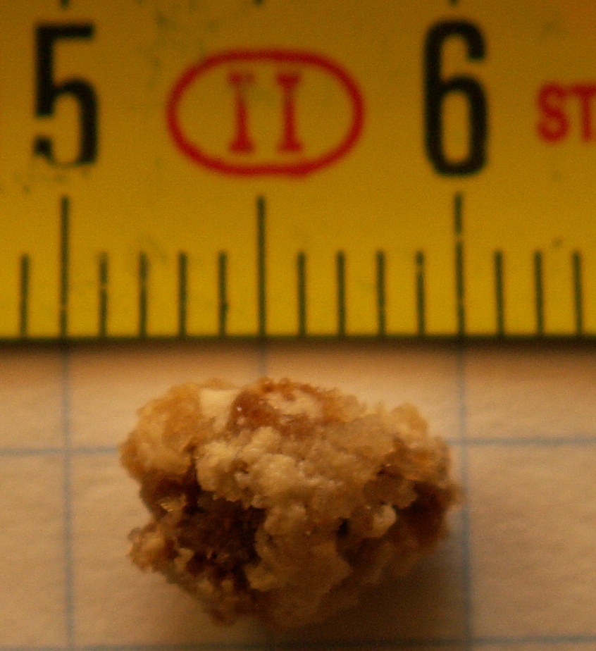
Nephrolithiasis is a multifaceted urological condition characterized by the formation of solid mineral deposits within the renal parenchyma or collecting system [1]A1c[8]D5. While often used interchangeably with urolithiasis, the term specifically refers to stones originating in the kidney, whereas urolithiasis encompasses the entire urinary tract, including the ureters and bladder [7]D5[8]D5. The condition is increasingly viewed as a systemic disorder rather than an isolated surgical event, frequently associated with chronic kidney disease (CKD) and metabolic bone disease (MBD) [1]A1c.
Synonyms and Abbreviations
- Calculi: Renal or urinary stones.
- Urolithiasis: Stones anywhere in the urinary tract.
- Nephrocalcinosis: Deposition of calcium salts in the renal parenchyma.
- Renal Colic: The acute pain syndrome caused by stone passage.
- CN: Calcium nephrolithiasis [1]A1c.
Classification Systems
Clinicians classify nephrolithiasis based on stone composition, anatomical location, and surgical complexity. Composition is the primary driver of long-term medical prevention, while size and location dictate acute surgical intervention [1]A1c[8]D5.
| Classification Axis | Categories / Variants | Clinical Significance |
|---|---|---|
| Composition | Calcium (Oxalate/Phosphate), Uric Acid, Struvite, Cystine | Dictates metabolic workup and medical prevention [1]A1c |
| Etiology | Idiopathic vs. Secondary (e.g., Hyperparathyroidism) | Identifies underlying systemic pathologies [1]A1c |
| Surgical Grade | Satava Classification (Grades 1-3) | Standardizes intraoperative complication reporting [6]C4 |
| Postoperative | Clavien Classification (Grades 1-5) | Categorizes the severity of surgical complications [4]C4 |
Operational Definitions
To standardize research and clinical triage, the following phenotypic definitions are utilized:
- Idiopathic Calcium Nephrolithiasis (CN): Calcium stone formation in the absence of systemic causes like or distal renal tubular acidosis [1]A1c.
- Clinical Activity: Defined by the frequency of stone events (formation of new stones or growth of existing ones) over a specific timeframe, used to assess treatment efficacy in randomized trials [1]A1c.
- Stone-Free Rate (SFR): The primary metric for surgical success, often defined as the complete absence of residual fragments or fragments <3 mm on follow-up imaging [6]C4.
Effective requires a multidisciplinary approach involving urologists for procedural intervention and nephrologists for metabolic evaluation to mitigate the risk of recurrence [1]A1c.
Pearl: Distinguish between nephrolithiasis and nephrocalcinosis; the former involves stones in the collecting system amenable to passage or extraction, while the latter involves parenchymal calcification that often signals underlying systemic metabolic disease [1]A1c.
| Term | Definition | Context |
|---|---|---|
| Idiopathic CN | Calcium stones without a known systemic cause | Most common phenotype [1]A1c |
| Struvite | Magnesium ammonium phosphate stones | Associated with infection [1]A1c |
| Satava 1-2 | Minor intraoperative complications | Used in RIRS protocols [6]C4 |
| Clavien 1-5 | Postoperative complication severity scale | Standard for surgical reporting [4]C4 |
Pathophysiology & Mechanism
- ▸Randall's plaques serve as the essential anchor for calcium oxalate stones in idiopathic formers, originating as interstitial apatite before eroding into the collecting system [22].
- ▸The NLRP3 inflammasome and mitochondrial oxidative stress are central mediators of the tubular injury and crystal retention that drive stone progression [17].
- ▸Genetic variants in SLC34A1 and other transport genes disrupt the calcium-phosphate balance, often manifesting as a triad of hypercalciuria, nephrocalcinosis, and bone demineralization [16, 34].
Supersaturation of urinary solutes serves as the primary driver for crystal nucleation, growth, and aggregation, a process governed by the balance between precipitating ions and endogenous inhibitors [23]D5. While luminal chemical composition is critical, the transformation from microscopic crystals to clinical nephrolithiasis involves complex interactions between the renal epithelium, the interstitial environment, and systemic metabolic pathways [17]D5[23]D5.
The Randall’s Plaque Hypothesis
In idiopathic calcium oxalate (CaOx) stone formers, the initial site of stone development is typically the renal papilla [22]C4. Interstitial calcium phosphate (apatite) deposits, known as Randall's plaques, originate in the basement membrane of the thin loops of Henle and migrate toward the papillary epithelium [22]C4[23]D5.
- Plaque Exposure: The interstitial plaque eventually erodes through the urothelium, exposing the calcium phosphate to the supersaturated pelvic urine [22]C4.
- Epitaxial Growth: CaOx crystals deposit directly onto the exposed apatite through epitaxial growth [22]C4.
- Stone Attachment: Micro-computed tomography (micro-CT) analysis of unattached stones in idiopathic formers reveals internal regions of calcium phosphate, suggesting that even free-floating stones often originate as attached structures that subsequently detached [22]C4.
Cellular and Molecular Mechanisms
Stone formation is not a passive chemical event but an active pathological process involving oxidative stress and tissue inflammation [17]D5[21]C4.
- Mitochondrial Dysfunction: Renal tubular epithelial cells exposed to CaOx crystals undergo mitochondrial stress, leading to the production of reactive oxygen species (ROS) [17]D5[38]D5.
- Inflammasome Activation: Oxidative stress triggers the NLRP3 inflammasome, which promotes the release of pro-inflammatory cytokines such as interleukin-6 (IL-6) and monocyte chemoattractant protein-1 (MCP-1) [17]D5[21]C4.
- Renal Tubular Damage: Increased intrarenal mRNA expression of MCP-1 and IL-6 correlates with the severity of tubular damage and compromised renal function [21]C4.
- Exosomal Signaling: Urinary small extracellular vesicles (sEVs) from stone formers show upregulation of miR-223-3p, which may further modulate inflammation and cell adhesion processes [32]C4.
Genetic and Metabolic Susceptibility
Primary , a polygenic trait affecting 5% to 10% of the general population, involves altered calcium transport across the intestine, kidney, and bone [16]D5[30]D5.
| Gene | Protein Product | Pathophysiologic Role |
|---|---|---|
| SLC34A1 | NaPi-IIa cotransporter | Mutations lead to phosphate wasting, hypercalciuria, and nephrocalcinosis [34]C4. |
| CLDN16 | Claudin-16 | Regulates paracellular calcium reabsorption in the thick ascending limb [16]D5. |
| VDR | Vitamin D receptor | Modulates intestinal calcium absorption and bone resorption [16]D5. |
| ADCY10 | Soluble adenylate cyclase | Involved in renal tubular calcium handling [16]D5. |
Mechanical and Obstructive Factors
Structural anomalies and physiological changes perturb urinary flow, facilitating crystal retention and stone growth [27]D5.
- Urinary Stasis: Congenital anomalies such as (UPJO), horseshoe kidney, and ectopic kidneys increase stone risk by promoting stasis and secondary infection [27]D5. In UPJO, up to 76% of patients also exhibit underlying metabolic abnormalities [27]D5.
- Intrarenal Pressure (IRP): During endoscopic intervention, IRP can reach maximums of 174 mmHg (median 28.5 mmHg) [19]C4. Elevated pressures may drive pyelovenous backflow and systemic absorption of bacteria or endotoxins [19]C4[35]C4.
- Calculous Anuria: Bilateral obstruction or obstruction in a solitary kidney leads to a rapid cessation of glomerular filtration, resulting in acute kidney injury and uremia [24]C4[35]C4.
Secondary Pathogenesis
Malabsorptive states, such as those following bariatric surgery or in inflammatory bowel disease (IBD), drive stone formation through enteric [28]D5. Reduced intestinal calcium binding allows for increased absorption of free oxalate, which is subsequently excreted in the urine [28]D5. Additionally, chronic diarrhea leads to hypocitraturia (loss of a key crystallization inhibitor) and low urine volume [28]D5.
Pearl: Nephrolithiasis is a systemic inflammatory disorder, not just a local mineral deposit; intrarenal expression of MCP-1 and IL-6 directly correlates with the degree of permanent renal impairment [21]C4.
| Category | Examples | Mechanism |
|---|---|---|
| Promoters | CD44 antigen, Galectin-3-binding protein, Kallikrein-1 | Enhance crystal-cell adhesion and aggregation [37]C4. |
| Inhibitors | Citrate, Magnesium, Protein AMBP | Complex with stone-forming ions or coat crystals to prevent growth [15]D5[37]C4. |
| Metabolic Modifiers | Caffeine (>300-360 mg) | Induces hypercalciuria but provides a compensatory diuretic effect [15]D5. |
Epidemiology, Etiology & Risk Factors
- ▸Nephrolithiasis prevalence is highest in North America (7-13%) and is increasing globally, particularly in pediatric populations.
- ▸Metabolic syndrome, hypertension, and vitamin D deficiency are significant systemic risk factors for stone formation.
- ▸Neurological impairment and indwelling catheter use dramatically increase the risk of calcium phosphate and struvite stones.
Nephrolithiasis prevalence varies globally, with rates ranging from 7% to 13% in North America, 5% to 9% in Europe, and 1% to 5% in Asia [70]D5. The incidence of pediatric stone disease has risen by approximately 6% to 10% annually over the last 25 years, reaching 50 per 100,000 adolescents [47]D5. While stone composition is similar across age groups, children are more likely to present with metabolic or genetic drivers [47]D5.
Demographic and Geographic Distribution
Risk profiles differ significantly by race and sex. In the southeastern United States, white men exhibit the highest risk, while no significant difference in risk is observed between black men and women [53]B2b. The age-adjusted incidence rate is 5.98 per 1000 person-years for white men compared to 2.19 for black men [53]B2b. Environmental factors also play a critical role; higher ambient temperatures are associated with increased urinary calcium excretion and supersaturation of calcium oxalate and phosphate [59]B2b. Meta-analysis indicates that higher ambient temperature is associated with an increased risk of urolithiasis presentation (pooled RR 1.31) [65]A1a.
Metabolic and Comorbid Risk Factors
Stone formation is strongly linked to systemic metabolic health and specific comorbidities. Metabolic syndrome is positively associated with nephrolithiasis (OR 1.30), with and serving as independent risk factors [52]B3b.
- Metabolic Syndrome: Prevalence of nephrolithiasis is approximately 31% in patients with metabolic syndrome [52]B3b.
- Vitamin D Deficiency: Stone formers have lower serum 25-hydroxyvitamin D levels than controls; deficiency (<20 ng/mL) is associated with an increased odds of nephrolithiasis (OR 2.29) [61]B3b.
- Neurological Disorders: Children with neurological disorders have a stone incidence of 5% to 54%, driven by immobilization, UTIs, and [43]B3a.
- Bladder : In patients with or spinal cord injury, the use of an indwelling catheter (OR 9.78) or intermittent catheterization (OR 3.50) significantly increases stone risk [55]B3b.
Etiological Risk Factors
| Factor | OR/RR/HR | Evidence Level |
|---|---|---|
| White Race (vs Black) | HR 2.23 [53]B2b | 2b |
| OR 18.18 [56]B3b | 3b | |
| Urinary pH < 6.0 | OR 15.63 [56]B3b | 3b |
| Metabolic Syndrome | OR 1.30 [52]B3b | 3b |
| Vitamin D Deficiency | OR 2.29 [61]B3b | 3b |
| Indwelling Catheter | OR 9.78 [55]B3b | 3b |
| High Oxidative Balance | HR 0.77 [67]B2b | 2b |
Iatrogenic and Genetic Drivers
Iatrogenic factors include surgical history and medications. Patients undergoing radical have a significantly higher incidence of postoperative stones (8.4%) compared to those undergoing (1.6%) [56]B3b. In pediatric populations, use is associated with urolithiasis in approximately 7% of patients [64]A1a. Genetic factors are also prominent; nephrolithiasis is roughly 50% heritable, and while often polygenic, single mutations in at least 27 genes can cause monogenic stone disease [45]D5. High genetic susceptibility interacts with lifestyle; a high Oxidative Balance Score (evaluating diet and lifestyle) reduces risk by 23% (HR 0.77) [67]B2b.
Pearl: Ambient temperature and metabolic health are potent modifiable drivers; every 10% increase in population compliance with high water intake could prevent thousands of stones and save millions in healthcare costs [58]B2b[65]A1a.
| Population | Calcium Oxalate | Calcium Phosphate | Struvite | Uric Acid | Cystine |
|---|---|---|---|---|---|
| General Pediatric [47]D5 | 75-80% | 5-10% | 10-20% | 5% | Rare |
| Multiple Sclerosis [55]B3b | 39% | 42% | 8% | NR | NR |
| Spinal Cord Injury [69]B2b | Common | Common | High Risk | NR | NR |
Clinical Presentation
- ▸Renal colic is characterized by paroxysmal flank pain and restlessness, often accompanied by nausea and hematuria.
- ▸Nonobstructing calyceal stones can cause significant pain, and their removal often leads to a durable reduction in pain scores.
- ▸Bilateral obstructing stones or obstruction in a solitary kidney can present as acute anuria, requiring emergent decompression.
The transition from metabolic lithogenesis to clinical syndrome typically occurs when a stone migrates into the ureter or obstructs a calyx, though many patients remain asymptomatic for years. While incidental identification of small renal calculi is rising due to high-resolution imaging, the natural history of these stones is variable; the risk of developing symptomatic episodes ranges from 0% to 59.4% [77]B2a. Once symptomatic, the presentation is dominated by acute renal colic, lower urinary tract symptoms (LUTS), and hematuria.
Presenting Symptoms
Renal colic is characterized by the sudden onset of severe, paroxysmal flank pain that often radiates to the groin or labia/scrotum as the stone descends. Unlike patients with peritonitis who remain still, those with nephrolithiasis are typically restless, frequently pacing or shifting positions to find relief.
- Pain Pattern: Flank pain is the most common symptom, but even nonobstructing calyceal stones can cause moderate to severe pain. Surgical removal of nonobstructing stones ≤10 mm has been shown to reduce mean pain scores from 5.5 to 1.8 and worst pain scores from 7.2 to 2.8 at 12 weeks post-procedure [79]B2b.
- Symptoms: Nausea and emesis are frequent, particularly in pediatric populations where they may be the primary complaint [24]C4.
- Lower Urinary Tract Symptoms (LUTS): As stones approach the ureterovesical junction, patients often report urgency, frequency, and dysuria [88]C4.
- Hematuria: Macroscopic hematuria is a hallmark sign, occurring in approximately 28% to 70% of symptomatic cases [82]B3b[88]C4.
Physical Examination Findings
The clinical examination focuses on localizing the obstruction and identifying complications such as infection or renal failure.
- Costovertebral Angle (CVA) Tenderness: Percussion of the flank typically elicits sharp pain on the affected side.
- Abdominal Distension: May be present in severe cases, particularly in children with bilateral obstruction [24]C4.
- Vital Signs: Tachycardia and are common due to pain. The presence of fever is a critical finding, suggesting obstructive pyelonephritis or urosepsis.
Phenotypic Variants
| Variant | Key Features | Frequency/Context |
|---|---|---|
| Asymptomatic Stones | Incidental finding; stone size does not reliably predict future symptoms [77]B2a. | Common in imaging |
| Staghorn Calculi | Large stones filling the renal pelvis; associated with chronic inflammation, fibrosis, and reduced creatinine clearance [84]B3b. | Chronic infection |
| Calculous Anuria | Sudden cessation of urine output due to bilateral obstruction or obstruction of a solitary kidney [24]C4. | Surgical emergency |
| Pediatric Presentation | Higher rates of metabolic abnormalities; often presents with emesis and abdominal pain rather than classic colic [24]C4[88]C4. | Increasing incidence |
Red Flags
Certain presentations require immediate surgical decompression or intensive care to prevent irreversible renal damage or death.
- Fever and Chills: Indicates infection proximal to the obstruction; requires urgent intervention.
- Anuria or Oliguria: Suggests bilateral obstruction or a solitary functioning kidney; can lead to rapid acute renal failure [24]C4[86]C4.
- Intractable Pain or Emesis: Inability to maintain oral hydration or achieve pain control with outpatient regimens.
Atypical Presentations
Clinicians must remain vigilant for rare conditions that mimic or coexist with nephrolithiasis. Primary adenocarcinoma of the renal pelvis can masquerade as chronic stone disease or xanthogranulomatous pyelonephritis [87]C4. Additionally, systemic diseases like may present with genitourinary complications including obstructive uropathy [85]C4. In rare instances, T-cell lymphoma has presented as bilateral nephrolithiasis and acute renal failure [86]C4. Metabolic disorders such as or Autosomal Dominant type 1 (ADH1) should be suspected in patients with recurrent stones and persistent mineral imbalances [92]A1a[95]C4.
Following the clinical assessment, the focus shifts to definitive to confirm stone location and composition.
Pearl: Stone size is a poor predictor of symptoms, but it strongly dictates the need for intervention; stones >5 mm are significantly more likely to require surgical than smaller fragments [77]B2a.
Diagnosis & Workup
- ▸Non-contrast CT is the gold standard for diagnosis, providing critical data on stone size, location, and Hounsfield density to predict treatment success.
- ▸Ultrasound is the preferred initial modality in pediatric and pregnant populations but carries a high risk of miscalculating stone size, leading to management errors in approximately 22% of cases.
- ▸Metabolic and genetic screening, including 24-hour urine collection and NHERF1/monogenic testing, is essential for recurrent or pediatric stone formers to identify underlying etiologies like cystinuria or phosphate wasting.
The diagnostic workup of suspected nephrolithiasis integrates clinical suspicion with rapid imaging to confirm the presence, location, and size of the calculus while assessing for obstructive complications. While the presentation of renal colic often suggests the diagnosis, imaging remains the definitive step to guide decisions between observation and surgical intervention [10]A1c[11]A1c.
History and Physical
Clinical evaluation focuses on the character of pain and the presence of systemic symptoms that may indicate urosepsis or impending renal failure.
- Pain Profile: Typically sudden onset of severe, unilateral flank pain radiating to the groin or labia/scrotum (renal colic).
- Associated Symptoms: Nausea, vomiting, and gross or microscopic hematuria.
- Red Flags: Fever, chills, or hypotension (suggesting infected hydronephrosis), and anuria or oliguria (suggesting bilateral obstruction or obstruction of a solitary kidney) [113]C4.
- Physical Signs: Costovertebral angle (CVA) tenderness is the hallmark finding; the abdomen is typically soft and non-tender, helping to differentiate from intraperitoneal pathology.
Imaging Modalities
Non-contrast computed tomography (CT) of the abdomen and pelvis is the gold-standard diagnostic test due to its high sensitivity and specificity for identifying stone size, location, and density (Hounsfield units) [104]D5[115]D5.
- Ultrasonography (US): Recommended as the initial imaging study in children and pregnant patients to minimize radiation [42]A1b[47]D5. However, US has a sensitivity of only 54% and a specificity of 91% compared to CT [106]B3b. It frequently overestimates the size of stones in the 0-10 mm range, potentially leading to inappropriate management recommendations in 22% of cases [106]B3b.
- Low-Dose CT: A preferred alternative to standard CT that maintains high diagnostic accuracy while reducing cumulative radiation exposure [104]D5.
- Plain Radiography (KUB): Useful for monitoring the progress of radiopaque stones (calcium oxalate, calcium phosphate, struvite, and cystine) but lacks the sensitivity to detect uric acid stones or small calculi [106]B3b.
Laboratory Studies
Initial laboratory evaluation assesses renal function, identifies metabolic risk factors, and screens for concurrent infection.
| Test | Finding / Significance | Sensitivity/Specificity |
|---|---|---|
| Urinalysis | Hematuria (micro or macro); pH <5.5 (uric acid) or >7.2 (struvite); Nitrites/LE (infection) | High sensitivity for hematuria |
| Serum Creatinine | Assesses for acute kidney injury (AKI) due to obstruction | Essential for baseline |
| Serum Calcium | Screens for | Standard screening |
| Lipid Profile | Dyslipidemia (high triglycerides/low HDL) is linked to uric acid stones [105]B3b | Metabolic marker |
| 24-Hour Urine | Measures volume, calcium, oxalate, citrate, uric acid, and sodium | Gold standard for prevention |
Genetic Testing
Genetic screening should be considered in pediatric patients, recurrent stone formers, or those with a strong family history. Monogenic causes may be responsible for a larger proportion of cases than previously recognized, with at least 27 genes identified as monogenic drivers of stone disease [45]D5. Specific mutations, such as those in the NHERF1 gene, can lead to renal phosphate loss and subsequent stone formation [109]B3b.
Diagnostic Algorithm
Pearl: While CT is the gold standard for adults, ultrasound is the mandatory first-line study in children to limit radiation, despite its 54% sensitivity and tendency to overestimate stone size by several millimeters [47]D5[106]B3b.
| Modality | Sensitivity | Specificity | Primary Utility |
|---|---|---|---|
| Non-contrast CT | >95% | >95% | Gold standard; surgical planning |
| Ultrasound | 54% | 91% | Initial screen in children/pregnancy |
| KUB (X-ray) | 44-77% | Variable | Monitoring radiopaque stone passage |
| Low-dose CT | High | High | Reducing cumulative radiation risk |
Severity, Staging & Risk Stratification
- ▸Surgical intervention for ureteric colic is predicted by symptom duration >3 days, stone density, and lack of alpha-blocker therapy.
- ▸Pure uric acid stones, suitable for chemolysis, typically present with a density of approximately 450 HU and are radiolucent in 76% of cases.
- ▸Family history is an independent predictor of hypercalciuria (OR 2.15) and calcium oxalate monohydrate stone composition (OR 2.05).
Risk stratification in nephrolithiasis converts diagnostic findings into a graded tier that determines the necessity of surgical intervention versus conservative surveillance. While acute obstructive uropathy is a medical emergency often requiring prompt decompression to avoid renal failure or sepsis [121]A1c, stable patients are stratified based on stone burden, location, and metabolic recurrence risk.
Predictors of Surgical Intervention
For patients presenting with acute ureteric colic, specific clinical and stone-related factors predict the failure of conservative . A validated nomogram (AUC 0.802) identifies the following independent predictors for the eventual need for surgical intervention [126]B3b:
- Symptom duration >3 days at presentation.
- Stone burden (size) and high stone density.
- Stone location (proximal vs. distal).
- Prior history of renal calculi.
- Lack of medical expulsive therapy (e.g., not receiving or other alpha-blockers) [126]B3b.
Anatomical and Compositional Staging
Stone location and composition significantly impact the success of definitive management. Lower pole stones (LPS) are particularly challenging due to the infundibulopelvic angle (IPA), infundibulopelvic width (IPW), and infundibular length [99]A1a. For LPS ≤20 mm, percutaneous nephrolithotomy (PNL) is more effective than shock wave lithotripsy (SWL) (RR 2.04) and retrograde intrarenal surgery (RIRS) (RR 1.31) [40]A1a.
Compositional stratification is essential for selecting candidates for oral chemolysis. A clinical nomogram (AUC 0.78) predicts "pure" uric acid stones (≥95% UA) using the following variables [125]B3b:
- Stone Density: Pure stones average 450 Hounsfield Units (HU) vs. 600 HU for mixed stones.
- Radiopacity: Only 24% of pure uric acid stones are radiopaque compared to 58% of mixed stones.
- Demographics: Older age and larger stone size further increase the probability of pure uric acid composition [125]B3b.
Metabolic and Recurrence Risk Stratification
Metabolic evaluation identifies patients at high risk for recurrent episodes, which is critical for initiating pharmacologic monotherapy with , , or [123]A1c.
| Risk Factor | Clinical Significance | Evidence |
|---|---|---|
| Family History | Predicts (OR 2.15) and Calcium Oxalate monohydrate stones (OR 2.05) [127]B3b. | [127]B3b |
| Ca:Citrate Ratio | In children, a mean ratio of 0.64 distinguishes recurrent formers from solitary formers (0.41, p=0.02) [120]B3b. | [120]B3b |
| Staghorn Calculi | Associated with robust inflammation, epithelial-mesenchymal transition (EMT), and renal fibrosis [84]B3b. | [84]B3b |
| Infectious Risk | Indwelling tubes or positive cultures require 7 days of preoperative for PNL to reduce sepsis (OR 3.1 for 2-day vs 7-day) [117]A1b. | [117]A1b |
Long-term Renal Impact
In pediatric populations, the choice of intervention (SWL, RIRS, or PNL) does not appear to impair long-term renal growth compared to non-treated kidneys [128]B3b. However, in adults with large staghorn calculi, renal fibrosis is inversely correlated with renal function (creatinine clearance), likely mediated by TGF-beta1-triggered EMT [84]B3b. Clinicians must also remain vigilant for upper tract urothelial carcinoma (UTUC) mimickers; a diagnosis of nephrolithiasis is associated with a delayed UTUC diagnosis (RR 1.23, 95% CI 1.06-1.44), particularly when managed by non-urologists [124]B2c.
Pearl: In patients with moderate-to-high infectious risk undergoing PNL, 7 days of preoperative antibiotics reduces sepsis risk by approximately 68% compared to a 2-day course (OR 3.1 for 2-day regimen) [117]A1b.
| Parameter | Threshold/Finding | Clinical Implication |
|---|---|---|
| Urine Ca:Citrate (Pediatric) | >0.64 | High risk for recurrence [120]B3b |
| Stone Density (HU) | <450 HU | Suggests pure uric acid stone; candidate for chemolysis [125]B3b |
| Antibiotic Duration (PNL) | 7 days | Required for moderate-high sepsis risk patients [117]A1b |
| Stone Size (Lower Pole) | >10 mm | PNL/RIRS significantly more effective than SWL [40]A1a |
Acute Management & Decompression
- ▸Obstructed nephrolithiasis with concomitant infection is a urologic emergency requiring immediate decompression via ureteral stent or percutaneous nephrostomy.
- ▸Non-opioid analgesia with [[ketorolac]] is non-inferior to opioids for stone-related pain and reduces the risk of long-term opioid misuse.
- ▸The CLAD-MB score effectively identifies patients at low risk for requiring surgery within 7 days of presentation.
Risk stratification following the assessment of severity determines the urgency of intervention, particularly when nephrolithiasis is complicated by infection or obstruction. While most patients with symptomatic calculi can be managed non-operatively, the presence of an obstructed, infected collecting system constitutes a surgical emergency requiring immediate decompression to prevent urosepsis and multi-organ failure [71]A1a[135]C4.
Step 1: Emergency Triage and Severity Classification
Clinicians must immediately identify "red flag" features that mandate inpatient admission and urgent procedural intervention. The CLAD-MB score (Complicated UroLithiasis and Alternative Diagnoses - Modified with Biology) provides a validated framework for predicting the need for surgery within 7 days [137]B2b.
- Indications for Urgent Decompression:
- Obstructed system with signs of infection (fever >37.8 °C, elevated CRP ≥50 mg/mL, or leukocytosis ≥12 G/L) [137]B2b.
- Intractable pain (score >7/10) or persistent vomiting [137]B2b.
- Acute kidney injury (elevated creatinine) or solitary kidney with obstruction [137]B2b.
- Bilateral obstructing stones.
Step 2: Initial Medical Stabilization
Pain and infection control are the priorities during the stabilization phase. The STONE Pathway demonstrates that standardized clinical protocols increase compliance with evidence-based practices, such as the use of , and reduce 30-day emergency department return rates from 13% to 2% (p=0.01) [143]B2b.
- : is non-inferior to opioids for postoperative pain management (mean pain score 3.20 vs 4.17, p=0.018) [132]A1b. Avoid routine opioid use where possible, as chronic opioid use predicts a 3.64-fold increased risk of unplanned ER visits for stent-related symptoms [29]B3b.
- : Prophylactic or therapeutic antibiotics must be initiated based on perioperative urine or stone cultures to prevent post-intervention urosepsis [138]D5.
Step 3: Surgical Decompression and Drainage
For the obstructed, infected system, the primary goal is drainage rather than definitive stone clearance. The AUA 2026 guidelines emphasize that the choice between retrograde placement and (PCN) should be guided by patient factors and resource availability [9]A1c[11]A1c.
- Ureteral Stenting: Silicone-hydrocoated stents are associated with 25% lower body pain scores at day 20 compared to standard polymer stents (p=0.015) and demonstrate significantly less mineral encrustation (p=0.0048) [74]A1b[133]A1b.
- Percutaneous Nephrostomy (PCN): Often preferred in cases of severe sepsis or when retrograde access is technically unfeasible. In pregnant patients, however, initial PCN is associated with significantly higher radiation exposure (286.9 mGy vs 3.7 mGy for stents) and a higher rate of tube dysfunction (>40%) [147]B3b.
Step 4: Monitoring and Definitive Planning
Following decompression, patients require close monitoring for the resolution of systemic inflammatory response syndrome (SIRS). Definitive stone clearance (e.g., or ) is typically deferred until the infection is eradicated [129]A1c. Pre-stenting before definitive retrograde intrarenal surgery (RIRS) has been shown to reduce operative time (58.8 vs 68.7 min, p=0.001) and increase stone-free rates (85.3% vs 64.7%) [140]A1b.
Controversies and Guideline Disagreement
| Question | Position A | Position B | Strength | Implication |
|---|---|---|---|---|
| Initial Imaging Modality | AUA/NEJM [42]A1b: Ultrasonography is a safe initial alternative to CT, reducing radiation without missing high-risk diagnoses. | Clinical Practice [131]A1b: Most patients (78%) still undergo CT before surgical intervention despite initial ultrasound. | Moderate | Ultrasound reduces radiation but often necessitates a second study (CT) for surgical planning. |
| Management in Octogenarians | Traditional View: Conservative management due to frailty. | Eredics et al. [139]B3b: Active stone removal is safe and improves survival compared to simple diversion. | Moderate | Fit elderly patients should be offered definitive treatment rather than permanent drainage. |
Pearl: Immediate decompression of an obstructed, infected kidney is mandatory to prevent a 20% mortality rate associated with septic multi-organ failure in this population [135]C4.
| Modality | Primary Indication | Key Advantage | Key Limitation | Evidence Level |
|---|---|---|---|---|
| Ureteral Stent | Routine obstruction | Internalized; lower radiation in pregnancy [147]B3b | Stent-related symptoms (USRS) in 20% [29]B3b | 1b |
| Percutaneous Nephrostomy | Severe urosepsis | Reliable drainage of pyonephrosis | Higher radiation; 40% dysfunction rate [147]B3b | 3b |
| Primary URS | Small distal stones | Definitive clearance in one session | Risk of sepsis if system is infected [71]A1a | 1a |
Long-term & Definitive Management: Medical vs Endourologic/Surgical
- ▸PCNL provides the highest stone-free rates for large (> 20 mm) stones but carries the highest risk of hemorrhage and longest hospital stay [111].
- ▸In pediatric patients with 10-20 mm stones, mini-PCNL achieves a 93.3% SFR compared to 33.3% for ESWL [60].
- ▸Intraoperative ketorolac reduces peri-operative narcotic requirements by 37% during ureteroscopy [78].
Definitive of nephrolithiasis transitions from acute relief to stone eradication and recurrence prevention, guided by stone size, location, and patient anatomy [9]A1c[10]A1c. While observation is appropriate for many asymptomatic stones, active intervention reduces subsequent stone-related surgery (OR 0.45, 95% CI 0.25-0.80) and stone growth (OR 0.24) in selected patients [14]A1a.
Step 1: Selection of Primary Intervention
The choice between (ESWL), (URS), and (PCNL) depends on the stone burden and location [110]A1a[111]A1a.
- Stones < 10 mm: Often managed with observation or ESWL. For lower pole stones (LPS) ≤ 10 mm, the benefit of RIRS or PCNL over ESWL is minimal [40]A1a.
- Stones 10-20 mm: URS or ESWL are standard. In the pediatric population (6 months to 6 years), mini-PCNL achieves a significantly higher stone-free rate (SFR) of 93.33% compared to 33.33% for ESWL (p < 0.001) [60]A1b.
- Stones > 20 mm: PCNL is the gold standard, achieving the highest SFR (RR 1.13 vs URS) but with higher transfusion rates and longer hospital stays [111]A1a. For 2-3 cm stones, Flexible and Navigable Suction Sheath RIRS (FANS-RIRS) may offer comparable SFRs to mini-PCNL (OR 0.80, 95% CI 0.51-1.24) with lower hemorrhage rates (OR 0.08) [101]A1a.
Step 2: Procedural Optimization and Technique
Surgical success is maximized through specific technical considerations and adjuncts.
- Ureteral Access Sheaths (UAS): UAS use increases irrigation flow and decreases intrapelvic pressure [98]D5. Larger sheaths (11/13-F or 12/14-F) lower intrarenal pressure more effectively than 10/12-F sheaths (P < 0.001) [19]C4 (4).
- Positioning: Increasing the angle of inclination (Trendelenburg) can reduce laser-induced stone retropulsion to < 1 cm [108]D5 (5).
- Pain Management: Intraoperative IV reduces peri-operative narcotic requirements by 37% [78]A1b. For PCNL, preoperative paravertebral block (PVB) significantly lowers postoperative opioid use and pain scores [150]A1b.
Step 3: Medical Prevention of Recurrence
Long-term medical management targets metabolic abnormalities to prevent the high rate of recurrence [41]A1b.
- Thiazide Diuretics: While widely used, recent data on (12.5 mg, 25 mg, or 50 mg daily) showed no significant difference in recurrence compared to placebo over 2.9 years (P = 0.66) [41]A1b.
- Alkali Therapy: (1 mEq/kg/day) may lower stone recurrence rates in children (RR 0.19, 95% CI 0.06-0.60), corresponding to 270 fewer recurrences per 1000 children [62]A1a.
- Dietary Modification: Increased water intake and salt reduction are universal recommendations for all stone formers [47]D5[159]A1c.
Step 4: Monitoring and Treatment Failure
Surgical recurrence occurs in approximately 24.5% of patients over 4 years [107]B3b. Risk factors for repeat surgery include age < 60 years, female gender, diabetes, and malabsorptive disease [107]B3b.
Controversies and Guideline Disagreement
| Question | Position A | Position B | Strength | Implication |
|---|---|---|---|---|
| Initial imaging for suspected stones | AUA/EAU, Non-contrast CT is the gold standard for diagnosis [159]A1c | Pediatric/General Guidelines, Ultrasound should be initial imaging to reduce radiation [42]A1b[47]D5 | Moderate | CT has higher accuracy but ultrasound reduces cumulative radiation without increasing 30-day complications [42]A1b. |
| Management of 10-20 mm stones | Traditional, ESWL or URS are first-line for most stones < 2 cm [110]A1a | Emerging Evidence, Mini-PCNL or RIRS with suction sheaths may offer superior SFR for 1-2 cm stones [60]A1b[101]A1a | Moderate | Shift toward more invasive but higher-efficacy endoscopic procedures in a single session. |
Pearl: For lower pole stones < 2 cm, active displacement to the upper pole during ureteroscopy significantly improves stone-free rates (RR 1.21) without increasing complications [99]A1a.
| Modality | Primary Indication | Stone-Free Rate (SFR) | Complication Profile | Evidence Level |
|---|---|---|---|---|
| ESWL | Stones < 10 mm; non-lower pole | Lowest (RR 0.67 vs PCNL) | Lowest; high retreatment risk | 1a [110]A1a[111]A1a |
| URS / RIRS | Ureteral stones; renal stones < 20 mm | Intermediate (80-89%) | Low; stent discomfort common | 1a [153]A1a[162]A1a |
| PCNL | Stones > 20 mm; complex stones | Highest (85-95%) | Highest; bleeding, sepsis risk | 1a [110]A1a[111]A1a |
| Mini-PCNL | Pediatric stones; 10-20 mm stones | Superior to ESWL in children | Higher pain than RIRS | 1b [60]A1b[151]A1b |
| Drug | Starting Dose | Target / Max Dose | Key Monitoring | Evidence |
|---|---|---|---|---|
| 10-20 mEq TID | 100 mEq/day | Serum K+, Urinary pH | 1a [62]A1a | |
| 12.5 mg daily | 50 mg daily | K+, Glucose, Uric acid | 1b [41]A1b | |
| 25 mg daily | 50 mg daily | Serum electrolytes | 1c [159]A1c |
History and Evolution of Treatment
- ▸Mini-PCNL provides significantly higher stone-free rates than SWL in pediatric patients (93.3% vs 33.3%) and superior results to RIRS in adults for stones up to 3 cm.
- ▸High fluid intake to maintain urine output > 2.5 L/d remains the most robust intervention for reducing stone recurrence (RR 0.39).
- ▸The NOSTONE trial (2023) found no significant dose-response benefit for hydrochlorothiazide in preventing calcium stone recurrence, challenging long-standing practice.
The therapeutic landscape for nephrolithiasis has transitioned from invasive open surgical procedures to minimally invasive endourologic techniques and metabolically targeted medical prevention. While historical figures like President Lyndon B. Johnson required open surgical removal of stones [178]D5, modern is dictated by stone composition, size, and patient-specific metabolic profiles [9]A1c[10]A1c. This evolution is grounded in landmark trials that established the efficacy of fluid intake, selective pharmacotherapy, and refined surgical modalities.
Evolution of Surgical Standards
Surgical intervention has shifted from open nephrotomy to and endoscopic procedures. The introduction of extracorporeal shock wave lithotripsy (SWL) initially revolutionized care, though recent evidence indicates that mini-percutaneous nephrolithotomy (mini-PCNL) provides superior stone-free rates (SFR) for specific populations. In pediatric patients with stones 10-20 mm, mini-PCNL achieved a 93.33% SFR compared to 33.33% for SWL (p < 0.001) [60]A1b. Similarly, flexible mini-PCNL (F-mPCNL) for stones 1.5-3 cm in adults demonstrated a superior SFR of 95.1% compared to 77.8% for retrograde intrarenal surgery (RIRS) (p < 0.001) [151]A1b.
Landmark Evidence in Medical Prevention
Medical management evolved from empirical dietary advice to evidence-based metabolic interventions. High fluid intake remains the cornerstone of secondary prevention, with trials showing that achieving a urine output > 2.5 L/d reduces recurrence risk (RR 0.39, 95% CI 0.19-0.80) [165]A1a.
- Thiazide Diuretics: Long considered the standard for hypercalciuric stone formers, recent data from the NOSTONE trial challenged the dose-response assumption. Hydrochlorothiazide at doses of 12.5 mg, 25 mg, or 50 mg did not show a significant difference in recurrence compared to placebo over 2.9 years (p = 0.66) [41]A1b.
- Urease Inhibitors: For struvite stones, (15 mg/kg/day) was established as an effective inhibitor of stone growth (p < 0.01), though its use is limited by a high prevalence of adverse effects such as tremulousness and phlebothrombosis [167]A1b.
- Alkalinization: Medical dissolution therapy using remains a non-invasive standard for uric acid stones, achieving complete resolution in 67% of patients in select cohorts [187]C4.
Shift in Pain Management and Imaging
The management of acute renal colic has moved away from routine opioid use due to the risk of prolonged dependency. Patients with a history of kidney stones have a significantly higher prevalence of current opioid use (10.9% vs 6.1%) [46]B2c. Trials such as TRUST demonstrated that intraoperative IV reduces perioperative milligram equivalent (MME) requirements by 37% [78]A1b. Diagnostic protocols have also evolved; initial ultrasonography is now preferred over CT to reduce cumulative radiation exposure without increasing the risk of missed high-risk diagnoses [42]A1b[47]D5.
Controversies and Guideline Disagreement
| Question | Position A | Position B | Strength | Implication |
|---|---|---|---|---|
| 24h Urine in PHPT | Routine measurement for surgical decisions [92]A1a. | Not a reliable independent predictor of stone risk in PHPT [179]B3b. | Moderate | Influences whether is prioritized for stone prevention. |
| Thiazide Efficacy | Strong recommendation for calcium stones [164]A1c. | No significant benefit over placebo in recent RCT [41]A1b. | High | Re-evaluating the role of hydrochlorothiazide in standard protocols. |
Pearl: The transition from SWL to mini-PCNL and RIRS has prioritized higher stone-free rates, while medical management is shifting from routine thiazide use toward personalized metabolic and genetic-directed therapies [45]D5[151]A1b.
| Procedure | Population | Stone Size | Primary Outcome (SFR) | Citation |
|---|---|---|---|---|
| Mini-PCNL | Pediatric | 1-2 cm | 93.33% | [60]A1b |
| SWL | Pediatric | 1-2 cm | 33.33% | [60]A1b |
| F-mPCNL | Adult | 1.5-3 cm | 95.1% | [151]A1b |
| RIRS | Adult | 1.5-3 cm | 77.8% | [151]A1b |
Endoscopic & Procedural Technique Considerations
- ▸Mini-PCNL provides a significantly higher stone-free rate (95.1%) than RIRS (77.8%) for stones 1.5-3 cm, albeit with longer hospital stays [151].
- ▸Intraoperative IV ketorolac reduces perioperative narcotic requirements by 37% during ureteroscopy [78].
- ▸Ureteral access sheaths (UAS) larger than 10/12-F are more effective at maintaining lower intrarenal pressures during flexible ureteroscopy [19].
Surgical success in nephrolithiasis is predicated on the selection of optimal treatment modalities based on patient factors, urinary tract anatomy, and stone characteristics [9]A1c, [10]A1c. While the evolution of treatment has moved toward less invasive options, the choice between (URS), (PCNL), and (ESWL) requires balancing stone-free rates (SFR) against procedural morbidity and the risk of retreatment [111]A1a.
Ureteroscopic Techniques and Access
Flexible ureteroscopy (FURS) has expanded to treat larger and more complex stones, often utilizing a standardized sequence of steps to maximize SFR [194]C4. The "Freiburg technique" emphasizes preoperative stenting, the use of dual hydrophilic wires, and semirigid ureteroscopy prior to FURS to facilitate access [194]C4.
- Ureteral Access Sheaths (UAS): UAS use increases irrigation flow and decreases intrapelvic pressure (IRP), potentially reducing infectious complications [98]D5. However, smaller 10/12-F sheaths may not lower IRP as effectively as 11/13-F or 12/14-F models [19]C4.
- Intrarenal Pressure (IRP) Monitoring: Modern digital ureteroscopes can now measure IRP in real-time [19]C4. During a median procedure time of 31.9 minutes, median IRP is typically 28.5 mmHg, though maximum pressures can reach 174 mmHg [19]C4. Pressures remain <60 mmHg for approximately 92% of the procedure [19]C4.
- Scope Diameter: Ultra-slim 6.3 Fr digital disposable ureteroscopes provide comparable visualization and maneuverability to 7.5 Fr models, with successful kidney entry in 100% of cases and potentially reduced ureteral trauma [102]A1b.
Percutaneous Access and Miniaturization
PCNL remains the most effective modality for high stone burdens but carries higher morbidity than URS [111]A1a. Miniaturized PCNL (mini-PCNL) and micro-percutaneous (microperc) techniques aim to bridge this gap.
- Mini-PCNL vs. RIRS: In stones 1.5-3 cm, mini-PCNL achieves a superior SFR of 95.1% compared to 77.8% for RIRS (p < 0.001) [151]A1b. Mini-PCNL also requires shorter operative times (47.6 vs 59.3 min) and less radiation exposure, though it is associated with longer hospital stays and higher postoperative pain [151]A1b, [153]A1a.
- Microperc: This technique is most effective for solitary stones with volumes <1000 mm³ and low Hounsfield density [160]C4.
- Tubeless PCNL: Omitting a nephrostomy tube at the conclusion of PCNL is safe in uneventful procedures and reduces postoperative pain, costs, and hospital stay [155]D5. Nephrostomy tubes should be retained if >2 access tracts are used or if significant bleeding occurs [155]D5.
Intraoperative Adjuncts and Safety
- Lithotripsy Modalities: laser lithotripsy is the most common intracorporeal tool [26]D5. To reduce stone retropulsion during ureteroscopic laser lithotripsy, increasing the patient's incline angle (Trendelenburg) can reduce migration to <1 cm [108]D5.
- Radiation Safety: The "as low as reasonably achievable" (ALARA) principle is mandatory [104]D5. Fluoroscopy time can be reduced through the use of laser-guided C-arms, tactile feedback for wire placement, and stent placement under direct vision [104]D5.
- Pain : Intraoperative IV (Toradol) reduces perioperative narcotic requirements by 37% (22.2 vs 30.4 combined MME, p < 0.02) and is an independent predictor of lower opioid use [78]A1b.
Controversies and Guideline Disagreement
| Question | Position A | Position B | Strength | Implication |
|---|---|---|---|---|
| UAS Use | Routine use to protect scope and lower IRP [98]D5. | Selective use due to risk of [98]D5. | Moderate | Surgeon preference dictates use based on ureteral caliber. |
| Lower Pole <1cm | ESWL as first-line due to non-invasiveness [110]A1a. | URS/mini-PCNL for higher single-session SFR [153]A1a. | High | Shared decision-making regarding retreatment risk. |
Pearl: For lower pole stones <2 cm, active displacement to the upper pole increases the stone-free rate by 21% (RR 1.21) with only a 6-minute increase in operative time [99]A1a.
| Modality | Stone-Free Rate (SFR) | Retreatment Rate | Complication Rate | Hospital Stay |
|---|---|---|---|---|
| ESWL | Lowest [111]A1a | Highest [111]A1a | Low [110]A1a | Shortest/Outpatient |
| URS/RIRS | Moderate (80.1%) [153]A1a | Moderate [111]A1a | Lowest [111]A1a | 2.5 days [153]A1a |
| Mini-PCNL | High (89.3%) [153]A1a | Low [151]A1b | Moderate [111]A1a | 4.0 days [153]A1a |
Complications
- ▸Cystinuria is associated with significantly lower creatinine clearance and a higher procedural burden compared to other stone types.
- ▸Single-session bilateral stone removal increases overall complication rates and operative time but does not increase high-grade (Clavien-Dindo ≥3) complications compared to staged procedures.
- ▸Thiazide-induced complications such as hypokalemia and new-onset diabetes occur frequently and are not clearly dose-dependent in stone prevention trials.
Complications of nephrolithiasis arise from both the natural history of stone disease and the sequelae of medical and surgical interventions. While many asymptomatic stones remain stable, a meta-analysis of randomized trials indicates that active intervention for asymptomatic stones reduces the pooled odds of subsequent surgery and stone growth compared to observation [14]A1a.
Disease-Related Complications
Chronic or recurrent stone formation can lead to progressive renal impairment. Patients with demonstrate significantly lower age- and gender-corrected creatinine clearance at entry compared to routine stone formers (91 L/day vs 160 L/day, P < 0.001) [198]B3b. These patients also require significantly more procedures over time [198]B3b. In pediatric populations, the risk of symptomatic recurrence is high, with approximately 50% of children presenting with a recurrence within 3 years of their first stone [48]B2b.
Metabolic and structural complications include:
- Renal Function Decline: Associated with recurrent obstruction, infection, or specific genetic etiologies [198]B3b.
- Infection: Recurrent urinary tract infections (UTIs) are associated with elevated urinary ammonium and an increased risk of postoperative complications [203]B3b[4]C4.
- Ureteral Stricture: May result from chronic stone impaction or iatrogenic injury during removal, sometimes requiring complex reconstruction such as appendiceal interposition [193]C4.
Treatment-Related Complications
Surgical interventions, while often definitive, carry specific risks. Single-session bilateral (SSB) procedures, though reducing anesthesia exposure, are associated with significantly higher overall complication rates compared to staged unilateral procedures (P = 0.007) and a higher need for unplanned additional interventions [196]A1a. However, the rate of severe complications (Clavien-Dindo grade 3 or higher) does not differ significantly between SSB and staged approaches [196]A1a.
Pharmacological prophylaxis also carries a burden of adverse effects. Thiazide diuretics, used to reduce recurrence, are associated with higher rates of hypokalemia, gout, new-onset diabetes mellitus, and plasma creatinine elevations exceeding 150% of baseline compared to placebo [41]A1b. Acetohydroxamic acid, used for infection-related stones, probably increases adverse events (moderate strength of evidence) [136]A1a.
Complication Summary Table
| Complication | Frequency/Risk | Prevention/Mitigation | |
|---|---|---|---|
| Symptomatic Recurrence | ~50% at 3 years (pediatric) [48]B2b | High fluid intake (>2 L/d), low sodium diet [165]A1a[136]A1a | Metabolic evaluation, pharmacotherapy [164]A1c |
| Postoperative Infection | Higher with positive pre-op culture [4]C4 | Preoperative urine culture and treatment [4]C4[159]A1c | , drainage if obstructed [159]A1c |
| Thiazide Metabolic Effects | Dose-independent risk [41]A1b | Monitoring electrolytes and glucose [41]A1b | Dose adjustment or discontinuation [41]A1b |
| Low with ultra-slim scopes [102]A1b | Use of ultra-slim (6.3 Fr) ureteroscopes [102]A1b | Stenting, surgical repair [193]C4 |
Controversies and Guideline Disagreement
| Question | Position A | Position B | Strength | Implication |
|---|---|---|---|---|
| Routine Thiazide Use | Recommended for calcium stone prevention [164]A1c[136]A1a | Evidence does not support routine use due to lack of dose-response [41]A1b[195]A1a | Moderate | Shift toward individualized metabolic therapy |
| Asymptomatic Stones | Active intervention to prevent future surgery [14]A1a | Surveillance and shared decision-making [14]A1a | Low | Case-by-case selection for surgery |
Pearl: Pediatric stone formers face a 50% recurrence risk within 3 years, necessitating aggressive metabolic follow-up, as completing a 24-hour urinalysis is associated with a 60% decreased risk of recurrence (HR 0.40) [48]B2b.
Prognosis & Natural History
- ▸Asymptomatic stones ≤10 mm carry a 12-35% risk of requiring future surgical intervention over 24 months.
- ▸Residual fragments >4 mm post-treatment have a 47% intervention rate and up to 88% progression rate at 50 months.
- ▸Clinical predictors of surgical recurrence include age <60, female gender, diabetes, and malabsorptive gastrointestinal disease.
Nephrolithiasis is characterized by a high risk of recurrence and variable symptomatic progression depending on stone size and location. While many stones remain stable, untreated or residual fragments often lead to pain, obstruction, or the need for surgical intervention [14]A1a. In low- and middle-income countries, late presentation with complications such as upper urinary tract obstruction can lead to chronic kidney disease or end-stage renal failure [18]D5.
Natural History of Asymptomatic Stones
The incidental detection of asymptomatic renal calculi has risen with high-resolution imaging, yet their trajectory remains unpredictable. For stones ≤10 mm, the risk of developing symptoms ranges from 0% to 59.4% over a mean follow-up of at least 24 months [77]B2a. While stone size is not a reliable predictor of future symptoms, it strongly dictates the likelihood of surgical intervention. The risk of emergency admission for these patients varies from 14% to 19%, with intervention rates between 12% and 35% [77]B2a. Active intervention in asymptomatic patients may reduce the odds of subsequent stone-related surgery (OR 0.45, 95% CI 0.25-0.80) and stone growth (OR 0.24) compared to observation [14]A1a.
Recurrence and Surgical Reintervention
Surgical recurrence, defined as repeat surgery on the same or contralateral renal unit, occurs in approximately 24.5% of patients over a median follow-up of 3.9 years [107]B3b. Approximately 82% of these repeat procedures are performed for symptomatic disease [107]B3b.
| Risk Factor for Repeat Surgery | Hazard Ratio (95% CI) | Significance |
|---|---|---|
| Age <60 years | 1.30 to 1.71 | Multivariate predictor [107]B3b |
| Female gender | 1.30 to 1.71 | Multivariate predictor [107]B3b |
| Diabetes mellitus | 1.30 to 1.71 | Multivariate predictor [107]B3b |
| Malabsorptive GI disease | 1.30 to 1.71 | Multivariate predictor [107]B3b |
| Personal history of stones | 1.30 to 1.71 | Multivariate predictor [107]B3b |
| Bilateral nephrolithiasis | 1.30 to 1.71 | Multivariate predictor [107]B3b |
Post-Treatment Residual Fragments
Residual fragments following or carry a significant risk of progression. Aggregate intervention rates for fragments ≤4 mm rise from 19% at 20 months to 22% at 50 months [166]A1a. For fragments >4 mm, the intervention rate increases from 22% to 47% over the same period, while disease progression rates (including stone growth) can reach 88% [166]A1a.
Long-term Functional Outcomes
In pediatric populations, urological interventions including (SWL), (URS), and (PCNL) do not appear to impair renal growth over a mean follow-up of 6.2 years [128]B3b. In the context of transplantation, ex vivo endourological stone surgery for donor kidneys achieves a 95.4% stone-free rate with a low (1.4%) risk of recurrent urolithiasis in the recipient [81]A1a.
Pearl: Stone size >5 mm is a primary driver for surgical intervention (NNT not calculable), but size alone does not accurately predict which asymptomatic patients will develop renal colic [77]B2a,[166]A1a.
| Factor | Association | Evidence |
|---|---|---|
| Age | <60 years | HR 1.3-1.7 [107]B3b |
| Gender | Female | HR 1.3-1.7 [107]B3b |
| Comorbidities | Diabetes, Malabsorptive GI disease | HR 1.3-1.7 [107]B3b |
| Stone Burden | Bilateral disease, Personal history | HR 1.3-1.7 [107]B3b |
Special Populations & Pregnancy
- ▸Pregnancy increases the risk of maternal complications like preeclampsia and gestational diabetes, though perinatal outcomes are typically preserved.
- ▸Pediatric stone disease is increasingly idiopathic, characterized by low urine volume and hypocitraturia rather than hypercalciuria.
- ▸Geriatric patients (≥80 years) undergoing PCNL have a nearly 3-fold higher risk of Clavien II-IV complications compared to younger adults.
Prognostic outcomes and strategies for nephrolithiasis are significantly altered by the physiologic demands of pregnancy, the developmental requirements of pediatrics, and the comorbid complexity of geriatric or anticoagulated patients. These factors necessitate a shift from standard protocols toward individualized diagnostic and therapeutic pathways [129]A1c[130]A1c.
Pregnancy
Symptomatic nephrolithiasis affects less than 1% of pregnancies but carries risks of preeclampsia, gestational diabetes, and cesarean delivery [141]D5[214]B3b. While maternal kidney stones are associated with recurrent abortions (OR 4.4, 95% CI 2.1-9.0 for obesity as a cofactor), they generally do not increase rates of preterm delivery or adverse perinatal outcomes like low birth weight [214]B3b.
- Diagnosis: remains the first-line imaging modality to minimize fetal ionizing radiation [141]D5.
- Management: Most patients (approximately 70-80%) respond to conservative management [141]D5. If intervention is required for obstruction, options include placement, (PCN), or (URS) [147]B3b.
- Radiation Considerations: Total mean radiation exposure is significantly higher with PCN (286.9 mGy) compared to stents (3.7 mGy) or URS (0.2 mGy, p < 0.001) [147]B3b. PCN is also associated with a higher number of procedures and a 40% dysfunction rate [147]B3b.
Pediatrics
Pediatric stone incidence is rising, often driven by dietary factors and metabolic abnormalities rather than purely genetic causes [100]B3b[209]C4. In a contemporary cohort, only 14% of children had an underlying systemic or genetic disease [209]C4.
- Metabolic Profile: Modern pediatric stone formers frequently exhibit low urine volume (89%) and hypocitraturia (68%), while is less common (11%) than historically reported [209]C4. Dietary risk factors include high sodium (OR 2.43), high calcium (OR 1.73), and low potassium (OR 0.31) [100]B3b.
- Surgical Efficacy: For stones 10-20 mm, mini-percutaneous nephrolithotomy (mini-PCNL) achieves a significantly higher stone-free rate (93.33%) compared to extracorporeal shock wave lithotripsy (33.33%, p < 0.001) [60]A1b. Retrograde intrarenal surgery (RIRS) is also effective for stones ≥1 cm, with an initial stone-free rate of 79% [162]A1a.
- Drug-Induced Stones: Ceftriaxone -induced urolithiasis occurs in approximately 7% of pediatric patients [64]A1a.
Geriatrics (Age ≥80)
Octogenarians present unique metabolic profiles, characterized by higher rates of hypocitraturia, low urine pH, and low urine volume compared to younger cohorts [215]C4.
- Surgical Risks: PCNL is feasible in patients aged 80 and older but is associated with higher rates of Clavien II-IV complications (28.8% vs 10.4% in younger adults, p = 0.02) and blood transfusions (10.2% vs 2.3%, p < 0.001) [217]B3b.
- Empiric Therapy: When 24-hour urine testing is unfeasible due to debility, empiric therapy should prioritize hydration and low-dose alkali therapy to address the common hypocitraturic profile [215]C4.
Anticoagulated and High-Risk Patients
Patients on anticoagulant (AC) or antiplatelet (AP) therapy require careful perioperative planning. In patients undergoing RIRS, the use of bridging anticoagulation is associated with lower surgical success rates (p = 0.026) and a higher need for auxiliary procedures (p = 0.009) compared to those who simply discontinue therapy [210]B3b. Additionally, patients with neurological disorders face a stone incidence of 5-54% due to immobilization and recurrent UTIs [43]B3a.
Pearl: In pregnant patients requiring intervention, primary ureteroscopy or stenting is preferred over percutaneous nephrostomy to minimize cumulative radiation exposure and the need for repeat procedures [147]B3b.
| Population | Primary Metabolic Risk | Preferred Intervention | Key Clinical Consideration |
|---|---|---|---|
| Pregnancy | Hypercalciuria (physiologic) | URS or Stent | Minimize fluoroscopy; PCN has highest radiation [147]B3b |
| Pediatrics | Low volume, Hypocitraturia | Mini-PCNL or RIRS | 50% recurrence rate within 3 years [48]B2b[60]A1b |
| Geriatrics | Low pH, Hypocitraturia | URS or PCNL | Higher transfusion (10.2%) and complication rates [217]B3b |
| Neurologic | Hypercalciuria, Stasis | RIRS or PCNL | High incidence (up to 54%) due to immobilization [43]B3a |
Prevention, Screening & Surveillance
- ▸Thiazide diuretics and potassium citrate are supported by high-certainty evidence for reducing recurrence in calcium stone formers.
- ▸Automated volumetric assessment via NCCT is superior to linear measurement for monitoring stone growth during surveillance due to 0% interobserver variability.
Secondary prevention strategies focus on mitigating the high rate of recurrence, which can affect up to 50% of patients over a 5-year period [58]B2b. While primary prevention is theoretically cost-effective if stone incidence exceeds 4.3% yearly or prevention costs remain below $23 per person annually, current evidence primarily supports interventions for those with established disease [218]B2c.
Dietary and Lifestyle Interventions
Increased fluid intake remains the cornerstone of recurrence prevention. Maintaining a urine output > 2.5 L/d reduces stone recurrence by approximately (RR 0.39, 95% CI 0.19-0.80) [165]A1a. In adolescents, a 1 L increase in daily water intake is associated with a 710 mL increase in 24-hour urine output [226]C4.
- Fluid Intake: High water intake is associated with annual cost savings of €273 million and 9265 fewer stones in a population of 65 million [58]B2b.
- Soft Drinks: Reducing intake in high consumers lowers the risk of renal colic (34% vs 41%, p=0.023) [165]A1a.
- Dietary Composition: A diet featuring normal to high calcium, low animal protein, and low sodium significantly reduces recurrence compared to standard advice (20% vs 38%, p=0.03) [165]A1a. Conversely, diets high in fruit and fiber but low in purine have shown heterogeneous results, with one trial reporting higher recurrence (30% vs 4%) [165]A1a.
- Oxalate: Patient health literacy regarding dietary oxalates is generally poor, though comprehensive counseling independently predicts higher disease-specific knowledge (p=0.032) [224]C4.
Pharmacologic Prophylaxis
Pharmacotherapy is indicated for patients with recurrent calcium stones or specific metabolic abnormalities identified on 24-hour urine collection, though the prevalence of such testing remains low at 7.4% [219]B2c.
- Alkali Therapy: reduces the risk of recurrence by 79% (GRADE: high level of evidence) [220]A1a. In children, 1 mEq/kg/day may lower recurrence rates (RR 0.19, 95% CI 0.06-0.60), preventing 270 recurrences per 1000 children [62]A1a.
- Urease Inhibitors: may reduce stone growth in infection-related stones but is associated with increased adverse events [136]A1a.
Surveillance of Asymptomatic Stones
Active intervention for asymptomatic stones reduces the odds of composite recurrence (OR 0.35, 95% CI 0.18-0.69) and subsequent surgery (OR 0.45, 95% CI 0.25-0.80) compared to observation [14]A1a. However, stone size is not a reliable predictor of symptoms (risk range 0% to 59.4%), though stones > 5 mm and > 10 mm carry a significantly higher risk of eventual intervention [77]B2a.
For longitudinal surveillance, automated volumetric assessment via noncontrast computed tomography (NCCT) is preferable to manual linear measurement, as it eliminates interobserver variability (0% vs 16.4-20.3%) and more accurately captures stone growth [225]B3b.
Controversies and Guideline Disagreement
| Question | Position A | Position B | Strength | Implication |
|---|---|---|---|---|
| Thiazide Dosing | Standard doses (e.g., 50 mg) are effective [195]A1a. | No evidence of dose-dependent effect (50 mg vs 12.5/25 mg) [195]A1a. | Low | Lower doses may be considered to limit adverse effects. |
| Asymptomatic Stones | Active intervention reduces subsequent surgery [14]A1a. | Surveillance is favored for many stones; size does not predict symptoms [77]B2a. | Moderate | Individualized shared decision-making is required. |
| Primary Prevention | Vitamin D and calcium supplements are ineffective [220]A1a. | Dietary calcium may provide benefit in secondary prevention [165]A1a. | High | Supplements should not be used for primary stone prevention. |
Pearl: High fluid intake to achieve > 2.5 L/d of urine output is the most cost-effective intervention, reducing recurrence risk by (RR 0.39) [165]A1a[58]B2b.
| Intervention | Effect Size (RR/OR) | 95% CI | Evidence Quality |
|---|---|---|---|
| 50 mg | OR 0.18 (Recurrence) | 0.04-0.88 | Moderate |
| 50 mg | OR 0.52 (Recurrence) | 0.29-0.93 | Moderate |
| 4 mg | OR 0.26 (Recurrence) | 0.10-0.68 | Moderate |
References
- [1]
Gambaro G, Croppi E, Coe F et al.. “Metabolic diagnosis and medical prevention of calcium nephrolithiasis and its systemic manifestations: a consensus statement.” Journal of nephrology (2016). PMID: 27456839 ↗
L1GUIDELINECited in: Definition, Classification & Nomenclature - [2]
Elhaie M, Koozari A, Albeshan SM et al.. “Applications of artificial intelligence algorithms in ultrasound-based kidney stone detection, classification, prediction, and management: a systematic review.” Abdominal radiology (New York) (2026). PMID: 41801379 ↗
L5SR_OBSCited in: Definition, Classification & Nomenclature, Diagnosis & Workup, Endoscopic & Procedural Technique Considerations - [3]
Arroyave JS, Restrepo M, Cohen D et al.. “Appraisal of Spanish-language online patient education resources for kidney stones.” World journal of urology (2025). PMID: 40259134 ↗
L4OTHERCited in: Definition, Classification & Nomenclature - [4]
Szydełko T, Kasprzak J, Apoznański W et al.. “Clavien classification of complications after 150 laparoscopic pyeloplasties.” Urology (2011). PMID: 21316094 ↗
L4OTHERCited in: Definition, Classification & Nomenclature, Complications - [5]
Li KD, Pearce RJ, Sui W et al.. “Renal Autotransplantation: Association Between Preoperative Disease Duration and Surgical Outcomes.” Urology (2024). PMID: 38942394 ↗
L3OTHERCited in: Definition, Classification & Nomenclature - [6]
Hein S, Schoenthaler M, Wilhelm K et al.. “Ultralow Radiation Exposure During Flexible Ureteroscopy in Patients With Nephrolithiasis-How Far Can We Go?” Urology (2017). PMID: 28648966 ↗
L4OTHERCited in: Definition, Classification & Nomenclature - [7]
Lu S, Cottone CM, Yoon R et al.. “Endockscope: A Disruptive Endoscopic Technology.” Journal of endourology (2019). PMID: 31195831 ↗
L5OTHERCited in: Definition, Classification & Nomenclature - [8]
Borofsky MS, Dauw CA, Cohen A et al.. “Integration and utilization of modern technologies in nephrolithiasis research.” Nature reviews. Urology (2016). PMID: 27549355 ↗
L5REVIEW_NARRATIVECited in: Definition, Classification & Nomenclature - [9]
Pearle MS, Matlaga BR, Antonelli JA et al.. “Surgical Management of Kidney and Ureteral Stones: AUA Guideline (2026). Part III: Treatment of Patients With Kidney and/or Ureteral Stones and Future Directions.” The Journal of urology (2025). PMID: 41263325 ↗
L1GUIDELINECited in: Pathophysiology & Mechanism, Diagnosis & Workup, Acute Management & Decompression, Long-term & Definitive Management: Medical vs Endourologic/Surgical, History and Evolution of Treatment, Endoscopic & Procedural Technique Considerations, Special Populations & Pregnancy - [10]
Pearle MS, Matlaga BR, Antonelli JA et al.. “Surgical Management of Kidney and Ureteral Stones: AUA Guideline (2026) Part I: Evaluation and Treatment of Patients With Kidney and/or Ureteral Stones.” The Journal of urology (2025). PMID: 41263323 ↗
L1GUIDELINECited in: Pathophysiology & Mechanism, Diagnosis & Workup, Acute Management & Decompression, Long-term & Definitive Management: Medical vs Endourologic/Surgical, History and Evolution of Treatment, Endoscopic & Procedural Technique Considerations, Special Populations & Pregnancy - [11]
Pearle MS, Matlaga BR, Antonelli JA et al.. “Surgical Management of Kidney and Ureteral Stones: AUA Guideline (2026). Part II: Evaluation and Treatment of Patients With Kidney and/or Ureteral Stones.” The Journal of urology (2025). PMID: 41263322 ↗
L1GUIDELINECited in: Pathophysiology & Mechanism, Diagnosis & Workup, Acute Management & Decompression, Long-term & Definitive Management: Medical vs Endourologic/Surgical, History and Evolution of Treatment, Endoscopic & Procedural Technique Considerations, Special Populations & Pregnancy - [12]
Robinson MR, Norris RD, Sur RL et al.. “Urolithiasis: not just a 2-legged animal disease.” The Journal of urology (2007). PMID: 17997446 ↗
L5SR_OBSCited in: Pathophysiology & Mechanism - [13]
Pais VM, Smith RE, Stedina EA et al.. “Does Omission of Ureteral Stents Increase Risk of Unplanned Return Visit? A Systematic Review and Meta-Analysis.” The Journal of urology (2016). PMID: 27287523 ↗
L1SR_OBSCited in: Pathophysiology & Mechanism, Clinical Presentation - [14]
Tuo X, Ma Y, Ma Y et al.. “Intervention versus observation for asymptomatic kidney stones: a systematic review and meta-analysis of randomized controlled trials.” World journal of urology (2026). PMID: 42289039 ↗
L1SR_MA_RCTCited in: Pathophysiology & Mechanism, Diagnosis & Workup, Long-term & Definitive Management: Medical vs Endourologic/Surgical, Complications, Prognosis & Natural History, Prevention, Screening & Surveillance - [15]
Barghouthy Y, Corrales M, Doizi S et al.. “Tea and coffee consumption and pathophysiology related to kidney stone formation: a systematic review.” World journal of urology (2020). PMID: 33052484 ↗
L5SR_OBSCited in: Pathophysiology & Mechanism - [16]
Vezzoli G, Soldati L, Gambaro G. “Update on primary hypercalciuria from a genetic perspective.” The Journal of urology (2008). PMID: 18343451 ↗
L5REVIEW_NARRATIVECited in: Pathophysiology & Mechanism - [17]
Su B, Ren Y, Yao W et al.. “Mitochondrial dysfunction and NLRP3 inflammasome: key players in kidney stone formation.” BJU international (2024). PMID: 38967108 ↗
L5REVIEW_NARRATIVECited in: Pathophysiology & Mechanism, History and Evolution of Treatment - [18]
Watson G, Payne SR, Kunitsky K et al.. “Stone disease in low- and middle-income countries: could augmented reality have a role in its management?” BJU international (2022). PMID: 35993671 ↗
L5REVIEW_NARRATIVECited in: Pathophysiology & Mechanism, Diagnosis & Workup, Long-term & Definitive Management: Medical vs Endourologic/Surgical, Endoscopic & Procedural Technique Considerations, Prognosis & Natural History, Special Populations & Pregnancy - [19]
Bhojani N, Koo KC, Bensaadi K et al.. “Retrospective first-in-human use of the LithoVue™ Elite ureteroscope to measure intrarenal pressure.” BJU international (2023). PMID: 37667553 ↗
L4OTHERCited in: Pathophysiology & Mechanism, Diagnosis & Workup, Long-term & Definitive Management: Medical vs Endourologic/Surgical, Endoscopic & Procedural Technique Considerations - [20]
Baccala A, Lee U, Hegarty N et al.. “Laparoscopic partial nephrectomy for tumour in the presence of nephrolithiasis or pelvi-ureteric junction obstruction.” BJU international (2008). PMID: 18990171 ↗
L4OTHERCited in: Pathophysiology & Mechanism - [21]
Boonla C, Hunapathed C, Bovornpadungkitti S et al.. “Messenger RNA expression of monocyte chemoattractant protein-1 and interleukin-6 in stone-containing kidneys.” BJU international (2008). PMID: 18241247 ↗
L4OTHERCited in: Pathophysiology & Mechanism - [22]
Miller NL, Williams JC, Evan AP et al.. “In idiopathic calcium oxalate stone-formers, unattached stones show evidence of having originated as attached stones on Randall's plaque.” BJU international (2009). PMID: 19549258 ↗
L4OTHERCited in: Pathophysiology & Mechanism, Long-term & Definitive Management: Medical vs Endourologic/Surgical - [23]
Moe OW. “Kidney stones: pathophysiology and medical management.” Lancet (London, England) (2006). PMID: 16443041 ↗
L5REVIEW_NARRATIVECited in: Pathophysiology & Mechanism - [24]
Chait AR, Hassig S, Cubillos J et al.. “Cystinuria Complicated by Anuria From Bilateral Obstructing Stones Requiring Bilateral Mini Percutaneous Nephrolithotomy in a 22-Month-Old.” Urology (2024). PMID: 38281667 ↗
L4CASE_REPORTCited in: Pathophysiology & Mechanism, Clinical Presentation, Special Populations & Pregnancy - [25]
Beland LE, Henry MA, Solomon T et al.. “Obstructing Nephrolithiasis in an 84-year-old Patient With a Diaphragmatic Herniated Ureter.” Urology (2018). PMID: 30472257 ↗
L4CASE_REPORTCited in: Pathophysiology & Mechanism - [26]
Scotland KB, Kroczak T, Pace KT et al.. “Stone technology: intracorporeal lithotripters.” World journal of urology (2017). PMID: 28608192 ↗
L5REVIEW_NARRATIVECited in: Pathophysiology & Mechanism, Long-term & Definitive Management: Medical vs Endourologic/Surgical, Endoscopic & Procedural Technique Considerations - [27]
Ergül RB, Ozervarli MF, Oba KC et al.. “Urinary tract anomalies and their role in pediatric stone formation.” World journal of urology (2025). PMID: 41083800 ↗
L5REVIEW_NARRATIVECited in: Pathophysiology & Mechanism, Endoscopic & Procedural Technique Considerations, Special Populations & Pregnancy - [28]
İbis MA, Oktar A, Gokce MI. “Dietary advice for patients with bowel-related conditions and malabsorption.” World journal of urology (2023). PMID: 36648528 ↗
L5REVIEW_NARRATIVECited in: Pathophysiology & Mechanism - [29]
Policastro C, Dispagna M, Smith G et al.. “Factors associated with unplanned clinical encounters for ureteral stent-related symptoms.” World journal of urology (2024). PMID: 38324162 ↗
L3OTHERCited in: Pathophysiology & Mechanism, Clinical Presentation, Acute Management & Decompression, History and Evolution of Treatment - [30]
Tasca A, Dalle Carbonare L, Nigro F et al.. “Bone disease in patients with primary hypercalciuria and calcium nephrolithiasis.” Urology (2009). PMID: 19428073 ↗
L5REVIEW_NARRATIVECited in: Pathophysiology & Mechanism - [31]
Cohen AJ, Adamsky MA, Nottingham CU et al.. “Impact of Statin Intake on Kidney Stone Formation.” Urology (2018). PMID: 29421299 ↗
L3OTHERCited in: Pathophysiology & Mechanism - [32]
Yang Y, Wang Q, Xun Y et al.. “The Preliminary Exploration of What Role miRNAs Derived From Urinary Exosomes Play in Kidney Stone Formation.” Urology (2022). PMID: 35636636 ↗
L4OTHERCited in: Pathophysiology & Mechanism - [33]
Cobb KD, Gomella PT, DiBianco JM et al.. “Are Emergently Placed Nephrostomy Tubes Suitable for Subsequent Percutaneous Endoscopic Renal Surgery?” Urology (2019). PMID: 30658069 ↗
L3OTHERCited in: Pathophysiology & Mechanism - [34]
Turan I, Atar M, Eltan M et al.. “Targeting Hypercalciuria in SLC34A1-Related Disorders: Impact of Oral Phosphate Therapy and Novel Genetic Insights in Pediatric Case Series.” Calcified tissue international (2026). PMID: 41543768 ↗
L4CASE_REPORTCited in: Pathophysiology & Mechanism - [35]
Chen H, Huang Z, Chen G et al.. “Case Report: Maternal near-miss-recovery from refractory septic shock with multiple organ dysfunction secondary to acute pyelonephritis in pregnancy.” Frontiers in medicine (2025). PMID: 41451099 ↗
L4CASE_REPORTCited in: Pathophysiology & Mechanism - [36]
Kalik SA, Abate EG, Cusano NE. “Normocalcemic Primary Hyperparathyroidism: An Evolving Understanding.” Endocrine practice : official journal of the American College of Endocrinology and the American Association of Clinical Endocrinologists (2026). PMID: 42419589 ↗
L5REVIEW_NARRATIVECited in: Pathophysiology & Mechanism - [37]
Rattananinsruang P, Peerapen P, Phuangkham S et al.. “Unveiling potential natural promoters of calcium oxalate kidney stones in the urine via anion-exchange chromatography, crystal assays, and proteomics.” Molecular and cellular biochemistry (2026). PMID: 42418121 ↗
L4OTHERCited in: Pathophysiology & Mechanism - [38]
Lahane GP, Bhat A, Kulkarni O et al.. “Nesfatin-1 mitigates calcium oxalate nephropathy in mice through GPR12 receptor modulation and PKCα/NADPH oxidase pathway inhibition.” Life sciences (2026). PMID: 42335987 ↗
L5OTHERCited in: Pathophysiology & Mechanism - [39]
Sur RL, Shore N, L'Esperance J et al.. “Silodosin to facilitate passage of ureteral stones: a multi-institutional, randomized, double-blinded, placebo-controlled trial.” European urology (2014). PMID: 25465978 ↗
L1RCTCited in: Epidemiology, Etiology & Risk Factors, Diagnosis & Workup, Acute Management & Decompression, Long-term & Definitive Management: Medical vs Endourologic/Surgical, History and Evolution of Treatment, Prognosis & Natural History - [40]
Donaldson JF, Lardas M, Scrimgeour D et al.. “Systematic review and meta-analysis of the clinical effectiveness of shock wave lithotripsy, retrograde intrarenal surgery, and percutaneous nephrolithotomy for lower-pole renal stones.” European urology (2014). PMID: 25449204 ↗
L1SR_OBSCited in: Epidemiology, Etiology & Risk Factors, Diagnosis & Workup, Severity, Staging & Risk Stratification, Long-term & Definitive Management: Medical vs Endourologic/Surgical, Prognosis & Natural History - [41]
Dhayat NA, Bonny O, Roth B et al.. “Hydrochlorothiazide and Prevention of Kidney-Stone Recurrence.” The New England journal of medicine (2023). PMID: 36856614 ↗
L1RCTCited in: Epidemiology, Etiology & Risk Factors, Long-term & Definitive Management: Medical vs Endourologic/Surgical, History and Evolution of Treatment, Complications, Prognosis & Natural History - [42]
Smith-Bindman R, Aubin C, Bailitz J et al.. “Ultrasonography versus computed tomography for suspected nephrolithiasis.” The New England journal of medicine (2014). PMID: 25229916 ↗
L1RCTCited in: Epidemiology, Etiology & Risk Factors, Diagnosis & Workup, Acute Management & Decompression, Long-term & Definitive Management: Medical vs Endourologic/Surgical, History and Evolution of Treatment - [43]
Tozsin A, Akdere H, Guven S et al.. “A systematic review on urolithiasis in children with neurological disorders.” World journal of urology (2024). PMID: 39522107 ↗
L3SR_OBSCited in: Epidemiology, Etiology & Risk Factors, Long-term & Definitive Management: Medical vs Endourologic/Surgical, Endoscopic & Procedural Technique Considerations, Special Populations & Pregnancy - [44]
Suarez Arbelaez MC, Monshine J, Porto JG et al.. “The emerging role of the urinary microbiome in benign noninfectious urological conditions: an up-to-date systematic review.” World journal of urology (2023). PMID: 37737900 ↗
L2SR_OBSCited in: Epidemiology, Etiology & Risk Factors, Complications - [45]
Policastro LJ, Saggi SJ, Goldfarb DS et al.. “Personalized Intervention in Monogenic Stone Formers.” The Journal of urology (2017). PMID: 29061541 ↗
L5REVIEW_NARRATIVECited in: Epidemiology, Etiology & Risk Factors, Diagnosis & Workup, History and Evolution of Treatment, Complications, Prognosis & Natural History, Special Populations & Pregnancy - [46]
Shoag JE, Patel N, Posada L et al.. “Kidney Stones and Risk of Narcotic Use.” The Journal of urology (2019). PMID: 30829133 ↗
L2OTHERCited in: Epidemiology, Etiology & Risk Factors, History and Evolution of Treatment - [47]
Tasian GE, Copelovitch L. “Evaluation and medical management of kidney stones in children.” The Journal of urology (2014). PMID: 24960469 ↗
L5REVIEW_NARRATIVECited in: Epidemiology, Etiology & Risk Factors, Diagnosis & Workup, Long-term & Definitive Management: Medical vs Endourologic/Surgical, History and Evolution of Treatment, Complications, Prognosis & Natural History - [48]
Tasian GE, Kabarriti AE, Kalmus A et al.. “Kidney Stone Recurrence among Children and Adolescents.” The Journal of urology (2016). PMID: 27521691 ↗
L2OTHERCited in: Epidemiology, Etiology & Risk Factors, Complications, Special Populations & Pregnancy - [49]
Shoag J, Halpern J, Goldfarb DS et al.. “Risk of chronic and end stage kidney disease in patients with nephrolithiasis.” The Journal of urology (2014). PMID: 24929140 ↗
L2OTHERCited in: Epidemiology, Etiology & Risk Factors - [50]
Askeland EJ, Arlen AM, Erickson BA et al.. “Urological manifestations of Duchenne muscular dystrophy.” The Journal of urology (2013). PMID: 23357214 ↗
L4OTHERCited in: Epidemiology, Etiology & Risk Factors - [51]
Fankhauser CD, Kranzbühler B, Poyet C et al.. “Long-term Adverse Effects of Extracorporeal Shock-wave Lithotripsy for Nephrolithiasis and Ureterolithiasis: A Systematic Review.” Urology (2015). PMID: 25917723 ↗
L1SR_OBSCited in: Epidemiology, Etiology & Risk Factors, Long-term & Definitive Management: Medical vs Endourologic/Surgical - [52]
Shojaei-Zarghani S, Safarpour AR, Askari H et al.. “Metabolic Syndrome and Nephrolithiasis; A Cross Sectional Population-based Study on the Baseline Data of the PERSIAN Kavar Cohort Study.” Urology (2022). PMID: 36435345 ↗
L3COHORTCited in: Epidemiology, Etiology & Risk Factors, History and Evolution of Treatment - [53]
Hsi RS, Kabagambe EK, Shu X et al.. “Race- and Sex-related Differences in Nephrolithiasis Risk Among Blacks and Whites in the Southern Community Cohort Study.” Urology (2018). PMID: 29753847 ↗
L2COHORTCited in: Epidemiology, Etiology & Risk Factors, History and Evolution of Treatment - [54]
Huang MM, Winoker JS, Allaf ME et al.. “Evidence-based quality and accuracy of YouTube videos about nephrolithiasis.” BJU international (2020). PMID: 32805761 ↗
L5OTHERCited in: Epidemiology, Etiology & Risk Factors, Clinical Presentation - [55]
Ganesan V, Chen WM, Jain R et al.. “Multiple sclerosis and nephrolithiasis: a matched-case comparative study.” BJU international (2017). PMID: 28220601 ↗
L3OTHERCited in: Epidemiology, Etiology & Risk Factors, Long-term & Definitive Management: Medical vs Endourologic/Surgical, History and Evolution of Treatment - [56]
Bagrodia A, Malcolm JB, Diblasio CJ et al.. “Variation in the incidence of and risk factors for the development of nephrolithiasis after radical or partial nephrectomy.” BJU international (2010). PMID: 20156212 ↗
L3OTHERCited in: Epidemiology, Etiology & Risk Factors, History and Evolution of Treatment - [57]
Chung SD, Liu SP, Keller JJ et al.. “Urinary calculi and an increased risk of stroke: a population-based follow-up study.” BJU international (2012). PMID: 22583934 ↗
L2OTHERCited in: Epidemiology, Etiology & Risk Factors, History and Evolution of Treatment - [58]
Lotan Y, Buendia Jiménez I, Lenoir-Wijnkoop I et al.. “Primary prevention of nephrolithiasis is cost-effective for a national healthcare system.” BJU international (2012). PMID: 22686216 ↗
L2OTHERCited in: Epidemiology, Etiology & Risk Factors, History and Evolution of Treatment, Special Populations & Pregnancy, Prevention, Screening & Surveillance - [59]
Eisner BH, Sheth S, Herrick B et al.. “The effects of ambient temperature, humidity and season of year on urine composition in patients with nephrolithiasis.” BJU international (2012). PMID: 22578009 ↗
L2OTHERCited in: Epidemiology, Etiology & Risk Factors - [60]
Alhefnawy MA, Selmy GI, Salah E et al.. “Mini-Percutaneous Nephrolithotomy vs Extracorporeal Shock Wave Lithotripsy for Management of Renal Stones in Pediatric Age Group Less Than 6 Years with Renal Stones Less Than 20 mm. A Prospective, Randomized Trial.” Journal of endourology (2026). PMID: 41814636 ↗
L1RCTCited in: Epidemiology, Etiology & Risk Factors, Diagnosis & Workup, Long-term & Definitive Management: Medical vs Endourologic/Surgical, History and Evolution of Treatment, Endoscopic & Procedural Technique Considerations, Complications, Prognosis & Natural History, Special Populations & Pregnancy - [61]
Ticinesi A, Nouvenne A, Ferraro PM et al.. “Idiopathic Calcium Nephrolithiasis and Hypovitaminosis D: A Case-control Study.” Urology (2015). PMID: 26494294 ↗
L3CASE_CONTROLCited in: Epidemiology, Etiology & Risk Factors, History and Evolution of Treatment - [62]
Kern A, Grimsby G, Mayo H et al.. “Medical and dietary interventions for preventing recurrent urinary stones in children.” The Cochrane database of systematic reviews (2017). PMID: 29117629 ↗
L1SR_OBSCited in: Epidemiology, Etiology & Risk Factors, Long-term & Definitive Management: Medical vs Endourologic/Surgical, Complications, Prognosis & Natural History, Prevention, Screening & Surveillance - [63]
Vosoughi F, Nikeghbali G, Yazdanmehr A et al.. “Lower Limb Fractures in Primary Hyperparathyroidism: A Systematic Review and Meta-Analysis of Incidence, Biochemical Risk Factors, and Surgical Management.” Endocrine practice : official journal of the American College of Endocrinology and the American Association of Clinical Endocrinologists (2026). PMID: 42413661 ↗
L1SR_OBSCited in: Epidemiology, Etiology & Risk Factors - [64]
Yaseen T, Afzal MU, Alhamaidah MA et al.. “Pooled frequency of ceftriaxone-induced urolithiasis in pediatric patients: a systematic review and meta-analysis.” Pediatric nephrology (Berlin, Germany) (2026). PMID: 42262580 ↗
L1SR_OBSCited in: Epidemiology, Etiology & Risk Factors, Diagnosis & Workup, Special Populations & Pregnancy - [65]
Villanueva-Congote J, Marin-Urrego JC, Bakbak H et al.. “Association Between Ambient Temperature and Urolithiasis: A Systematic Review and Meta-analysis.” Current urology reports (2026). PMID: 42176144 ↗
L1SR_OBSCited in: Epidemiology, Etiology & Risk Factors, Diagnosis & Workup, Acute Management & Decompression - [66]
Kim SJ, Mock S, Stock JA. “Cystine nephrolithiasis.” Urology (2013). PMID: 23688378 ↗
L4CASE_REPORTCited in: Epidemiology, Etiology & Risk Factors, Diagnosis & Workup, Special Populations & Pregnancy - [67]
Li Z, Liu Y, Xu K et al.. “Association between oxidative balance score, genetic susceptibility and nephrolithiasis: a cohort study based on the UK Biobank.” European journal of nutrition (2026). PMID: 42371152 ↗
L2COHORTCited in: Epidemiology, Etiology & Risk Factors - [68]
Levin Iaina N, Elshami H, Asali M. “Risk Factors and Prediction of Acute Kidney Injury in Hospitalized Urology Patients: A Retrospective Cohort Study.” Journal of clinical medicine (2026). PMID: 42123226 ↗
L3COHORTCited in: Epidemiology, Etiology & Risk Factors - [69]
Kasabwala K, Borofsky M, Grove S et al.. “Association of stone surgery with patient-reported complications after spinal cord injury.” Neurourology and urodynamics (2022). PMID: 35114016 ↗
L2OTHERCited in: Epidemiology, Etiology & Risk Factors, History and Evolution of Treatment, Prevention, Screening & Surveillance - [70]
Sorokin I, Mamoulakis C, Miyazawa K et al.. “Epidemiology of stone disease across the world.” World journal of urology (2017). PMID: 28213860 ↗
L5REVIEW_NARRATIVECited in: Epidemiology, Etiology & Risk Factors - [71]
De Coninck V, Keller EX, Somani B et al.. “Complications of ureteroscopy: a complete overview.” World journal of urology (2019). PMID: 31748953 ↗
L1REVIEW_NARRATIVECited in: Epidemiology, Etiology & Risk Factors, Diagnosis & Workup, Acute Management & Decompression, Long-term & Definitive Management: Medical vs Endourologic/Surgical - [72]
Canvasser NE, Alken P, Lipkin M et al.. “The economics of stone disease.” World journal of urology (2017). PMID: 28108799 ↗
L5OTHERCited in: Epidemiology, Etiology & Risk Factors, Diagnosis & Workup, Acute Management & Decompression - [73]
He H, Lei X, Zou X et al.. “Unravelling new mechanisms of occurrence and recurrence in calcium nephrolithiasis: the role of epigenetics.” World journal of urology (2026). PMID: 41944902 ↗
L5REVIEW_NARRATIVECited in: Epidemiology, Etiology & Risk Factors - [74]
Wiseman O, Ventimiglia E, Doizi S et al.. “Effects of Silicone Hydrocoated Double Loop Ureteral Stent on Symptoms and Quality of Life in Patients Undergoing Flexible Ureteroscopy for Kidney Stone: A Randomized Multicenter Clinical Study.” The Journal of urology (2020). PMID: 32364838 ↗
L1RCTCited in: Clinical Presentation, Acute Management & Decompression - [75]
Sivalingam S, Streeper NM, Sehgal PD et al.. “Does Combination Therapy with Tamsulosin and Tolterodine Improve Ureteral Stent Discomfort Compared with Tamsulosin Alone? A Double-Blind, Randomized, Controlled Trial.” The Journal of urology (2015). PMID: 26393904 ↗
L1RCTCited in: Clinical Presentation - [76]
Wood LN, Markowitz MA, Parameshwar PS et al.. “Is it Safe to Reduce Water Intake in the Overactive Bladder Population? A Systematic Review.” The Journal of urology (2018). PMID: 29499207 ↗
L5SR_OBSCited in: Clinical Presentation, Complications, Prognosis & Natural History - [77]
Lovegrove CE, Geraghty RM, Yang B et al.. “Natural history of small asymptomatic kidney and residual stones over a long-term follow-up: systematic review over 25 years.” BJU international (2021). PMID: 34157218 ↗
L2SR_OBSCited in: Clinical Presentation, Diagnosis & Workup, Acute Management & Decompression, History and Evolution of Treatment, Prognosis & Natural History, Prevention, Screening & Surveillance - [78]
Syed JS, Khan A, Van-Ryn MG et al.. “Toradol to Reduce Ureteroscopy Symptoms Trial (TRUST).” Urology (2022). PMID: 35314184 ↗
L1RCTCited in: Clinical Presentation, Diagnosis & Workup, Long-term & Definitive Management: Medical vs Endourologic/Surgical, History and Evolution of Treatment, Endoscopic & Procedural Technique Considerations - [79]
Bhojani N, Wollin DA, El Tayeb MM et al.. “Prospective Multicenter Evaluation of Pain Before and After Removal of Nonobstructing Renal Calculi: A CoRE Initiative.” The Journal of urology (2023). PMID: 38100842 ↗
L2OTHERCited in: Clinical Presentation - [80]
Aksenov LI, Fairchild RJ, Kaplan SJ et al.. “Behavioral Economics in Urology: A Scoping Review.” The Journal of urology (2022). PMID: 35344397 ↗
L5REVIEW_NARRATIVECited in: Clinical Presentation, Complications - [81]
Haberal HB, Tonyali S, Piana A et al.. “Current Perspectives on Endourological Ex Vivo Stone Interventions in Kidney Transplantation: A Systematic Review.” Urology (2024). PMID: 38878828 ↗
L1SR_OBSCited in: Clinical Presentation, Diagnosis & Workup, Long-term & Definitive Management: Medical vs Endourologic/Surgical, Prognosis & Natural History - [82]
Carvalho M, Martin RL, Passos RC et al.. “Nephrectomy as a cause of chronic kidney disease in the treatment of urolithiasis: a case-control study.” World journal of urology (2012). PMID: 22373698 ↗
L3CASE_CONTROLCited in: Clinical Presentation, Special Populations & Pregnancy - [83]
Umekawa T, Iguchi M, Uemura H et al.. “Oxalate ions and calcium oxalate crystal-induced up-regulation of osteopontin and monocyte chemoattractant protein-1 in renal fibroblasts.” BJU international (2006). PMID: 16925768 ↗
L5OTHERCited in: Clinical Presentation - [84]
Boonla C, Krieglstein K, Bovornpadungkitti S et al.. “Fibrosis and evidence for epithelial-mesenchymal transition in the kidneys of patients with staghorn calculi.” BJU international (2011). PMID: 21410631 ↗
L3OTHERCited in: Clinical Presentation, Severity, Staging & Risk Stratification - [85]
Weinberg AE, Hazard FK, Hsieh MH. “A case of genitourinary Crohn's disease.” Urology (2012). PMID: 22999453 ↗
L4CASE_REPORTCited in: Clinical Presentation - [86]
Cochran D, Cook G, Rensing A et al.. “A Rare Case of T-Cell Lymphoma Presenting With Bilateral Nephrolithiasis and Acute Renal Failure.” Urology (2020). PMID: 32592766 ↗
L4CASE_REPORTCited in: Clinical Presentation - [87]
Chang CP, Wang SS, Wen MC et al.. “Mucinous adenocarcinoma of the renal pelvis masquerading as xanthogranulomatous pyelonephritis.” Urology (2013). PMID: 23622771 ↗
L4CASE_REPORTCited in: Clinical Presentation - [88]
Acar B, Inci Arikan F, Emeksiz S et al.. “Risk factors for nephrolithiasis in children.” World journal of urology (2008). PMID: 18810456 ↗
L4OTHERCited in: Clinical Presentation - [89]
Martínez-Quintana E, Rodríguez-González F. “Crystalluria in adolescent and adult patients with congenital heart disease.” World journal of urology (2023). PMID: 37552266 ↗
L3OTHERCited in: Clinical Presentation - [90]
Giulioni C, Castellani D, Somani BK et al.. “The efficacy of retrograde intra-renal surgery (RIRS) for lower pole stones: results from 2946 patients.” World journal of urology (2023). PMID: 36930255 ↗
L4OTHERCited in: Clinical Presentation - [91]
Ortolini M, Breu B, Masnada A et al.. “Prolonged preoperative double J stenting increases post-ureteroscopy infectious complications.” World journal of urology (2025). PMID: 41160126 ↗
L3OTHERCited in: Clinical Presentation - [92]
Jahrreiss V, Yurdakul O, Veser J et al.. “Effect of parathyroidectomy on stone recurrence in primary hyperparathyroidism : A systematic review.” Wiener klinische Wochenschrift (2026). PMID: 41874646 ↗
L1SR_OBSCited in: Clinical Presentation, History and Evolution of Treatment - [93]
Hansen NF, Khizir L, Lay AH et al.. “Real-world efficacy and safety of the CVAC 2.0 ureteroscope: results from a single-center retrospective study.” Urolithiasis (2026). PMID: 42417984 ↗
L4COHORTCited in: Clinical Presentation - [94]
Manfredi G, Turisani A, Piasentier A et al.. “A Case of Tumor-Induced Osteomalacia Masked by Parathyroid Carcinoma.” Journal of clinical medicine (2026). PMID: 42279228 ↗
L4CASE_REPORTCited in: Clinical Presentation - [95]
Della Valentina S, Pierotti L, Sardella C et al.. “Clinical and genetic insights into Autosomal Dominant Hypocalcemia type 1: a single-center case series including genotype-phenotype correlations, pregnancy outcomes, and novel CASR variants.” Journal of endocrinological investigation (2026). PMID: 42223909 ↗
L4CASE_REPORTCited in: Clinical Presentation, Special Populations & Pregnancy - [96]
Bey A. “Primary hyperparathyroidism in pregnancy successfully treated with microwave ablation: a case report.” Frontiers in medicine (2026). PMID: 41657575 ↗
L4CASE_REPORTCited in: Clinical Presentation, Special Populations & Pregnancy - [97]
Eid MT, de Mul A, Muresan-Vintila L et al.. “Enamel renal syndrome due to FAM20A mutations: challenging kidney management in view of nephrocalcinosis, hypophosphatemia and hypocalciuria.” Orphanet journal of rare diseases (2026). PMID: 41645214 ↗
L4CASE_REPORTCited in: Clinical Presentation - [98]
De Coninck V, Keller EX, Rodríguez-Monsalve M et al.. “Systematic review of ureteral access sheaths: facts and myths.” BJU international (2018). PMID: 29752769 ↗
L5SR_OBSCited in: Diagnosis & Workup, Long-term & Definitive Management: Medical vs Endourologic/Surgical, Endoscopic & Procedural Technique Considerations - [99]
Dingwall A, Leighton J, Luk A et al.. “Ureteroscopy and lasertripsy for lower pole stones <2 cm, in situ vs displacement? A systematic review and meta-analysis.” BJU international (2024). PMID: 39400510 ↗
L1SR_OBSCited in: Diagnosis & Workup, Severity, Staging & Risk Stratification, Long-term & Definitive Management: Medical vs Endourologic/Surgical, Endoscopic & Procedural Technique Considerations, Prognosis & Natural History - [100]
Wang HS, Panagides J, Cahill D et al.. “Dietary Risk Factors for Pediatric Kidney Stones: A Case-Control Study.” The Journal of urology (2022). PMID: 35377774 ↗
L3CASE_CONTROLCited in: Diagnosis & Workup, Special Populations & Pregnancy - [101]
Li Y, Yao Z, Zhou Z et al.. “Efficacy and safety of retrograde intrarenal surgery with a flexible and navigable suction sheath (FANS-RIRS) versus mini-percutaneous nephrolithotomy (mPCNL) for 2-3 cm renal stones: a systematic review and meta-analysis of randomized controlled trials.” World journal of urology (2026). PMID: 41817775 ↗
L1SR_MA_RCTCited in: Diagnosis & Workup, Long-term & Definitive Management: Medical vs Endourologic/Surgical, Prognosis & Natural History - [102]
Krajewski W, Nowak Ł, Tomczak W et al.. “Feasibility and safety of 6.3 Fr vs. 7.5 Fr digital disposable ureteroscopes in retrograde intrarenal surgery: a prospective randomised trial.” World journal of urology (2025). PMID: 40853384 ↗
L1RCTCited in: Diagnosis & Workup, History and Evolution of Treatment, Endoscopic & Procedural Technique Considerations, Complications - [103]
Saglam R, Muslumanoglu AY, Tokatlı Z et al.. “A new robot for flexible ureteroscopy: development and early clinical results (IDEAL stage 1-2b).” European urology (2014). PMID: 25059998 ↗
L4OTHERCited in: Diagnosis & Workup, Long-term & Definitive Management: Medical vs Endourologic/Surgical, Endoscopic & Procedural Technique Considerations, Prognosis & Natural History - [104]
Chen TT, Wang C, Ferrandino MN et al.. “Radiation Exposure during the Evaluation and Management of Nephrolithiasis.” The Journal of urology (2015). PMID: 26055822 ↗
L5REVIEW_NARRATIVECited in: Diagnosis & Workup, Long-term & Definitive Management: Medical vs Endourologic/Surgical, Endoscopic & Procedural Technique Considerations - [105]
Torricelli FC, De SK, Gebreselassie S et al.. “Dyslipidemia and kidney stone risk.” The Journal of urology (2013). PMID: 24055417 ↗
L3OTHERCited in: Diagnosis & Workup - [106]
Ganesan V, De S, Greene D et al.. “Accuracy of ultrasonography for renal stone detection and size determination: is it good enough for management decisions?” BJU international (2016). PMID: 27459091 ↗
L3OTHERCited in: Diagnosis & Workup - [107]
Iremashvili V, Li S, Best SL et al.. “Clinical and demographic predictors of repeat stone surgery.” BJU international (2019). PMID: 31166648 ↗
L3OTHERCited in: Diagnosis & Workup, Long-term & Definitive Management: Medical vs Endourologic/Surgical, History and Evolution of Treatment, Endoscopic & Procedural Technique Considerations, Complications, Prognosis & Natural History - [108]
Patel RM, Walia AS, Grohs E et al.. “Effect of positioning on ureteric stone retropulsion: 'gravity works'.” BJU international (2018). PMID: 30098120 ↗
L5OTHERCited in: Diagnosis & Workup, Long-term & Definitive Management: Medical vs Endourologic/Surgical, Endoscopic & Procedural Technique Considerations - [109]
Karim Z, Gérard B, Bakouh N et al.. “NHERF1 mutations and responsiveness of renal parathyroid hormone.” The New England journal of medicine (2008). PMID: 18784102 ↗
L3OTHERCited in: Diagnosis & Workup - [110]
Setthawong V, Srisubat A, Potisat S et al.. “Extracorporeal shock wave lithotripsy (ESWL) versus percutaneous nephrolithotomy (PCNL) or retrograde intrarenal surgery (RIRS) for kidney stones.” The Cochrane database of systematic reviews (2023). PMID: 37526261 ↗
L1SR_OBSCited in: Diagnosis & Workup, Long-term & Definitive Management: Medical vs Endourologic/Surgical, Endoscopic & Procedural Technique Considerations, Prognosis & Natural History - [111]
Terro K, Vanthoor J, Wiseman O. “Evaluation of outcomes in kidney stone management: stone-free rate vs. cost vs. quality of life: a systematic review from EAU endourology.” World journal of urology (2026). PMID: 42001348 ↗
L1SR_OBSCited in: Diagnosis & Workup, Long-term & Definitive Management: Medical vs Endourologic/Surgical, Endoscopic & Procedural Technique Considerations, Prognosis & Natural History - [112]
Machado Filho C, Lepine HL, Lima MCMDS et al.. “One Session or Two? A Meta-Analysis of Same-Session Versus Staged Bilateral Percutaneous Nephrolithotomy.” Journal of endourology (2026). PMID: 41810589 ↗
L1SR_OBSCited in: Diagnosis & Workup, Long-term & Definitive Management: Medical vs Endourologic/Surgical, Complications, Prognosis & Natural History - [113]
Rapoport MJ, Sadah AY. “Gas-containing renal stones.” Urology (2006). PMID: 17070381 ↗
L4CASE_REPORTCited in: Diagnosis & Workup - [114]
Schulz AE, Green BW, Gupta K et al.. “Management of large kidney stones in the geriatric population.” World journal of urology (2023). PMID: 36856833 ↗
L5REVIEW_NARRATIVECited in: Diagnosis & Workup, Long-term & Definitive Management: Medical vs Endourologic/Surgical, Prognosis & Natural History, Special Populations & Pregnancy - [115]
Kroczak T, Scotland KB, Chew B et al.. “Shockwave lithotripsy: techniques for improving outcomes.” World journal of urology (2017). PMID: 28608191 ↗
L5REVIEW_NARRATIVECited in: Diagnosis & Workup, Long-term & Definitive Management: Medical vs Endourologic/Surgical, Endoscopic & Procedural Technique Considerations - [116]
Patel RM, Jefferson FA, Owyong M et al.. “Characterization of intracalyceal pressure during ureteroscopy.” World journal of urology (2020). PMID: 32462302 ↗
L4OTHERCited in: Diagnosis & Workup - [117]
Sur RL, Krambeck AE, Large T et al.. “A Randomized Controlled Trial of Preoperative Prophylactic Antibiotics for Percutaneous Nephrolithotomy in Moderate to High Infectious Risk Population: A Report from the EDGE Consortium.” The Journal of urology (2020). PMID: 33369488 ↗
L1RCTCited in: Severity, Staging & Risk Stratification - [118]
Wong C, Xu P, Dean N et al.. “A prospective survey evaluating the visual quality of KARL STORZ fiberoptic, digital, and disposable flexible ureteroscopes.” World journal of urology (2025). PMID: 39937281 ↗
L1RCTCited in: Severity, Staging & Risk Stratification - [119]
La Rochelle JC, Coogan CL. “Urological manifestations of sarcoidosis.” The Journal of urology (2011). PMID: 22088341 ↗
L5REVIEW_NARRATIVECited in: Severity, Staging & Risk Stratification - [120]
DeFoor W, Jackson E, Schulte M et al.. “Calcium-to-Citrate Ratio Distinguishes Solitary and Recurrent Urinary Stone Forming Children.” The Journal of urology (2017). PMID: 28365270 ↗
L3OTHERCited in: Severity, Staging & Risk Stratification - [121]
Scheidt MJ, Hohenwalter EJ, Pinchot JW et al.. “ACR Appropriateness Criteria® Radiologic Management of Urinary Tract Obstruction.” Journal of the American College of Radiology : JACR (2020). PMID: 32370972 ↗
L1GUIDELINECited in: Severity, Staging & Risk Stratification - [122]
Dillman JR, Rigsby CK, Iyer RS et al.. “ACR Appropriateness Criteria® Hematuria-Child.” Journal of the American College of Radiology : JACR (2018). PMID: 29724430 ↗
L1GUIDELINECited in: Severity, Staging & Risk Stratification - [123]
Qaseem A, Dallas P, Forciea MA et al.. “Dietary and pharmacologic management to prevent recurrent nephrolithiasis in adults: a clinical practice guideline from the American College of Physicians.” Annals of internal medicine (2014). PMID: 25364887 ↗
L1GUIDELINECited in: Severity, Staging & Risk Stratification - [124]
Chappidi MR, Kates M, Tosoian JJ et al.. “Evaluation of gender-based disparities in time from initial haematuria presentation to upper tract urothelial carcinoma diagnosis: analysis of a nationwide insurance claims database.” BJU international (2017). PMID: 28418183 ↗
L2OTHERCited in: Severity, Staging & Risk Stratification - [125]
Zieber L, Creiderman G, Krenawi M et al.. “A nomogram to predict "pure" vs. "mixed" uric acid urinary stones.” World journal of urology (2024). PMID: 39480595 ↗
L3OTHERCited in: Severity, Staging & Risk Stratification - [126]
Bajaj M, Yuan L, Holmes LC et al.. “Predictors of surgical intervention following initial surveillance for acute ureteric colic.” World journal of urology (2018). PMID: 29600333 ↗
L3OTHERCited in: Severity, Staging & Risk Stratification, Prevention, Screening & Surveillance - [127]
Golomb D, Confino I, Avda Y et al.. “Predictive value of family history of kidney stones for metabolic abnormalities and stone composition.” World journal of urology (2026). PMID: 42360497 ↗
L3OTHERCited in: Severity, Staging & Risk Stratification - [128]
Reisiger K, Vardi I, Yan Y et al.. “Pediatric nephrolithiasis: does treatment affect renal growth?” Urology (2007). PMID: 17572213 ↗
L3OTHERCited in: Severity, Staging & Risk Stratification, Prognosis & Natural History, Special Populations & Pregnancy - [129]
Assimos D, Krambeck A, Miller NL et al.. “Surgical Management of Stones: American Urological Association/Endourological Society Guideline, PART I.” The Journal of urology (2016). PMID: 27238616 ↗
L1GUIDELINECited in: Acute Management & Decompression, Long-term & Definitive Management: Medical vs Endourologic/Surgical, History and Evolution of Treatment, Special Populations & Pregnancy - [130]
Assimos D, Krambeck A, Miller NL et al.. “Surgical Management of Stones: American Urological Association/Endourological Society Guideline, PART II.” The Journal of urology (2016). PMID: 27238615 ↗
L1GUIDELINECited in: Acute Management & Decompression, History and Evolution of Treatment, Special Populations & Pregnancy - [131]
Metzler IS, Smith-Bindman R, Moghadassi M et al.. “Emergency Department Imaging Modality Effect on Surgical Management of Nephrolithiasis: A Multicenter, Randomized Clinical Trial.” The Journal of urology (2016). PMID: 27773846 ↗
L1RCTCited in: Acute Management & Decompression - [132]
Demasi M, Segall M, Mengotto A et al.. “Optimizing pain management following kidney stone surgery: can we avoid narcotics?” World journal of urology (2022). PMID: 36371742 ↗
L1RCTCited in: Acute Management & Decompression, History and Evolution of Treatment - [133]
Barghouthy Y, Wiseman O, Ventimiglia E et al.. “Silicone-hydrocoated ureteral stents encrustation and biofilm formation after 3-week dwell time: results of a prospective randomized multicenter clinical study.” World journal of urology (2021). PMID: 33688992 ↗
L1RCTCited in: Acute Management & Decompression - [134]
Alexander CE, Gowland S, Cadwallader J et al.. “Shock wave lithotripsy (SWL): outcomes from a national SWL database in New Zealand.” BJU international (2016). PMID: 26923107 ↗
L2OTHERCited in: Acute Management & Decompression - [135]
Berger I, Wildhofen S, Lee A et al.. “Emergency nephrectomy due to severe urosepsis: a retrospective, multicentre analysis of 65 cases.” BJU international (2009). PMID: 19338556 ↗
L4OTHERCited in: Acute Management & Decompression - [136]
Asher GN, Viprakasit DP, Aymes SE et al.. “Prevention of Recurrent Nephrolithiasis in Adults and Children : A Systematic Review.” Annals of internal medicine (2026). PMID: 41871357 ↗
L1SR_OBSCited in: Acute Management & Decompression, Complications, Prognosis & Natural History, Prevention, Screening & Surveillance - [137]
Balen F, Cideron C, Noizet M et al.. “Predicting surgery within one week of emergency department presentation for renal colic using the CLAD-MB score: A prospective cohort study.” The American journal of emergency medicine (2025). PMID: 41175442 ↗
L2COHORTCited in: Acute Management & Decompression, History and Evolution of Treatment - [138]
Wollin DA, Joyce AD, Gupta M et al.. “Antibiotic use and the prevention and management of infectious complications in stone disease.” World journal of urology (2017). PMID: 28160088 ↗
L5REVIEW_NARRATIVECited in: Acute Management & Decompression - [139]
Eredics K, Drerup M, Özsoy M et al.. “Active stone removal is a safe option for ocotogenarians and nonagenarians with nephrolithiasis.” World journal of urology (2023). PMID: 36754879 ↗
L3OTHERCited in: Acute Management & Decompression - [140]
Ansari MI, Khan SA, Thakur DK et al.. “Comparative efficacy of pre-stented versus non-stented retrograde intrarenal surgery: A randomized controlled trial.” Medicine (2025). PMID: 40441236 ↗
L1RCTCited in: Acute Management & Decompression - [141]
Dai JC, Nicholson TM, Chang HC et al.. “Nephrolithiasis in Pregnancy: Treating for Two.” Urology (2020). PMID: 32866511 ↗
L5REVIEW_NARRATIVECited in: Acute Management & Decompression, Special Populations & Pregnancy - [142]
Roth JD, Pariser JJ, Stout TE et al.. “Presentation and Management Patterns of Lower Urinary Tract Symptoms in Adults Due to Rare Inherited Neuromuscular Diseases.” Urology (2019). PMID: 31626855 ↗
L4REVIEW_NARRATIVECited in: Acute Management & Decompression, Complications, Prognosis & Natural History - [143]
Raskolnikov D, Hall MK, Ngo SD et al.. “Strategies to Optimize Nephrolithiasis Emergency Care (STONE): Prospective Evaluation of an Emergency Department Clinical Pathway.” Urology (2021). PMID: 34757049 ↗
L2OTHERCited in: Acute Management & Decompression - [144]
Hsiang WR, Yousman L, Kim D et al.. “Access to Urologic Care at Urgent Care Centers.” Urology (2021). PMID: 34181971 ↗
L2OTHERCited in: Acute Management & Decompression, Endoscopic & Procedural Technique Considerations - [145]
DeWitt-Foy ME, Gao T, Schold J et al.. “Stones and Moans: Higher Number of Nephrolithiasis Related Encounters Increases the Odds of Opioid Misuse.” Urology (2021). PMID: 34653430 ↗
L3OTHERCited in: Acute Management & Decompression - [146]
Ziemba JB, Jones A, Lin G et al.. “Postoperative Recovery of Quality-of-Life Following Ureteroscopy for Nephrolithiasis: The Impact on Pain Intensity and Interference and the Ability to Participate in Social Roles.” Urology (2024). PMID: 38508532 ↗
L2OTHERCited in: Acute Management & Decompression - [147]
Lyon M, Sun A, Shah A et al.. “Comparison of Radiation Exposure for Pregnant Patients Requiring Intervention for Suspected Obstructing Nephrolithiasis.” Urology (2023). PMID: 37783398 ↗
L3OTHERCited in: Acute Management & Decompression, Special Populations & Pregnancy - [148]
Dean NS, Millan B, Uy M et al.. “Ureteral Wall Thickness Is an Effective Predictor of Ureteral Stone Impaction and Management Outcomes: A Systematic Review and Meta-analysis.” The Journal of urology (2023). PMID: 37232694 ↗
L1SR_OBSCited in: Long-term & Definitive Management: Medical vs Endourologic/Surgical, Prognosis & Natural History - [149]
Leder BZ, Tsai JN, Uihlein AV et al.. “Denosumab and teriparatide transitions in postmenopausal osteoporosis (the DATA-Switch study): extension of a randomised controlled trial.” Lancet (London, England) (2015). PMID: 26144908 ↗
L1RCTCited in: Long-term & Definitive Management: Medical vs Endourologic/Surgical, History and Evolution of Treatment - [150]
Baldea KG, Patel PM, Delos Santos G et al.. “Paravertebral block for percutaneous nephrolithotomy: a prospective, randomized, double-blind placebo-controlled study.” World journal of urology (2020). PMID: 31982963 ↗
L1RCTCited in: Long-term & Definitive Management: Medical vs Endourologic/Surgical - [151]
Higazy A, Kandil M, Elshafei A et al.. “Flexible mini-percutaneous nephrolithotomy versus retrograde intra-renal surgery in the management of renal stones: a randomized controlled trial.” World journal of urology (2025). PMID: 40418257 ↗
L1RCTCited in: Long-term & Definitive Management: Medical vs Endourologic/Surgical, History and Evolution of Treatment, Endoscopic & Procedural Technique Considerations, Prognosis & Natural History - [152]
El-Shaer W, Kandeel W, Abdel-Lateef S et al.. “Complete Ultrasound-guided Percutaneous Nephrolithotomy in Prone and Supine Positions: A Randomized Controlled Study.” Urology (2019). PMID: 30902696 ↗
L1RCTCited in: Long-term & Definitive Management: Medical vs Endourologic/Surgical, History and Evolution of Treatment - [153]
Davis NF, Quinlan MR, Poyet C et al.. “Miniaturised percutaneous nephrolithotomy versus flexible ureteropyeloscopy: a systematic review and meta-analysis comparing clinical efficacy and safety profile.” World journal of urology (2018). PMID: 29450733 ↗
L1SR_OBSCited in: Long-term & Definitive Management: Medical vs Endourologic/Surgical, Endoscopic & Procedural Technique Considerations, Prognosis & Natural History - [154]
Talyshinskii A, Juliebø-Jones P, Tzelves L et al.. “Current state of AI for shockwave lithotripsy: a systematic review from YAU and EAU endourology.” World journal of urology (2025). PMID: 40643681 ↗
L5SR_OBSCited in: Long-term & Definitive Management: Medical vs Endourologic/Surgical - [155]
Zilberman DE, Lipkin ME, de la Rosette JJ et al.. “Tubeless percutaneous nephrolithotomy--the new standard of care?” The Journal of urology (2010). PMID: 20723920 ↗
L5REVIEW_NARRATIVECited in: Long-term & Definitive Management: Medical vs Endourologic/Surgical, Endoscopic & Procedural Technique Considerations - [156]
DiBianco JM, Daignault-Newton S, Fernandez Moncaleano G et al.. “Ureteroscopy vs Shock Wave Lithotripsy for Lower Pole Renal Stones: Treatment Variation and Outcomes in a Surgical Collaborative.” The Journal of urology (2025). PMID: 40489579 ↗
L3OTHERCited in: Long-term & Definitive Management: Medical vs Endourologic/Surgical - [157]
Bloom J, Matthews G, Phillips J. “Factors Influencing Readmission after Elective Ureteroscopy.” The Journal of urology (2015). PMID: 26602889 ↗
L4OTHERCited in: Long-term & Definitive Management: Medical vs Endourologic/Surgical, History and Evolution of Treatment, Endoscopic & Procedural Technique Considerations - [158]
Smaldone MC, Corcoran AT, Docimo SG et al.. “Endourological management of pediatric stone disease: present status.” The Journal of urology (2008). PMID: 19012920 ↗
L5REVIEW_NARRATIVECited in: Long-term & Definitive Management: Medical vs Endourologic/Surgical, Endoscopic & Procedural Technique Considerations - [159]
Ziemba JB, Matlaga BR. “Guideline of guidelines: kidney stones.” BJU international (2015). PMID: 25684222 ↗
L1OTHERCited in: Long-term & Definitive Management: Medical vs Endourologic/Surgical, Complications, Prognosis & Natural History - [160]
Ganpule A, Chhabra JS, Kore V et al.. “Factors predicting outcomes of micropercutaneous nephrolithotomy: results from a large single-centre experience.” BJU international (2015). PMID: 26331966 ↗
L4OTHERCited in: Long-term & Definitive Management: Medical vs Endourologic/Surgical, Endoscopic & Procedural Technique Considerations - [161]
Randell RL, Maharaj A, Laughon M et al.. “Population Pharmacokinetics and Exposure-Safety Analysis of Furosemide in Preterm Infants.” Journal of clinical pharmacology (2026). PMID: 42003090 ↗
L2RCTCited in: Long-term & Definitive Management: Medical vs Endourologic/Surgical, History and Evolution of Treatment, Special Populations & Pregnancy - [162]
Talyshinskii A, Khairley G, Tur AB et al.. “Role of pediatric ureteroscopy for large renal stones: a systematic review and meta-analysis from EAU endourology.” World journal of urology (2025). PMID: 41176750 ↗
L1SR_OBSCited in: Long-term & Definitive Management: Medical vs Endourologic/Surgical, Complications, Special Populations & Pregnancy - [163]
Jung M, Rai A, Wang L et al.. “Nephrolithiasis in a 17-Year-Old Male With Seckel Syndrome and Horseshoe Kidneys: Case Report and Review of the Literature.” Urology (2018). PMID: 29894776 ↗
L4CASE_REPORTCited in: Long-term & Definitive Management: Medical vs Endourologic/Surgical, History and Evolution of Treatment, Endoscopic & Procedural Technique Considerations - [164]
Pearle MS, Goldfarb DS, Assimos DG et al.. “Medical management of kidney stones: AUA guideline.” The Journal of urology (2014). PMID: 24857648 ↗
L1GUIDELINECited in: History and Evolution of Treatment, Complications, Prognosis & Natural History - [165]
Fink HA, Akornor JW, Garimella PS et al.. “Diet, fluid, or supplements for secondary prevention of nephrolithiasis: a systematic review and meta-analysis of randomized trials.” European urology (2009). PMID: 19321253 ↗
L1SR_OBSCited in: History and Evolution of Treatment, Complications, Prognosis & Natural History, Prevention, Screening & Surveillance - [166]
Brain E, Geraghty RM, Lovegrove CE et al.. “Natural History of Post-Treatment Kidney Stone Fragments: A Systematic Review and Meta-Analysis.” The Journal of urology (2021). PMID: 33904756 ↗
L1SR_OBSCited in: History and Evolution of Treatment, Prognosis & Natural History - [167]
Williams JJ, Rodman JS, Peterson CM. “A randomized double-blind study of acetohydroxamic acid in struvite nephrolithiasis.” The New England journal of medicine (1984). PMID: 6472365 ↗
L1RCTCited in: History and Evolution of Treatment - [168]
Lu P, Chen K, Wang Z et al.. “Clinical efficacy and safety of flexible ureteroscopic lithotripsy using 365 μm holmium laser for nephrolithiasis: a prospective, randomized, controlled trial.” World journal of urology (2019). PMID: 31030229 ↗
L1RCTCited in: History and Evolution of Treatment - [169]
Ozkaya O, Söylemezoğlu O, Misirlioğlu M et al.. “Polymorphisms in the vitamin D receptor gene and the risk of calcium nephrolithiasis in children.” European urology (2003). PMID: 12814692 ↗
L3OTHERCited in: History and Evolution of Treatment - [170]
Pagano F, Tasca A, Oliva G. “The evolution of renal function after surgical treatment of malignant lithiasis with severely compromised renal function.” European urology (1982). PMID: 7060603 ↗
L4OTHERCited in: History and Evolution of Treatment - [171]
Donsimoni R, Hennequin C, Fellahi S et al.. “New aspects of urolithiasis in France. GERBAP: Groupe d'Evaluation et de Recherche des Biologistes de l'Assistance Publique des Hôpitaux de Paris.” European urology (1997). PMID: 9032529 ↗
L2OTHERCited in: History and Evolution of Treatment - [172]
Taylor EN, Chan AT, Giovannucci EL et al.. “Cholelithiasis and the risk of nephrolithiasis.” The Journal of urology (2011). PMID: 21944091 ↗
L2OTHERCited in: History and Evolution of Treatment - [173]
Krautschick A, Esen T. “The role of the papilla in idiopathic calcium oxalate nephrolithiasis.” World journal of urology (1997). PMID: 9280049 ↗
L5REVIEW_NARRATIVECited in: History and Evolution of Treatment - [174]
Srirangapatanam S, Wiener S, Stoller ML. “Role of core body temperature in nephrolithiasis.” BJU international (2020). PMID: 32750202 ↗
L3OTHERCited in: History and Evolution of Treatment - [175]
Parr JM, Desai D, Winkle D. “Natural history and quality of life in patients with cystine urolithiasis: a single centre study.” BJU international (2015). PMID: 26204884 ↗
L4OTHERCited in: History and Evolution of Treatment - [176]
Penniston KL, Jones AN, Nakada SY et al.. “Vitamin D repletion does not alter urinary calcium excretion in healthy postmenopausal women.” BJU international (2009). PMID: 19389005 ↗
L2OTHERCited in: History and Evolution of Treatment - [177]
Baggio B, Gambaro G, Marchini F et al.. “An inheritable anomaly of red-cell oxalate transport in "primary" calcium nephrolithiasis correctable with diuretics.” The New England journal of medicine (1986). PMID: 3945245 ↗
L3OTHERCited in: History and Evolution of Treatment - [178]
Husmann DA, Pappas TN, Preminger GM. “Nephrolithiasis on the National Stage: The Kidney Stones of President Lyndon B. Johnson.” Urology (2024). PMID: 39293672 ↗
L5REVIEW_NARRATIVECited in: History and Evolution of Treatment, Endoscopic & Procedural Technique Considerations - [179]
Bhan A, Simon R, Yaseen A et al.. “The predictive value of 24-hour urinary calcium for kidney stone risk in primary hyperparathyroidism: insight from a retrospective study of parathyroid adenoma cases.” Frontiers in endocrinology (2025). PMID: 41377934 ↗
L3COHORTCited in: History and Evolution of Treatment - [180]
Lieske JC, Groothoff JW, Frishberg Y et al.. “Natural History of Advanced Primary Hyperoxaluria Type 1: A Retrospective Study.” Kidney medicine (2025). PMID: 41209157 ↗
L3COHORTCited in: History and Evolution of Treatment - [181]
Qi H, Chen H, Dalbeth N et al.. “Two-decade trajectories of gout: a multicentre cohort study of 10 658 Chinese patients with gout.” Rheumatology (Oxford, England) (2026). PMID: 41137675 ↗
L2COHORTCited in: History and Evolution of Treatment - [182]
Liu N, Feng Y, Li J et al.. “Relationship between the dietary inflammatory index and kidney stone prevalence.” World journal of urology (2022). PMID: 35396944 ↗
L2OTHERCited in: History and Evolution of Treatment - [183]
Green BW, Labagnara K, Macdonald E et al.. “Evaluating the association between food insecurity and risk of nephrolithiasis: an analysis of the National Health and Nutrition Examination Survey.” World journal of urology (2022). PMID: 36125503 ↗
L2OTHERCited in: History and Evolution of Treatment - [184]
Ricapito A, Gupta K, Zipkin J et al.. “Comparison of metabolic parameters between pure-uric acid and mixed-uric acid kidney stone formers.” World journal of urology (2024). PMID: 38478092 ↗
L3OTHERCited in: History and Evolution of Treatment - [185]
Cotta BH, Nguyen V, Sur RL et al.. “Opiates prescribed for acute renal colic are associated with prolonged use.” World journal of urology (2020). PMID: 32740804 ↗
L3OTHERCited in: History and Evolution of Treatment - [186]
Liu CJ, Li WH, Li CH et al.. “Nephrolithiasis is associated with the severity of coronary artery calcification, but not with coronary artery stenosis.” World journal of urology (2023). PMID: 37284843 ↗
L3OTHERCited in: History and Evolution of Treatment - [187]
Gridley CM, Sourial MW, Lehman A et al.. “Medical dissolution therapy for the treatment of uric acid nephrolithiasis.” World journal of urology (2019). PMID: 30810833 ↗
L4OTHERCited in: History and Evolution of Treatment - [188]
Ganesan C, Frassetto L, Pao AC. “Nephrolithiasis: A Paleolithic Perspective and Physiological Approach to Prevention.” Annual review of medicine (2026). PMID: 41592925 ↗
L5REVIEW_NARRATIVECited in: History and Evolution of Treatment - [189]
Bacchetta J, Acquaviva-Bourdain C, Abid N et al.. “Primary hyperoxaluria(s): from trials to real-life data and pipeline therapies.” Kidney international (2026). PMID: 41579959 ↗
L5REVIEW_NARRATIVECited in: History and Evolution of Treatment - [190]
Abukhalil M, Mehjez O, Aladdam M et al.. “The radiological evaluation process of the potential live kidney donor assessment programme at al-Shifa Hospital: study and clinical audit.” Lancet (London, England) (2018). PMID: 29553430 ↗
L4OTHERCited in: Endoscopic & Procedural Technique Considerations - [191]
Moon H, Kim YC, Park HC et al.. “Association between nephrolithiasis and kidney disease progression in patients with autosomal dominant polycystic kidney disease: a prospective cohort study.” Clinical kidney journal (2026). PMID: 42428517 ↗
L2COHORTCited in: Endoscopic & Procedural Technique Considerations - [192]
Wymer KM, Boddu SP, Choudry M et al.. “Access to Care and Health Care Utilization Among Patients With Nephrolithiasis.” Urology (2024). PMID: 38432429 ↗
L3OTHERCited in: Endoscopic & Procedural Technique Considerations - [193]
Burns ZR, Sawyer KN, Selph JP. “Appendiceal Interposition for Ureteral Stricture Disease: Technique and Surgical Outcomes.” Urology (2020). PMID: 32961223 ↗
L4OTHERCited in: Endoscopic & Procedural Technique Considerations, Complications - [194]
Miernik A, Wilhelm K, Ardelt PU et al.. “Standardized flexible ureteroscopic technique to improve stone-free rates.” Urology (2012). PMID: 23206763 ↗
L4OTHERCited in: Endoscopic & Procedural Technique Considerations - [195]
Oliveira AV, Sampaio ALN, Mascarenhas RW et al.. “Thiazide and thiazide-like diuretics for kidney stones recurrence: a systematic review and network meta-analysis of randomised controlled trials.” World journal of urology (2025). PMID: 41396435 ↗
L1SR_MA_RCTCited in: Complications, Prognosis & Natural History, Prevention, Screening & Surveillance - [196]
Andrade MA, Godinho NJS, de Amorim LGCR et al.. “Single-session bilateral vs. staged unilateral renal stone removal: comparative outcomes in a systematic review and meta-analysis.” World journal of urology (2025). PMID: 41021030 ↗
L1SR_OBSCited in: Complications - [197]
Garbens A, Pearle MS. “Causes and prevention of kidney stones: separating myth from fact.” BJU international (2021). PMID: 34192414 ↗
L5REVIEW_NARRATIVECited in: Complications, Prognosis & Natural History - [198]
Worcester EM, Coe FL, Evan AP et al.. “Reduced renal function and benefits of treatment in cystinuria vs other forms of nephrolithiasis.” BJU international (2006). PMID: 16686727 ↗
L3OTHERCited in: Complications - [199]
Yang JYC, Sarwal RD, Ky K et al.. “Non-radiological assessment of kidney stones using the kidney injury test (KIT), a spot urine assay.” BJU international (2020). PMID: 31869527 ↗
L3OTHERCited in: Complications - [200]
Sui W, Yang H, Velasquez MC et al.. “Re-defining the interpretation of 24-h urine studies for stone formers.” BJU international (2025). PMID: 39846125 ↗
L3OTHERCited in: Complications - [201]
Freise J, Nunez M, Chi T et al.. “Therapeutic Living Donor Nephrectomy for Proximal Ureteral Pathology: A Longitudinal Case Series.” Urology (2022). PMID: 35550384 ↗
L4CASE_REPORTCited in: Complications, Prognosis & Natural History - [202]
Svihra J, Sopilko I, Blichova T et al.. “The comparison of synchronous bilateral and unilateral percutaneous nephrolithotomy: Meta-analysis.” Central European journal of urology (2026). PMID: 42375725 ↗
L1SR_OBSCited in: Complications - [203]
Sui W, Hancock J, Asplin JR et al.. “Nephrolithiasis and Elevated Urinary Ammonium: A Matched Comparative Study.” Urology (2020). PMID: 32544550 ↗
L3OTHERCited in: Complications, Prognosis & Natural History - [204]
Turner EN, Talwar R, Familusi OO et al.. “Race/Ethnicity and Insurance's Impact on Delays to Kidney Stone Surgery Scheduling.” Urology (2022). PMID: 35469809 ↗
L3OTHERCited in: Complications - [205]
Anderson A, Singh JA. “Pegloticase for chronic gout.” The Cochrane database of systematic reviews (2010). PMID: 20238366 ↗
L1SR_OBSCited in: Prognosis & Natural History - [206]
Bjelakovic G, Gluud LL, Nikolova D et al.. “Vitamin D supplementation for prevention of mortality in adults.” The Cochrane database of systematic reviews (2011). PMID: 21735411 ↗
L1SR_OBSCited in: Prognosis & Natural History, Special Populations & Pregnancy - [207]
Bjelakovic G, Gluud LL, Nikolova D et al.. “Vitamin D supplementation for prevention of cancer in adults.” The Cochrane database of systematic reviews (2014). PMID: 24953955 ↗
L1SR_OBSCited in: Prognosis & Natural History, Special Populations & Pregnancy - [208]
Bjelakovic G, Gluud LL, Nikolova D et al.. “Vitamin D supplementation for prevention of mortality in adults.” The Cochrane database of systematic reviews (2014). PMID: 24414552 ↗
L1SR_OBSCited in: Prognosis & Natural History, Special Populations & Pregnancy - [209]
Bevill M, Kattula A, Cooper CS et al.. “The Modern Metabolic Stone Evaluation in Children.” Urology (2016). PMID: 27838366 ↗
L4OTHERCited in: Prognosis & Natural History, Special Populations & Pregnancy - [210]
Simsekoglu MF, Ozman O, Sahin MF et al.. “Outcomes of retrograde intrarenal surgery in patients on anticoagulant or antiplatelet therapy: a multicenter matched case-control study by the RIRSearch Study Group.” World journal of urology (2025). PMID: 41148357 ↗
L3CASE_CONTROLCited in: Special Populations & Pregnancy - [211]
Tong CMC, Bicknell BT, Ellison JS et al.. “Influence of a Prospective Multicenter Clinical Trial on Adherence to Imaging Acquisition and Office Visit Follow-Up After Kidney Stone Surgery: A Bi-Institutional Experience.” Journal of endourology (2026). PMID: 42051168 ↗
L3TRIAL_NONRANDOMCited in: Special Populations & Pregnancy - [212]
Jha V, Venkateswarlu M, Shaharyar A et al.. “Thiazide therapy in chronic hypoparathyroidism: effects on hypercalciuria and renal function-a systematic review and exploratory meta-analysis.” Journal of endocrinological investigation (2026). PMID: 42287530 ↗
L1SR_OBSCited in: Special Populations & Pregnancy - [213]
Mazumder H, Gain EP, Shimul MH et al.. “Ambient Temperature and Risk of Renal Colic: A Systematic Review and Meta-analysis.” Kidney medicine (2025). PMID: 41531667 ↗
L2SR_OBSCited in: Special Populations & Pregnancy - [214]
Rosenberg E, Sergienko R, Abu-Ghanem S et al.. “Nephrolithiasis during pregnancy: characteristics, complications, and pregnancy outcome.” World journal of urology (2011). PMID: 21691721 ↗
L3OTHERCited in: Special Populations & Pregnancy - [215]
Khargi R, Blake RM, Yaghoubian AJ et al.. “Drivers of calcium oxalate stone formation in the octogenarian population.” World journal of urology (2023). PMID: 37847263 ↗
L4OTHERCited in: Special Populations & Pregnancy - [216]
Lee AS, McGarry L, Bowen DK et al.. “Patient Characteristics Associated With Completion of 24-hour Urine Analyses Among Children and Adolescents With Nephrolithiasis.” Urology (2019). PMID: 30796989 ↗
L2OTHERCited in: Special Populations & Pregnancy - [217]
Abedali ZA, Large T, Heiman JM et al.. “Percutaneous Nephrolithotomy in the 80 Years of Age and Older Population.” Urology (2019). PMID: 31536740 ↗
L3OTHERCited in: Special Populations & Pregnancy - [218]
Lotan Y, Pearle MS. “Cost-effectiveness of primary prevention strategies for nephrolithiasis.” The Journal of urology (2011). PMID: 21683379 ↗
L2OTHERCited in: Prevention, Screening & Surveillance - [219]
Milose JC, Kaufman SR, Hollenbeck BK et al.. “Prevalence of 24-hour urine collection in high risk stone formers.” The Journal of urology (2013). PMID: 24018242 ↗
L2OTHERCited in: Prevention, Screening & Surveillance - [220]
Veronese N, Ciriminna S, Errera CM et al.. “Preventing and treating kidney stones: an umbrella review of meta-analyses of non-surgical randomized controlled trials.” Minerva urology and nephrology (2025). PMID: 40891477 ↗
L1SR_MA_RCTCited in: Prevention, Screening & Surveillance - [221]
Porto BC, Terada BD, Gonçalves FG et al.. “Thiazide diuretics for preventing calcium oxalate recurrent kidney stones: an updated systematic review, meta-analysis and trial sequential analysis of randomized controlled trials.” Minerva urology and nephrology (2025). PMID: 40528770 ↗
L1SR_MA_RCTCited in: Prevention, Screening & Surveillance - [222]
Boissier R, Rodriguez-Faba O, Zakri RH et al.. “Evaluation of the Effectiveness of Interventions on Nephrolithiasis in Transplanted Kidney.” European urology focus (2022). PMID: 36567234 ↗
L1SR_OBSCited in: Prevention, Screening & Surveillance - [223]
Anagnostis P, Vaitsi K, Veneti S et al.. “Efficacy of parathyroidectomy compared with active surveillance in patients with mild asymptomatic primary hyperparathyroidism: a systematic review and meta-analysis of randomized-controlled studies.” Journal of endocrinological investigation (2020). PMID: 33074457 ↗
L1SR_OBSCited in: Prevention, Screening & Surveillance - [224]
Sui W, Gupta L, Azzawi AS et al.. “The association between quality of life, intensity of counseling and health literacy amongst patients with nephrolithiasis.” World journal of urology (2026). PMID: 41843185 ↗
L4OTHERCited in: Prevention, Screening & Surveillance - [225]
Patel SR, Wells S, Ruma J et al.. “Automated volumetric assessment by noncontrast computed tomography in the surveillance of nephrolithiasis.” Urology (2012). PMID: 22578829 ↗
L3OTHERCited in: Prevention, Screening & Surveillance - [226]
Bernard J, Song L, Henderson B et al.. “Association Between Daily Water Intake and 24-hour Urine Volume Among Adolescents With Kidney Stones.” Urology (2020). PMID: 32004558 ↗
L4OTHERCited in: Prevention, Screening & Surveillance