On this page
Quick Reference
Overview and Recommendations
Background
- •Primary open-angle glaucoma (POAG) is a chronic, progressive optic neuropathy defined by characteristic structural damage to the optic nerve head and corresponding visual field loss, typically with an open anterior chamber angle. It is the leading cause of irreversible blindness globally, affecting an estimated 57.5 million people in 2015, projected to rise to 65.5 million by 2020. Prevalence is highest in African-descent populations (OR 2.80 versus European ancestry) and increases with age (OR 1.32 per 5-year increase).
- •The central pathophysiologic driver is increased resistance to aqueous humor outflow through the trabecular meshwork (TM), leading to elevated intraocular pressure (IOP). This resistance is driven by transforming growth factor beta-2 (TGF-β2)-mediated extracellular matrix remodeling, oxidative stress, and genetic susceptibility (e.g., MYOC mutations in ~5% of cases). Elevated IOP, along with vascular dysregulation and low cerebrospinal fluid pressure, mechanically and biochemically damages retinal ganglion cells (RGCs) at the lamina cribrosa, culminating in RGC apoptosis.
- •POAG is subclassified into high-tension (IOP >21 mmHg at diagnosis) and normal-tension (NTG; IOP ≤21 mmHg) forms. NTG accounts for 30-50% of POAG in East Asian populations and is associated with vascular risk factors. Juvenile-onset open-angle glaucoma (JOAG) presents before age 40 years, often with IOPs of 30-50 mmHg and a strong family history; MYOC mutations account for 10-20% of cases.
- •Key modifiable risk factors include higher systolic blood pressure (HR 1.16 per 10 mmHg increase), low body mass index (HR 1.30), and evening light exposure >1000 lux (HR 1.47). Protective factors include SGLT2 inhibitor use (HR 0.64), GLP-1 receptor agonist use (HR 0.58), nicotinamide supplementation (HR 0.34; NNT = 18), and omega-3 fatty acid supplementation (HR 0.45).
- •The heritability of POAG is estimated at 0.30-0.50. A polygenic risk score (PRS) in the highest decile is associated with 7.4% prevalence versus 1.3% in the lowest decile (OR 1.74 per 1-SD increase). However, PRS performance varies by ancestry and is not yet ready for routine clinical use.
Evaluation
- •Suspect POAG in any patient with asymptomatic visual field loss, difficulty with night vision, bumping into objects, or a family history of glaucoma. Remember that 50-90% of cases are undiagnosed because early disease is asymptomatic.
- •Ask about age, race, family history of glaucoma, history of ocular trauma or surgery, systemic conditions (hypertension, diabetes, sleep apnea), and medication use (especially corticosteroids). Inquire about symptoms of angle closure (acute eye pain, headache, halos) to exclude acute angle-closure glaucoma.
- •Examine visual acuity, pupillary reactions, and perform a thorough slit-lamp examination including anterior chamber depth, lens status, and signs of pseudoexfoliation or pigment dispersion. Measure intraocular pressure (IOP) with Goldmann applanation tonometry; note that a single reading is insufficient, consider diurnal variation and medication effects.
- •Perform gonioscopy to confirm an open anterior chamber angle and exclude angle closure, pseudoexfoliation, or pigment dispersion. Direct gonioscopy (Goldmann or Zeiss lens) is essential for differentiating open-angle from angle-closure glaucoma.
- •Examine the optic disc with a 78D or 90D lens for vertical cup-to-disc ratio (VCDR) >0.5, focal rim notching (especially inferotemporal and superotemporal), optic disc hemorrhages (splinter-shaped), and retinal nerve fiber layer (RNFL) defects. Asymmetry between eyes (VCDR difference ≥0.2) is suspicious.
- •Measure central corneal thickness (CCT) with pachymetry; thinner CCT is a risk factor for POAG development and progression. In the Ocular Hypertension Treatment Study (OHTS), CCT was a significant predictor of conversion to POAG.
- •Order optical coherence tomography (OCT) of the peripapillary RNFL and macular ganglion cell-inner plexiform layer (GCIPL) to quantify structural damage. Average and inferior RNFL parameters achieve diagnostic accuracy of 0.77. OCT angiography (OCTA) can assess peripapillary vessel density, which adds independent prognostic information (each 1% decrease in VD increases risk of VF progression by 5%).
- •Perform standard automated perimetry (24-2 Swedish Interactive Thresholding Algorithm) to document functional visual field loss. The 10-2 test is more sensitive for central defects. Diagnostic criteria require a reproducible visual field defect compatible with glaucomatous optic neuropathy in the presence of an open angle.
- •Diagnostic criteria for POAG include evidence of glaucomatous optic neuropathy (structural damage) and/or reproducible visual field loss (functional damage) with an open angle. IOP is a risk factor, not a criterion. The OHTS risk calculator integrates age, IOP, CCT, pattern standard deviation (PSD), and VCDR to estimate 5-year risk of POAG in ocular hypertensive patients.
- •Consider genetic testing for MYOC, OPTN, and WDR36 mutations in early-onset or familial cases (e.g., JOAG), but routine genetic testing is not recommended for typical POAG due to low diagnostic yield (~8.9%).
- •Differential diagnosis includes other causes of optic neuropathy and visual field loss: compressive lesions, ischemic optic neuropathy (NAION), autoimmune retinopathy, vitreomacular traction, and secondary glaucomas (pseudoexfoliative, pigmentary, uveitic). In normal-tension glaucoma, consider non-glaucomatous optic atrophy (e.g., compressive lesion, ischemic optic neuropathy).
- •Red flags for urgent evaluation include rapid progression (visual field MD worsening ≥36%/year), recurrent disc hemorrhages, unilateral or highly asymmetric cupping, IOP >30 mmHg in a young patient, family history of blindness from glaucoma, and inferior hemifield defect encroaching within 5° of fixation.
Management
- •Establish an individualized target IOP, typically a 20-30% reduction from untreated baseline, with lower targets (e.g., <15 mmHg) for advanced disease. The goal is to prevent progressive optic neuropathy and visual field loss.
- •Initiate first-line medical therapy with a prostaglandin F2α analog (PGFA): latanoprost 0.005%, travoprost 0.004%, or bimatoprost 0.01%, one drop in the affected eye(s) once daily in the evening. PGFAs are the most effective monotherapy class, with a mean IOP reduction of -4.75 mmHg (high certainty).
- •Alternative first-line options include the EP2 receptor agonist omidenepag isopropyl 0.002% once daily (noninferior to latanoprost) or selective laser trabeculoplasty (SLT). The LiGHT trial supports SLT as first-line due to similar efficacy, lower cost, and reduced medication burden.
- •If target IOP is not achieved with monotherapy, add a second agent. The most effective dual combination is PGFA plus a carbonic anhydrase inhibitor (CAI) (e.g., brinzolamide 1% or dorzolamide 2% TID). Avoid using two prostaglandin analogs simultaneously, no additive effect, increased side effects. Avoid beta-blockers (timolol) in patients with asthma, COPD, or bradycardia.
- •Consider fixed combinations to improve adherence, such as latanoprost/timolol, dorzolamide/timolol, or brinzolamide/brimonidine. Non-PGFA triple combinations (alpha-agonist + beta-blocker + CAI) achieve -7.22 mmHg IOP reduction.
- •Offer selective laser trabeculoplasty (SLT) as a first-line or add-on therapy. SLT achieves a mean IOP reduction of 20-30% and can be repeated. If IOP reduction <20% at 3 months, consider repeat SLT or proceed to incisional surgery.
- •When medical and laser therapy fail to achieve target IOP or disease progresses, proceed to incisional surgery. Trabeculectomy remains the gold standard, achieving a mean IOP of 10-12 mmHg in successful cases. However, it carries a higher risk of hypotony (51.1% at 2 years), bleb leaks, and infection.
- •Minimally invasive glaucoma surgery (MIGS) is appropriate for mild-to-moderate POAG, especially when combined with cataract surgery. Options include trabecular microstents (iStent inject, Hydrus microstent), ab interno trabeculotomy (Kahook Dual Blade), and subconjunctival implants (Xen, MicroShunt). The Hydrus microstent combined with cataract surgery reduced the need for incisional glaucoma surgery (2.4% vs 6.2% at 5 years; NNT = 26).
- •Glaucoma drainage devices (tube shunts) are used after failed trabeculectomy or in eyes with conjunctival scarring. The Ahmed valve and Baerveldt implant are common options.
- •Monitor patients lifelong with regular IOP checks, OCT imaging, and visual field testing. The OHTS 20-year follow-up showed that even with treatment, the cumulative incidence of POAG was 41.9% in the medication group. Visual field progression occurs at a mean rate of -0.40 dB/year after diagnosis, with 21% of eyes progressing at ≥-1.0 dB/year.
- •Escalate therapy if there is confirmed progression on perimetry or OCT, IOP consistently above target, or intolerance to medications. Consider switching from medical to laser or surgical therapy. The Fast-PACE study demonstrated that clustered testing over 6 months can identify fast progressors with 93% sensitivity.
- •What NOT to do: Do not administer laser peripheral iridotomy in a patient with confirmed open angles, it is ineffective and may cause complications. Do not rely solely on a single medication in an acute setting; combination therapy is necessary. Do not use two prostaglandin analogs simultaneously. Avoid beta-blockers in patients with asthma or bradycardia. Avoid brimonidine in young children (risk of CNS depression).
- •Refer to a glaucoma specialist when disease is advanced (severe visual field loss, IOP >30 mmHg despite maximal therapy, rapid progression), when surgical intervention is indicated, or when the diagnosis is uncertain (e.g., atypical optic disc appearance, normal-tension glaucoma with possible non-glaucomatous etiology).
- •Discharge criteria for acute IOP elevation: IOP reduced to a safe level (<25 mmHg in severe disease), corneal edema resolved, no optic disc edema, and patient tolerating medications. Transition to long-term management with established IOP targets and follow-up interval.
Board Review — High Yield
- •Silent thief of sight, POAG is asymptomatic until moderate-to-severe damage; 50-90% of cases are undiagnosed.
- •Gold standard diagnostic combination, Optic disc cupping (VCDR >0.5) + RNFL thinning on OCT + reproducible visual field defect on perimetry.
- •Prostaglandin analogs are first-line, Latanoprost 0.005% once daily reduces IOP by ~30% and has the longest treatment persistence.
- •LiGHT trial, Selective laser trabeculoplasty (SLT) is noninferior to topical medication as first-line therapy and more cost-effective.
- •Normal-tension glaucoma (NTG), IOP ≤21 mmHg; more common in Asian populations; consider vascular risk factors and OPTN/TBK1 mutations.
- •Optic disc hemorrhage, Strong predictor of progression (OR 4.51); prompt intensified IOP-lowering and closer follow-up.
- •Trabeculectomy vs MIGS, Trabeculectomy achieves lower mean IOP (10-12 mmHg) but with higher hypotony risk (51%); MIGS is safer for mild-moderate disease.
- •Central corneal thickness (CCT), Thinner CCT is a risk factor for POAG development and progression; corrects IOP measurement.
- •OCT angiography, Each 1% decrease in peripapillary vessel density increases risk of VF progression by 5%.
- •Disc hemorrhage + peak RNFL thinning >4.5 µm/year, Two strongest actionable predictors of rapid progression; escalate therapy.
Deep Dive — Evidence Details
Definition, Classification & Nomenclature
- ▸POAG is defined by characteristic optic nerve damage and visual field loss, not solely by IOP elevation.
- ▸Subtypes include high-tension POAG, normal-tension glaucoma, and juvenile-onset POAG, each with distinct clinical features.
- ▸There is no universally accepted diagnostic definition; the ISGEO classification is the most widely used for epidemiologic studies.
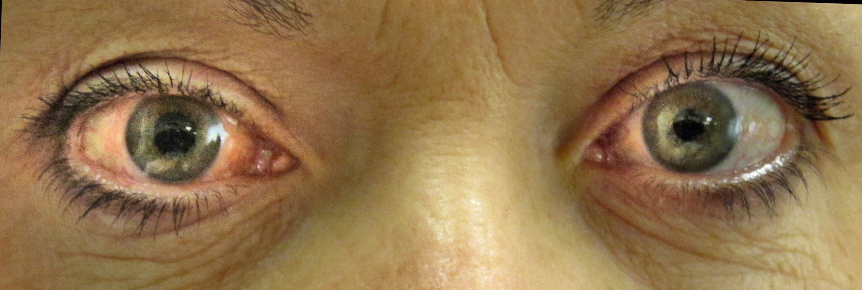
Primary open-angle glaucoma (POAG) is a chronic, progressive optic neuropathy defined by characteristic structural damage to the optic nerve and corresponding visual field loss, typically in the presence of an open anterior chamber angle and often (but not invariably) associated with elevated intraocular pressure (IOP) [19]B3b[30]A1a.
Synonyms and Abbreviations
- Primary open-angle glaucoma (POAG), the most common subtype.
- Open-angle glaucoma (OAG), broader term sometimes used interchangeably.
- Chronic open-angle glaucoma (COAG), historical term.
- Simple glaucoma, older term, rarely used.
- Normal-tension glaucoma (NTG), a POAG subtype with IOP consistently ≤21 mmHg.
- Juvenile-onset open-angle glaucoma (JOAG), onset before age 40 years, often more aggressive [21]C4.
Clinical Significance
POAG is the leading cause of irreversible blindness worldwide, affecting an estimated 57.5 million people in 2015 and projected to rise to 65.5 million by 2020 [5]B2a. It accounts for a substantial proportion of global visual impairment, with prevalence varying markedly by race, age, and geography [5]B2a[7]B2a.
Classification and Subtypes
POAG is subclassified into several clinical phenotypes, each with distinct demographic and prognostic features:
| Subtype | Key Distinguishing Feature | Associated Characteristics |
|---|---|---|
| High-tension POAG | IOP >21 mmHg at diagnosis | Classic risk profile; strong IOP-disease progression correlation [12]C4 |
| Normal-tension glaucoma (NTG) | IOP ≤21 mmHg at diagnosis | More common in Asian populations; vascular risk factors implicated [25]D5 |
| Juvenile-onset POAG (JOAG) | Onset <40 years | Often autosomal dominant; frequently requires early surgical intervention; cluster analysis identifies four phenotypes (e.g., normal iris crypts with featureless angle) [21]C4 |
Staging Systems
Functional staging of POAG severity relies on standard automated perimetry. The modified Glaucoma Staging System (mGSS) and Hodapp-Parrish-Anderson (HPA) classification show substantial agreement with the Advanced Glaucoma Intervention Study (AGIS) score, with mGSS recommended for clinic use due to its simplicity [35]C4. Structural assessment (e.g., vertical cup-to-disc ratio, retinal nerve fiber layer thickness) complements functional staging but is not a substitute for perimetry [30]A1a.
Diagnostic Criteria Heterogeneity
Despite decades of research, there is no universally accepted single definition of POAG across clinical trials, leading to 57 unique operational definitions in recent randomized controlled trials [30]A1a. The International Society of Geographical and Epidemiological Ophthalmology (ISGEO) classification provides a standardized framework for population-based studies, requiring both structural optic nerve damage and compatible visual field defects [9]C4[30]A1a.
From this foundational definition, the next section examines the pathophysiology and mechanisms that drive the progressive neurodegeneration central to POAG.
Pearl: There is no universally accepted diagnostic definition; the ISGEO classification is the most widely used for epidemiologic studies.
Pathophysiology & Mechanism
- ▸Elevated IOP in POAG stems from increased trabecular outflow resistance, primarily due to TGF-β2-driven extracellular matrix remodeling and cellular loss in the juxtacanalicular tissue.
- ▸Myocilin mutations (5% of cases) cause ER stress in TM cells; CAV1/2 variants impair mechanotransduction; NOS3 polymorphisms reduce NO bioavailability, all converging on reduced outflow facility.
- ▸RGC death results from combined mechanical injury at the lamina cribrosa, oxidative stress, neuroinflammation, and possibly altered cerebrospinal fluid pressure and microvascular insufficiency.
The disease originates from increased resistance to aqueous humor outflow through the conventional (trabecular) pathway [74]D5[73]D5. This resistance, generated primarily in the juxtacanalicular region of the trabecular meshwork (TM) and the inner wall of Schlemm's canal, is the dominant mechanism of intraocular pressure (IOP) elevation in primary open-angle glaucoma (POAG). The resulting IOP rise, along with other pathogenic factors, initiates a cascade of events that culminate in progressive retinal ganglion cell (RGC) death.
Outflow Obstruction: The Primary Defect
Aqueous humor leaves the eye through two principal routes. The conventional (trabecular) pathway accounts for approximately 80% of outflow and is the site of pathologic resistance in POAG [73]D5. The uveoscleral pathway drains the remaining 20% and typically remains unaffected. Morphologic studies show that the extracellular matrix (ECM) in the juxtacanalicular tissue (JCT) is increased in quantity and altered in composition in POAG eyes, narrowing the outflow channels and increasing hydraulic resistance [74]D5. This ECM remodeling is driven largely by transforming growth factor beta-2 (TGF-β2), which is consistently elevated in the aqueous humor of POAG patients [74]D5[97]D5. TGF-β2 binds its receptor, phosphorylates SMAD2/3, and translocates to the nucleus to upregulate pro-fibrotic genes such as connective tissue growth factor (CTGF), while simultaneously downregulating matrix-degrading enzymes [52]B2a[97]D5. The result is a net accumulation of fibronectin, collagen, and other ECM components in the JCT, stiffening the TM and increasing outflow resistance [74]D5[82]D5.
Molecular Drivers of Trabecular Meshwork Dysfunction
TGF-β2 signaling is not the only contributor. Thrombospondin-1 (TSP-1), which activates latent TGF-β2, is also elevated in POAG aqueous humor and potentiates the fibrotic phenotype [74]D5. Connective tissue growth factor (CTGF) acts as a downstream mediator of TGF-β2, directly inducing ECM synthesis [74]D5. In contrast, bone morphogenetic protein-7 (BMP-7) normally counteracts TGF-β2 signaling; its expression is reduced in POAG, tipping the balance toward fibrosis [74]D5.
Nitric oxide (NO) pathway dysregulation further impairs outflow. Endothelial NO synthase (eNOS) in Schlemm's canal (SC) endothelium produces NO in response to shear stress caused by IOP elevation; NO relaxes TM cells and increases SC permeability, reducing outflow resistance [54]D5. Decreased eNOS activity or NO bioavailability disrupts this homeostatic feedback loop, contributing to ocular [54]D5. Polymorphisms in NOS3 (the eNOS gene) modify the relationship between environmental exposures and glaucoma risk [54]D5.
Myocilin mutations account for approximately 5% of POAG cases, almost all in the olfactomedin domain [50]D5[86]D5. Mutant misfolds, aggregates within the endoplasmic reticulum (ER) of TM cells, and is not secreted; this triggers ER stress, activates the unfolded protein response, and leads to TM cell loss [50]D5[86]D5. The gain-of-toxic-function mechanism explains why heterozygotes develop disease and why the pathology is confined to the anterior segment [50]D5.
Caveolin dysfunction is implicated through genome-wide association studies (GWAS) linking the CAV1/CAV2 locus to POAG, particularly in women and those with early paracentral visual field loss [46]B3b[53]D5. Caveolae are plasma membrane invaginations that mediate mechanotransduction, endocytosis, and signaling. In the eye, caveolin-1 is highly expressed in TM and SC endothelium; its deficiency impairs outflow facility and alters cellular responses to mechanical stretch [51]D5[53]D5.
Age-related integrin dysregulation also drives TM fibrosis. With aging, α5β1 integrin expression declines in TM cells, while αvβ3 integrin becomes activated; this shift increases fibronectin assembly, α-smooth muscle actin expression, and cellular contractility, stiffening the tissue and restricting outflow [82]D5.
Genetic and Environmental Modulators of Susceptibility
GWAS have identified over 100 loci associated with POAG risk [88]D5[56]D5. Several are linked to IOP-independent pathways and may influence optic nerve vulnerability. Key loci include CDKN2B (cell-cycle regulation, associated with vertical cup-disc ratio), ATOH7 (neurodevelopment), SIX1, GAS7, AFAP1, and FNDC3B [47]B3b[68]B3b. The table below summarizes selected loci with known or proposed pathogenic mechanisms.
| Locus / Gene | Proposed Mechanism | Evidence |
|---|---|---|
| MYOC | Misfolded myocilin → ER stress → TM cell death | [50]D5[86]D5 |
| CAV1/CAV2 | Impaired mechanotransduction, outflow regulation | [46]B3b[53]D5 |
| CDKN2B | Cell-cycle dysregulation, altered cup-to-disc ratio | [47]B3b |
| ATOH7 | Neurodevelopmental defects, reduced RGC resilience | [47]B3b |
| GAS7, AFAP1, FNDC3B | Actin dynamics, TGF-β signaling (HTG-specific) | [68]B3b |
| NOS3 | eNOS activity → NO bioavailability → outflow tone | [54]D5 |
Oxidative stress and mitochondrial dysfunction are central to TM aging and damage. Aqueous humor from POAG patients has reduced antioxidant capacity, including lower ascorbate and total reactive antioxidant potential [62]C4[81]B3b. In TM tissue, especially from Black patients, nuclear factor erythroid 2-related factor 2 (NRF2) and catalase expression are lower than in matched lens epithelium, suggesting a tissue-specific antioxidant deficit [81]B3b. Mitochondrial dysfunction promotes the generation of reactive oxygen species (ROS), which in turn activate TGF-β2, induce ECM synthesis, and trigger TM cell senescence [49]D5[77]D5. Metabolomic profiling of POAG aqueous humor reveals dysregulation of glycolysis, fatty acid oxidation, and the electron transport chain, with decreased levels of the neuroprotective metabolites agmatine and thiamine [78]B3b.
Inflammatory para-inflammation is also dysregulated. Normally, the eye maintains a low-grade para-inflammatory state that clears debris and maintains homeostasis; in POAG, this balance shifts toward a pro-inflammatory phenotype [49]D5. Elevated levels of interleukin-8 (IL-8), macrophage inflammatory protein-1α (MIP-1α), fractalkine, and Flt3 ligand have been measured in the aqueous humor [44]B3b[83]B3b. Chronic inflammation in the TM and optic nerve contributes to tissue damage and may accelerate RGC loss [49]D5.
Vascular and perfusion mechanisms further modulate risk. Low diastolic ocular perfusion pressure, particularly in patients on systemic antihypertensive therapy, is associated with POAG [64]C4. Optic disc hemorrhages, a strong risk factor for progression, likely reflect microvascular insufficiency [67]D5. OCT angiography demonstrates reduced optic disc vessel density that correlates with visual field progression, even after adjusting for retinal nerve fiber layer thinning [65]C4.
Low cerebrospinal fluid (CSF) pressure may contribute to optic nerve head damage by increasing the translaminar pressure difference (IOP minus CSF pressure). In POAG patients, CSF pressure is significantly lower than in age-matched controls (mean 9.2 vs 13.0 mm Hg), and a larger translaminar pressure difference correlates with greater cup-to-disc ratio [43]B3b. This mechanical stress concentrates at the lamina cribrosa, where axonal bundles are compressed, impairing axoplasmic transport and triggering RGC apoptosis [43]B3b[67]D5.
Mechanisms of Optic Nerve Head Damage and RGC Loss
The final common pathway is RGC death. Mechanical compression at the lamina cribrosa obstructs anterograde and retrograde axonal transport, depriving RGCs of neurotrophic factors from the lateral geniculate nucleus and superior colliculus [49]D5[67]D5. Elevated IOP also induces strain on the optic nerve head, activating glial cells (astrocytes, microglia) that release inflammatory cytokines and reactive oxygen species, amplifying the injury [49]D5. Mitochondrial dysfunction and impaired mitophagy render RGCs vulnerable to energy failure and apoptosis [77]D5. Neurodegeneration extends beyond the eye: functional MRI and EEG studies show early cortical atrophy and altered activity in visual processing and attention networks, suggesting that POAG is a brain disease with ocular manifestations [80]B3b[94]B3b.
Pearl: The unifying pathogenic concept in POAG is that trabecular outflow obstruction, driven by TGF-β2-mediated fibrosis, oxidative stress, and genetic susceptibility, elevates IOP, which, together with vascular dysregulation and low CSF pressure, mechanically and biochemically damages the optic nerve, culminating in RGC death.
Epidemiology, Etiology & Risk Factors
- ▸Global POAG prevalence is 3.54% (111.8 million projected by 2040), with African ancestry conferring the highest risk (OR 2.80 vs. European).
- ▸More than half of European cases are undiagnosed, underscoring the need for targeted screening in high-risk groups.
- ▸Modifiable risk factors include SBP >130 mmHg, low BMI, evening light exposure, and favorable associations with SGLT2i, GLP-1RA, nicotinamide, metformin, and omega-3s.
From the pathophysiologic cascade of trabecular meshwork dysfunction and retinal ganglion cell loss, we now turn to the population-level determinants of who develops POAG. The global prevalence of glaucoma in adults aged 40-80 years is 3.54% (95% credible interval [CrI], 2.09-5.82), translating to 64.3 million affected individuals in 2013, projected to rise to 76.0 million by 2020 and 111.8 million by 2040 [101]B2a. In Europe, age-standardized prevalence is 2.99% (95% CI, 2.86-3.12%), with no temporal trend identified over the past three decades [102]B2a. More than half (56.4%) of European cases were previously undiagnosed, a proportion exceeding 80% in those under 55 years [102]B2a. In Asia, prevalence varies by subregion, with POAG predominating over primary angle-closure glaucoma; normal-tension glaucoma constitutes 70% of POAG in Chinese populations [103]B2a[4]B2a.
Demographic Risk Factors
Age is the strongest non-modifiable risk factor: the odds ratio per 5-year increase is 1.32 (95% CI, 1.29-1.36) [102]B2a. Men are more likely to have POAG than women (OR, 1.36; 95% CrI, 1.23-1.52) [101]B2a. African ancestry confers the highest risk: OR, 2.80 (95% CrI, 1.83-4.06) compared with European ancestry [101]B2a. In Africa, POAG prevalence at age 80 years reaches 12.2% (95% CrI, 8.9-16.6%) [5]B2a.
Modifiable and Systemic Risk Factors
| Factor | Association (OR/RR) | Evidence Source |
|---|---|---|
| Higher systolic BP (SBP 130-150 mmHg vs. 120-130 mmHg) | HR 1.16 (95% CI, 1.01-1.33) for incident POAG | [133]B2b |
| Low adult body mass index (vs. average) | HR 1.30 (95% CI, 1.19-1.43) | [106]B2b |
| Outdoor daylight exposure >1 hour/day | HR 1.10 (95% CI, 1.02-1.18) per additional hour | [125]B2b |
| Evening light >1000 lux (20:00-23:30) | HR 1.47 (95% CI, 1.07-2.03) for POAG | [142]B2b |
| SGLT2 inhibitor use (vs. ) | HR 0.64 (95% CI, 0.51-0.81) for POAG | [107]B2b |
| GLP-1 receptor agonist use (vs. insulin) | HR 0.58 (95% CI, 0.45-0.76) at 3 years | [111]B2b |
| Nicotinamide supplementation | HR 0.34 (95% CI, 0.25-0.47); absolute risk reduction 5.5%; NNT = 18 | [120]B2b |
| Metformin use (in diabetes) | HR 0.81 (95% CI, 0.67-0.97); NNT not calculable from reported data | [138]B2b |
| Omega-3 fatty acid supplementation | HR 0.45 (95% CI, 0.33-0.63) for POAG | [144]B2b |
Modifiable risk factors also include higher comorbidity burden (Charlson Comorbidity Index; OR 1.06-1.09 per point) [143]B2b and long-term systolic blood pressure variability (HR 1.12 per 1% increase in variability) [145]B2b.
Genetic Risk Factors
Heritability of POAG is estimated at 0.30-0.50 [75]D5. A polygenic risk score (PRS) in the highest decile is associated with a 7.4% POAG prevalence vs. 1.3% in the lowest decile (OR 1.74 per 1-SD increase) [118]B3b. A PRS threshold below the 48th percentile confers a 1.49-times higher likelihood of disease-free status at 20 years (95% CI, 1.04-2.15) [115]A1b. In African-ancestry populations, PRS performance is weaker, with ORs for the highest quintile ranging from 1.68 to 7.05 depending on ancestry composition [117]B3b.
Pearl: A single modifiable factor, evening light exposure >1000 lux, carries a hazard ratio of 1.47 (95% CI, 1.07-2.03) for POAG, comparable to the effect of a 10-mmHg IOP increase; advising patients to dim indoor lighting after 8 PM may represent a simple, low-cost preventive strategy [142]B2b.
| Factor | Association (OR/RR) | Evidence Source |
|---|---|---|
| Age (per 5-year increase) | OR 1.32 (95% CI, 1.29-1.36) | [102]B2a |
| Male sex (vs. female) | OR 1.36 (95% CrI, 1.23-1.52) | [101]B2a |
| African ancestry (vs. European) | OR 2.80 (95% CrI, 1.83-4.06) | [101]B2a |
| Higher systolic BP (130-150 mmHg) | HR 1.16 (95% CI, 1.01-1.33) | [133]B2b |
| Low adult BMI (vs. average) | HR 1.30 (95% CI, 1.19-1.43) | [106]B2b |
| Outdoor daylight (per +1 hr/day) | HR 1.10 (95% CI, 1.02-1.18) | [125]B2b |
| Evening light >1000 lux | HR 1.47 (95% CI, 1.07-2.03) | [142]B2b |
| SGLT2 inhibitor (vs. metformin) | HR 0.64 (95% CI, 0.51-0.81) | [107]B2b |
| GLP-1RA (vs. insulin) | HR 0.58 (95% CI, 0.45-0.76) | [111]B2b |
| Nicotinamide supplementation | HR 0.34 (95% CI, 0.25-0.47); NNT=18 | [120]B2b |
| Metformin (in diabetes) | HR 0.81 (95% CI, 0.67-0.97) | [138]B2b |
| Omega-3 fatty acids | HR 0.45 (95% CI, 0.33-0.63) | [144]B2b |
Clinical Presentation
- ▸POAG is asymptomatic until moderate-to-advanced stages; 50-90% of cases are undiagnosed in population surveys.
- ▸The cardinal sign is optic disc cupping with vertical cup-to-disc ratio >0.6, often asymmetric; disc hemorrhages mark active disease.
- ▸Normal-tension glaucoma (NTG) and juvenile open-angle glaucoma (JOAG) are distinct phenotypes with different genetic bases and clinical trajectories.
The transition from epidemiologic risk to clinical disease is silent in most patients. Population-based studies consistently report that 50-90% of POAG cases are undiagnosed in the community [171]B2c[173]B2c[176]B2c, reflecting the absence of early symptoms.
Presenting Symptoms
Patients typically perceive no visual loss until moderate-to-severe damage has accrued. The early deficits are peripheral, scotomatous, and binocularly overlapping. Symptoms emerge insidiously: difficulty with night vision, bumping into doorframes, or trouble reading. Inferior hemifield defects are particularly disabling, they impair near activities and peripheral vision and are associated with worse self-reported vision-related quality of life than superior defects of similar depth [170]B3b. In advanced disease, patients may report reduced contrast sensitivity, slower reading speed (by 19% compared with age-matched controls [184]B2c), and an increased risk of motor vehicle collisions (25% of severe POAG patients had crashed in a 10-year period vs 3.5% of controls [181]B3b). Even before visual field loss is detectable on perimetry, patients with preperimetric POAG demonstrate disrupted saccadic eye movements, lower peak velocity, hypometric saccades, and more antisaccade errors, suggesting subcortical or cortical compensatory dysfunction [80]B3b.
Optic Disc and Retinal Signs
The cardinal sign on examination is optic disc cupping. The vertical cup-to-disc ratio (VCDR) is typically >0.6, and often asymmetric between eyes (≥0.2 difference is suspicious). Focal neural rim thinning (notching), particularly at the inferotemporal and superotemporal poles, is characteristic. Disc hemorrhages, splinter-shaped, located at the rim, are a marker of active disease and predict progression. The retinal nerve fiber layer (RNFL) shows focal or diffuse atrophy, visible as slit defects on red-free ophthalmoscopy. In moderate-to-advanced disease, the optic disc may appear pale due to extensive cupping, and the retinal vessels may shift nasally. Macular involvement, detected on optical coherence tomography (OCT) as ganglion cell-inner plexiform layer (GCIPL) loss, correlates with visual acuity in moderate-advanced glaucoma: a thinner foveal ganglion cell complex and a larger foveal avascular zone on OCT angiography are associated with worse logMAR VA [177]B2c. Diffuse macular GCIPL loss, rather than focal loss, is a stronger predictor of diminished vision-related quality of life [172]B3b.
Variants and Phenotypic Subtypes
POAG encompasses a spectrum of presentations. High-tension POAG (IOP >21 mmHg at diagnosis) is the most common form in African-descent populations [166]B3b. Normal-tension glaucoma (NTG), defined by IOP ≤21 mmHg, accounts for 30-50% of POAG in East Asian populations; approximately 1-2% of NTG cases are caused by mutations in OPTN, TBK1, or MYOC [174]D5. Juvenile open-angle glaucoma (JOAG) presents before age 40 years, often with IOPs of 30-50 mmHg and a strong family history; MYOC mutations account for 10-20% of cases. Patients with higher mitochondrial genetic risk scores (TXNRD2 and ME3) tend to have higher treated IOP and a greater prevalence of paracentral visual field loss [169]B3b.
Red Flags and Atypical Presentations
The following features should prompt more urgent evaluation or aggressive :
- Rapid progression: Visual field mean deviation (MD) worsening at ≥36%/year. Risk factors include older age, larger baseline VCDR, worse baseline MD, and African American race [185]B2b.
- Recurrent disc hemorrhages, associated with faster progression.
- Unilateral or highly asymmetric cupping, suggests a secondary cause (e.g., angle closure, uveitic, traumatic) or a structural anomaly.
- IOP >30 mmHg at presentation in a young patient, consider JOAG with MYOC mutation.
- Family history of blindness from glaucoma, first-degree relative with POAG confers an odds ratio of 3.4 for disease [175]B3b.
- Inferior hemifield defect encroaching within 5° of fixation, threatens central vision.
Atypical presentations include NTG masquerading as non-glaucomatous optic atrophy (e.g., compressive lesion, ischemic optic neuropathy) and POAG in highly myopic eyes where fundus examination is challenging. Late presentation is more common in men, in patients with angle-closure glaucoma (vs POAG), and in those with elevated baseline IOP [189]B3b.
Pearl: The patient with POAG is typically asymptomatic until 30-50% of retinal ganglion cells are lost; the most common “symptom” is no symptom at all, so diagnosis depends entirely on the examiner’s diligence in detecting optic disc cupping and performing perimetry on at-risk individuals.
| Variant | Key Features | Frequency |
|---|---|---|
| High-tension POAG | IOP >21 mmHg at diagnosis; typical onset after age 50; progressive optic neuropathy | Most common in African descent populations [166]B3b |
| Normal-tension glaucoma (NTG) | IOP ≤21 mmHg; more common in East Asian populations; associated with disc hemorrhages, peripapillary atrophy, nocturnal hypotension | ~30-50% of POAG in East Asia; 1-2% due to OPTN, TBK1, or MYOC mutations [174]D5 |
| Juvenile open-angle glaucoma (JOAG) | Onset before age 40; often high IOP (30-50 mmHg); strong family history; MYOC mutations in 10-20% | Rare |
Clinical Examination and Diagnostic Workup
- ▸The diagnosis of POAG requires evidence of glaucomatous optic neuropathy and/or reproducible visual field loss, not merely elevated IOP.
- ▸Goldmann applanation tonometry, gonioscopy, and slit-lamp optic disc examination are the essential bedside tools; CCT and corneal hysteresis add prognostic value.
- ▸The OHTS risk calculator integrates IOP, CCT, age, PSD, and CDR to estimate 5-year conversion risk in ocular hypertensive patients.
The clinical presentation of POAG, often asymptomatic until moderate to advanced stages, places the burden of detection on the structured ophthalmic examination. The bedside evaluation combines tonometry, gonioscopy, and optic disc assessment to raise suspicion, while formal diagnosis requires confirmation with structural and functional testing (covered in the next section).
Bedside Ophthalmic Examination Battery
Tonometry. Goldmann applanation tonometry remains the gold standard for intraocular pressure (IOP) measurement. IOP >21 mmHg is a major risk factor but not diagnostic; many patients with POAG have IOP in the normal range (normal-tension glaucoma). The OHTS demonstrated that baseline IOP, central corneal thickness (CCT), age, pattern standard deviation (PSD), and cup-to-disc ratio (CDR) predict conversion to POAG [99]B2b. A single IOP reading is insufficient, diurnal variation and the effect of medications must be considered. In the medication washout study, discontinuation of 1, 2, and 3 medications was associated with mean IOP increases of 5.4, 6.9, and 9.0 mmHg, respectively [100]B2b.
Gonioscopy. Direct gonioscopy (e.g., Goldmann or Zeiss lens) is essential to differentiate open-angle from angle-closure glaucoma. In POAG, the angle is open without peripheral anterior synechiae. Gonioscopy also identifies pseudoexfoliation material, pigment dispersion, and angle dysgenesis (in juvenile-onset cases [21]C4).
Optic Disc Examination. Slit-lamp biomicroscopy with a 78D or 90D lens is the standard. Key findings: vertical CDR >0.5, focal rim notching, optic disc hemorrhage, retinal nerve fiber layer (RNFL) defects, and asymmetry between eyes. A deep learning model applied to OHTS fundus photographs achieved an area under the receiver operating characteristic curve (AUROC) of 0.88 for detecting POAG based on optic disc or visual field changes [208]B3b. Predicted RNFL thickness from photographs is also a risk factor for conversion (HR 1.97 per 10 μm thinner) [99]B2b.
Central Corneal Thickness. Measured by pachymetry; thinner CCT is associated with higher risk of POAG development and progression. In OHTS, CCT was a significant predictor of conversion [99]B2b.
Corneal Hysteresis (CH). Lower CH is independently associated with glaucoma and with an increased risk of progression, even in patients with apparently well-controlled IOP [203]B2a. CH measurement complements structural and functional assessments, though it is not yet universally adopted.
Slit-Lamp Examination. Assess anterior chamber depth, lens status, and signs of pseudoexfoliation or pigment dispersion. The presence of pseudoexfoliation material increases the risk of glaucoma progression and blindness [109]B2a.
Systemic Examination. Blood pressure measurement is relevant: is associated with higher IOP (pooled increase of 0.26 mmHg per 10 mmHg increase in systolic BP) [197]B2a, and higher systolic blood pressure and pulse pressure are linked to incident POAG [133]B2b. Low adult body size is also a risk factor (HR 1.35 for low-low trajectory) [106]B2b.
Diagnostic Algorithm
The diagnosis of POAG requires evidence of glaucomatous optic neuropathy (structural damage) and/or reproducible visual field loss (functional damage) in the presence of an open angle. IOP is a risk factor, not a criterion. The OHTS risk calculator integrates age, IOP, CCT, PSD, and CDR to estimate 5-year risk of POAG in ocular hypertensive patients [99]B2b.
Gold standard for diagnosis is a combination of:
- Optic disc appearance (CDR ≥0.6, asymmetry, notching, hemorrhage)
- RNFL thinning on OCT (or predicted from photographs [99]B2b)
- Reproducible visual field defect on standard automated perimetry (24-2 or 10-2)
The Fast-PACE study demonstrated that clustered testing over 6 months identifies fast progressors with 93% sensitivity (95% CI 66%-100%) and 85% specificity [201]B2b.
Genetic Testing
Routine genetic testing is not recommended for POAG. The diagnostic yield of whole-exome sequencing in POAG is low (8.9%) [199]B2a. However, testing for MYOC, OPTN, and WDR36 mutations is justified in early-onset or familial cases (e.g., juvenile open-angle glaucoma) [219]D5. Polygenic risk scores for IOP and vertical cup-to-disc ratio explain 12.9% and 22.0% of phenotypic variance, respectively [15]B2b, but are not yet ready for clinical use [224]D5. A multitrait PRS can stratify risk in pseudoexfoliation syndrome (OR 4.22 for glaucoma diagnosis) [204]B3b.
Differential Diagnosis
Must exclude other causes of optic neuropathy and visual field loss: compressive lesions, ischemic optic neuropathy, non-arteritic anterior ischemic optic neuropathy (NAION), autoimmune retinopathy [207]C4, vitreomacular traction [48]C4, and secondary glaucomas (e.g., pseudoexfoliative, pigmentary, uveitic).
Pearl: The clinical examination for POAG must never rely on IOP alone, more than half of diagnosed cases have IOP <21 mmHg at presentation. The combination of optic disc assessment, gonioscopy, and pachymetry, with selective use of corneal hysteresis, provides the most informative bedside evaluation.
| Component | Tool | Key Finding | Diagnostic Performance |
|---|---|---|---|
| Tonometry | Goldmann applanation | IOP >21 mmHg (risk factor) | Sensitivity/specificity limited; alone not diagnostic |
| Gonioscopy | Goldmann/Zeiss lens | Open angle, no PAS | Essential for differential diagnosis |
| Optic disc exam | 78D/90D lens | CDR >0.5, notch, hemorrhage, RNFL defect | Deep learning AUROC 0.88 for detecting POAG [208]B3b |
| Pachymetry | Ultrasound or optical | CCT | Predictor of conversion (HR 1.97 per 10 μm thinner) [99]B2b |
| Corneal hysteresis | Ocular Response Analyzer | Lower CH | Associated with progression risk [203]B2a |
Ophthalmic Imaging and Functional Testing
- ▸OCT RNFL thickness has diagnostic accuracy ~0.77 but is lower in African descent, highlighting the need for race-specific normative data.
- ▸OCTA peripapillary vessel density is a strong predictor of VF progression (HR 1.05 per 1% decrease); fast VD loss (≥0.75%/yr) doubles the risk of progression.
- ▸Clustered testing (Fast-PACE protocol) identifies fast progressors with 93% sensitivity over 6 months, enabling early intervention.
From the clinical examination, the quantitative, serially-tracked diagnostic arsenal of optical coherence tomography (OCT), OCT angiography (OCTA), and standard automated perimetry (SAP) provides the objective measurements on which glaucoma staging, treatment thresholds, and progression monitoring are built.
Optical Coherence Tomography (OCT)
Peripapillary retinal nerve fiber layer (RNFL) thickness remains the most widely used structural parameter. In network meta-analysis, average and inferior RNFL parameters achieve a diagnostic accuracy of 0.77 [228]B3a. However, accuracy is significantly lower in eyes of African descent (area under the receiver operating curve [AUROC] 0.85 vs 0.91 for European descent, P=0.04) [251]B3b. The ISNT rule (inferior > superior > nasal > temporal rim width) is maintained in about two-thirds of normal eyes but is not reliable for individual diagnosis [275]B3b.
The macular ganglion cell-inner plexiform layer (GCIPL) is particularly useful for detecting early damage. In preperimetric glaucoma, GCIPL thickness loss (4.72%) is similar to vessel density loss (4.97%), but in early perimetric glaucoma, GCIPL loss (9.86%) exceeds vessel density loss (6.93%) [246]C4. Diffuse rather than focal GCIPL loss is associated with a 6.15-point lower NEI VFQ-25 composite score (P=0.03), indicating worse vision-related quality of life [172]B3b.
Lamina cribrosa (LC) morphology provides prognostic information. The adjusted LC curvature index (aLCCI) above 4.12 is associated with faster subsequent visual field (VF) deterioration (P<0.001) [236]B2b. Eyes with acquired optic disc pits have faster global RNFL thinning (-1.44 vs -0.93 μm/yr, P=0.008) [244]B3b. The laminar dot sign on stereophotography indicates thinner prelaminar tissue and a higher risk of structural progression (84.9% vs 25.8%) [276]B2b.
Optical Coherence Tomography Angiography (OCTA)
OCTA measures vessel density (VD) without the floor effect that limits OCT in advanced glaucoma [268]A1a. In meta-analysis, each 1% decrease in baseline peripapillary VD was associated with a 5% increased risk of VF progression (HR 1.05, 95% CI 1.02-1.07) [268]A1a. Eyes with fast initial VD loss (≥0.75% per year) had a VF progression rate of -0.43 dB/yr vs -0.15 dB/yr for slow progressors (P=0.006) [238]B2b. Fast OCTA progressors had a 1.96-fold higher risk of VF progression [243]B2b.
Choroidal microvasculature dropout (MvD) is more prevalent in POAG than in pseudoexfoliation glaucoma (79.5% vs 46.2%) [134]C4. Eyes with MvD have faster ganglion cell complex thinning (mean difference -0.50 μm/yr, P=0.003) [262]B2b. MvD enlargement is associated with greater intraocular pressure fluctuation and worse baseline VF mean deviation [216]B3b.
Foveal avascular zone (FAZ) enlargement is associated with faster GCIPL thinning (-0.7 μm/yr, P=0.026) and VF MD loss (-0.3 dB/yr, P=0.017) [260]B2b. However, FAZ area does not differ between healthy and glaucomatous eyes after axial length adjustment [215]B3b.
Standard Automated Perimetry (SAP)
The 24-2 Swedish Interactive Thresholding Algorithm is the standard for functional assessment. The Fast-PACE study demonstrated that clustered testing (5 weekly visits repeated after 6 months) can identify fast progressors with 93% sensitivity and 85% specificity over 6 months [201]B2b. The 10-2 test is more sensitive for central defects. Lower peripapillary VD on OCTA is associated with faster VF loss on both 24-2 and 10-2 testing [238]B2b.
Emerging Measures
Contrast sensitivity function is impaired across all spatial frequencies in POAG, with low spatial frequencies most affected [258]C4. Epigenetic age acceleration (Horvath clock) is associated with 15% higher odds of fast progression per year of acceleration (OR 1.15) [205]B3b. The macular naso-temporal ratio distinguishes POAG from chiasmal compression with AUC 95.3% (95% CI 90-100%) [255]B3b.
These structural and functional measures are integrated into severity staging systems and risk stratification algorithms, which are discussed in the next section.
Pearl: The combination of OCT RNFL thickness and peripapillary vessel density provides complementary information for predicting progression; eyes with fast OCTA vessel density loss (≥0.75%/yr) have a nearly 2-fold increased risk of VF progression [238]B2b[243]B2b.
Severity, Staging & Risk Stratification
- ▸Staging with mGSS or HPA provides substantial agreement with AGIS and is recommended for clinical use.
- ▸Thinner RNFL, lower CCT, lower corneal hysteresis, and presence of disc hemorrhage are the most validated risk factors for progression.
- ▸Polygenic risk scores show promise but have limited discriminative accuracy in non-European populations and are not yet ready for population screening.
Building on the diagnostic data from imaging and perimetry, the clinician's next task is to assign a validated severity tier, a classification that directly governs treatment targets, follow-up intervals, and surgical thresholds.
Staging Systems for Visual Field Severity
Several perimetric staging systems translate mean deviation (MD) and pattern deviation into ordinal categories. The Hodapp-Parrish-Anderson (HPA) classification and the modified Glaucoma Staging System (mGSS) show substantial agreement with the Advanced Glaucoma Intervention Study (AGIS) score (Cohen κ = 0.686-0.687 and 0.687, respectively) [35]C4. The enhanced Glaucoma Staging System (eGSS) shows only slight agreement (κ = 0.103) [35]C4. The mGSS is recommended for routine clinical use owing to its simplicity and clarity [35]C4.
In the Ocular Treatment Study (OHTS), eyes that developed POAG had a mean post-diagnosis MD slope of -0.40 ± 0.64 dB/year for all POAG eyes; 41% of VF POAG eyes progressed at ≤ -0.5 dB/year and 21% at ≤ -1.0 dB/year [195]B2b. These rates underscore the need for early detection of fast progressors to prevent vision loss.
| System | Stages | Basis | Agreement with AGIS (κ) |
|---|---|---|---|
| HPA | Early, Moderate, Severe | MD and pattern deviation | 0.686 |
| mGSS | 1-5 | MD and pattern deviation | 0.687 |
| eGSS | 0-5 | MD and pattern deviation | 0.103 |
| AGIS | 0-20 | Scoring of six test points | Reference |
Structural Staging with OCT
Retinal nerve fiber layer (RNFL) thickness and ganglion cell complex (GCC) thickness provide structural severity. In the OHTS, predicted baseline RNFL thickness (from deep learning) was a predictor of conversion to POAG: **HR 1.97 per 10-μm thinner RNFL ** [99]B2b. Longitudinal change in predicted RNFL (per 1-μm/year faster loss) had an HR of **6.01 ** [99]B2b.
OCT angiography parameters add prognostic value. Lower baseline peripapillary vessel density (VD) is associated with increased risk of VF progression (HR 1.05 per 1% decrease, 95% CI 1.02-1.07) [268]A1a. The presence of choroidal microvascular dropout (MvD) is associated with faster GCC thinning (mean difference -0.50 μm/year, 95% CI -0.83 to -0.17) [262]B2b.
Risk Stratification for Progression
Multiple baseline factors stratify risk:
- Central corneal thickness (CCT): Strong evidence that thinner CCT is a risk factor for progression from ocular hypertension to POAG [285]A1c.
- Corneal hysteresis (CH): Lower CH is associated with increased risk of progression, even in eyes with apparently well-controlled IOP [203]B2a.
- Myopia: Myopic refractive error is associated with increased POAG risk (OR 2.95 for moderate/high myopia) [3]B2a. However, in established POAG, high myopia may be a protective factor for structural progression (HR 0.323, P = 0.031) [295]B2b, possibly due to difficulty in detecting change.
- Blood pressure variability: Higher systolic BP variability is associated with faster VF progression (coefficient -0.127 dB/year per 1% increase, 95% CI -0.247 to -0.008) [145]B2b.
- Systemic comorbidity: Higher age-adjusted Charlson Comorbidity Index (age-CCI) is associated with increased odds of progression: OR 1.06 per 1-point increase for early-to-moderate stage, OR 1.09 for progression to severe stage [143]B2b.
Genetic Risk Scores
Polygenic risk scores (PRS) can stratify POAG risk. In the UK Biobank, a 1-SD increase in PRS was associated with 1.74-fold higher odds of POAG (95% CI 1.71-1.77) [118]B3b. POAG prevalence in the highest decile was 7.4% vs 1.3% in the lowest decile [118]B3b. However, performance varies by ancestry: in African-ancestry populations, ORs for the highest quintile range from 1.68 to 7.24 depending on the PRS [117]B3b. Genetic risk scores currently show moderate discriminative accuracy (AUC 0.56-0.65) [113]B3b and are not yet ready for population screening [224]D5.
Pearl: The presence of optic disc hemorrhage at baseline confers a 4.5-fold increased odds of progression (OR 4.51, 95% CI 3.24-6.26) and should prompt intensified IOP-lowering and closer follow-up [299]B2a.
Acute & Vision-Threatening Management
- ▸Acute management is triggered by IOP >30 mmHg, disc edema, vision loss, or rapid progression despite therapy; gonioscopy must rule out angle closure before treatment.
- ▸Immediate combination therapy with topical aqueous suppressants and a prostaglandin analog, plus oral acetazolamide if needed, achieve rapid IOP reduction.
- ▸SLT and trabeculectomy provide surgically effective IOP control when medical therapy fails or as early intervention in advanced disease.
Once severity is staged, patients with advanced damage or rapid progression require urgent intervention to prevent irreversible vision loss. While primary open-angle glaucoma is typically a chronic disease, acute IOP elevation in the setting of already compromised optic nerve function constitutes a vision-threatening emergency demanding immediate treatment.
Step 1: Identify the Acute or Vision-Threatening Scenario
Urgent is indicated when any of the following are present: IOP >30 mmHg, optic disc edema, corneal edema, acute vision loss, or documented rapid progression of visual field defects despite maximal medical therapy [300]A1c[301]A1c. The first critical step is to confirm open angles on gonioscopy, acute angle-closure glaucoma must be ruled out before initiating therapy. Once the angle is confirmed open, proceed with immediate IOP reduction.
Step 2: Initial Medical Therapy for Urgent IOP Reduction
Initiate topical therapy with a combination of aqueous suppressants and a prostaglandin analog [300]A1c. The AAO Preferred Practice Pattern recommends a stepped approach using one or more of the following classes: prostaglandin analogs, beta-blockers, alpha-2 agonists, and carbonic anhydrase inhibitors [300]A1c. For acute IOP spikes, add oral acetazolamide (if not contraindicated) to achieve rapid systemic aqueous suppression [300]A1c. If corneal edema is present and topical penetration may be impaired, consider intravenous acetazolamide or mannitol [300]A1c. Monitor IOP hourly or as needed until it falls below a safe threshold (typically <25 mmHg in severe disease) [301]A1c.
Step 3: Early Procedural Intervention
If medical therapy fails to adequately lower IOP or if the clinical scenario demands rapid sustained control, proceed with laser or incisional surgery. Selective laser trabeculoplasty (SLT) has been shown to achieve a mean IOP reduction of 7.3 to 8.3 mmHg (34-39%) within 12 months in treatment-naïve patients (308) (2b). In a cohort of African-derived patients in the developing world, the 12-month Kaplan-Meier survival rate (≥10% IOP reduction from baseline) was 77.7% [308]C4. For eyes with advanced damage or in resource-limited settings, early trabeculectomy provides better IOP control than medical therapy alone, mean IOP at 6 months was 18.5 mmHg vs 22.8 mmHg in medically treated patients (P<0.001) [160]A1b (1b).
| Indication for Urgent Intervention | Recommended First Step | Second-Line / Escalation | Evidence Level |
|---|---|---|---|
| IOP >30 mmHg with advanced damage | Combination topical therapy + oral acetazolamide | IV acetazolamide or mannitol; urgent SLT or trabeculectomy | 1c [300]A1c |
| Optic disc edema or acute vision loss | Topical aqueous suppressants + prostaglandin analog | Proceed to incisional surgery if no response within hours | 5 [300]A1c[321]D5 |
| Rapid progression despite maximal medical therapy | Offer SLT (308) (2b) or trabeculectomy [160]A1b (1b) | Glaucoma drainage device or cyclodestruction | 1b-2b |
Step 4: Monitoring and Titration
After acute reduction, re-evaluate IOP at intervals appropriate to the initial severity, hourly until stable, then daily, then weekly as the eye stabilizes [300]A1c. Assess visual fields and optic nerve imaging at baseline and regularly thereafter to guide further titration of therapy [301]A1c. If IOP remains above the individualized target (e.g., ≤15 mmHg for advanced disease), escalate therapy by adding another medication class or proceeding with laser/surgery.
Step 5: Transition to Long-Term Management
Once the acute threat is controlled, transition the patient to a structured long-term care plan. Refer to the section for guidance on selecting sustained medical therapy, repeat SLT, trabeculectomy, or tube shunt surgery. Document the baseline damage and establish clear IOP targets and follow-up intervals to prevent future acute episodes [300]A1c[321]D5.
What NOT to Do: Do not administer laser peripheral iridotomy in a patient with confirmed open angles, it is ineffective and may cause complications. Do not defer treatment when IOP is very high and the optic disc is at risk, assuming the patient will tolerate the pressure chronically. Do not rely solely on a single medication in an acute setting; combination therapy is necessary [300]A1c.
Pearl: In the acute vision-threatening setting, confirm open angles first, then rapidly lower IOP with combination topical therapy and oral acetazolamide; once IOP is controlled, proceed to SLT or trabeculectomy without delay to prevent further irreversible damage [300]A1c[308]C4[160]A1b.
Long-term & Definitive Management
- ▸First-line therapy is a prostaglandin analog (latanoprost, travoprost, bimatoprost) or selective laser trabeculoplasty (SLT); both achieve ~25-30% IOP reduction.
- ▸Trabeculectomy provides the greatest IOP reduction (mean 10.7 mmHg at 2 years) but carries higher hypotony risk; MIGS offers a safer alternative for mild‑moderate disease.
- ▸Long-term monitoring with perimetry and OCT every 6-12 months is essential; fast progressors can be identified with clustered testing over 6 months.
Once acute IOP elevation has been controlled, the focus shifts to sustained IOP reduction to prevent progressive optic neuropathy. The target IOP is individualized based on disease severity, baseline IOP, and rate of progression, typically a 20-30% reduction from untreated baseline, with lower targets (e.g., <15 mmHg) for advanced disease. follows a stepwise ladder: medical therapy, laser trabeculoplasty, and incisional surgery.
Step 1: First-Line Medical Therapy
A prostaglandin F2α analog (PGFA) is the recommended first-line agent. A 2025 component network meta-analysis of 166 trials (36 494 participants) ranked PGFAs as the most effective monotherapy class, with a mean IOP reduction of -4.75 mmHg (95% CI -5.19 to -4.31; high certainty) [330]A1a. Nitric oxide-donating prostaglandin analogs (NO-PGA) showed slightly greater reduction (-5.15 mmHg) but with low certainty [330]A1a. Among PGFAs, latanoprost, travoprost, and bimatoprost are similarly effective; a 24‑hour meta-analysis found bimatoprost (29%) and travoprost (27%) produced the greatest diurnal reduction [332]A1a. Latanoprost has the longest treatment persistence (mean 52 months) [346]B2b.
Dosing: Latanoprost 0.005%, travoprost 0.004%, or bimatoprost 0.01%, one drop in the affected eye(s) once daily in the evening. Bimatoprost 0.03% is also available but associated with more conjunctival hyperemia.
Alternative first-line options: The EP2 receptor agonist omidenepag isopropyl 0.002% once daily was noninferior to latanoprost in the phase 3 AYAME study (mean IOP reduction -5.93 vs -6.56 mmHg; 95% CI for difference 0.01-1.26) [334]A1b. Rho‑kinase inhibitors (ripasudil 0.4%, netarsudil 0.02%) are approved as second-line add-on therapy; netarsudil added to latanoprost provided an additional -2.36 mmHg reduction at week 4 (J‑ROCKET‑2) [218]A1b.
What NOT to do: Do not use two prostaglandin analogs simultaneously, no additive effect, increased side effects. Avoid beta‑blockers (timolol) in patients with asthma, , or bradycardia. Avoid brimonidine in young children (risk of CNS depression).
Step 2: Combination Therapy and Laser Trabeculoplasty
If target IOP is not achieved with monotherapy, options include adding a second agent or proceeding to laser trabeculoplasty.
Add-on medical therapy: The component network meta-analysis identified PGFA plus carbonic anhydrase inhibitor (CAI) as the most effective dual combination, with synergy (-2.05 mmHg beyond additive) [330]A1a. PGFA plus beta‑blocker showed antagonism (+1.26 mmHg) [330]A1a. Non‑PGFA triple combinations (alpha‑agonist + beta‑blocker + CAI) achieved -7.22 mmHg (95% CI -9.04 to -5.40) [330]A1a. Fixed combinations improve adherence.
Selective laser trabeculoplasty (SLT): The LiGHT trial (718 patients) compared initial SLT vs topical medication and found SLT noninferior for IOP control and quality of life at 3 years, with lower treatment burden and cost [155]A1b[153]A1b. SLT increases tonographic outflow facility by 37-41% [337]A1b. A meta-analysis of unilateral SLT showed a contralateral IOP reduction of 1.85 mmHg (95% CI 1.23-2.47) at 3-6 months, suggesting a systemic or neurogenic effect [110]B2a. SLT can be repeated; 360° treatment is more effective than 180° but with similar safety [337]A1b.
Treatment failure protocol for SLT: If IOP reduction <20% at 3 months, consider repeat SLT or proceed to incisional surgery.
Step 3: Incisional Surgery
When medical and laser therapy fail to achieve target IOP or disease progresses, incisional surgery is indicated.
Trabeculectomy remains the gold standard. The MicroShunt versus trabeculectomy randomized trial (527 patients) reported 2‑year success (≥20% IOP reduction without medication increase) of 64.4% for trabeculectomy vs 50.6% for MicroShunt (P = 0.005) [326]A1b. Mean IOP at 2 years was 10.7 mmHg (trabeculectomy) vs 13.9 mmHg (MicroShunt) [326]A1b. Hypotony was more common with trabeculectomy (51.1% vs 30.9%) [326]A1b. The Treatment of Advanced Glaucoma Study economic model found trabeculectomy cost‑effective over a lifetime (incremental cost per QALY £9679) [348]B2c.
Glaucoma drainage devices (tube shunts): Used when trabeculectomy fails or in eyes with conjunctival scarring. The IRIS® Registry analysis showed tube shunts accounted for 39% of first surgeries in POAG [167]B2b.
Minimally invasive glaucoma surgery (MIGS): Options include trabecular microstents (iStent inject, Hydrus), ab interno trabeculotomy (Kahook Dual Blade, Tanito microhook), and subconjunctival implants (Xen, MicroShunt). The HORIZON trial (5‑year data) showed that Hydrus microstent combined with cataract surgery achieved a higher proportion of eyes with IOP ≤18 mmHg without medications vs cataract surgery alone (49.5% vs 33.8%; P = 0.003; NNT = 7) and reduced need for subsequent incisional glaucoma surgery (2.4% vs 6.2%; P = 0.027) [148]A1b. The Swedish MIGS study found combined cataract surgery with iStent Inject W or Kahook Dual Blade Glide achieved higher surgical success than cataract surgery alone (87.5% vs 52.5%; P < 0.001) [350]A1b. A systematic review of 30 RCTs concluded MIGS is safe and effective for mild‑to‑moderate POAG [366]A1a.
Cyclodestructive procedures (e.g., transscleral cyclophotocoagulation) are reserved for refractory cases or eyes with poor visual potential due to higher complication rates [57]A1a.
Step 4: Monitoring and Escalation
Patients require lifelong monitoring. The OHTS 20‑year follow-up showed that even with treatment, the cumulative incidence of POAG was 41.9% in the medication group [309]B2b. Visual field progression occurs at a mean rate of -0.40 dB/year after POAG diagnosis, with 21% of eyes progressing at ≥-1.0 dB/year [195]B2b. The Fast‑PACE study demonstrated that clustered testing over 6 months can identify fast progressors with 93% sensitivity [201]B2b.
Escalation triggers: Confirmed progression on perimetry or OCT, IOP consistently above target, or intolerance to medications. Consider switching from medical to laser or surgical therapy.
Controversies and Guideline Disagreement
| Question | Position A | Position B | Strength | Implication for practice |
|---|---|---|---|---|
| First-line treatment: SLT vs medication | LiGHT trial supports SLT as first-line due to similar efficacy, lower cost, and reduced medication burden [155]A1b | AAO Preferred Practice Pattern recommends prostaglandin analog as first-line medical therapy | Moderate | SLT is a reasonable first-line option, especially in patients with adherence concerns or medication intolerance. |
| MIGS vs trabeculectomy for mild‑moderate POAG | MIGS preferred by 20.3% of AGS members for themselves (ab interno trabeculotomy) [365]D5 | Trabeculectomy remains more effective for IOP reduction (mean 10.7 vs 13.9 mmHg) [326]A1b | Strong | MIGS is appropriate for mild‑moderate disease with lower target IOP; trabeculectomy for advanced disease or lower targets. |
Pearl: Initiate therapy with a prostaglandin analog or SLT; escalate to combination medical therapy, then to trabeculectomy or MIGS based on disease severity and progression rate, the goal is a sustained IOP reduction of ≥20% from baseline, with lower targets for advanced disease (OHTS, LiGHT, HORIZON) [309]B2b[155]A1b[148]A1b.
| Drug Class | Examples | Starting Dose | Target/Max Dose | Key Monitoring |
|---|---|---|---|---|
| Prostaglandin analog | Latanoprost 0.005% | 1 drop qHS | 1 drop qHS | IOP, conjunctival hyperemia, iris color change |
| Beta-blocker | Timolol 0.5% | 1 drop BID | 1 drop BID | Heart rate, blood pressure, asthma/COPD |
| Alpha-agonist | Brimonidine 0.2% | 1 drop TID | 1 drop TID | CNS depression (children), dry mouth |
| Carbonic anhydrase inhibitor | Dorzolamide 2% | 1 drop TID | 1 drop TID | Corneal edema, sulfa allergy |
| Rho-kinase inhibitor | Ripasudil 0.4% | 1 drop BID | 1 drop BID | Conjunctival hyperemia (common, transient) |
| EP2 agonist | Omidenepag 0.002% | 1 drop qHS | 1 drop qHS | Conjunctival hyperemia, corneal thickening |
| Fixed combination | Latanoprost/timolol | 1 drop qAM | 1 drop qAM | As per components |
| Procedure | Mean IOP at 1-2 yr | Medication Reduction | Success Rate | Key Adverse Events | Evidence |
|---|---|---|---|---|---|
| Trabeculectomy | 10.7 mmHg [326]A1b | -2.5 meds [326]A1b | 64.4% at 2 yr [326]A1b | Hypotony 51%, bleb leak, infection | 1b |
| MicroShunt | 13.9 mmHg [326]A1b | -2.2 meds [326]A1b | 50.6% at 2 yr [326]A1b | Hypotony 31%, implant reposition 6.8% | 1b |
| Hydrus microstent + cataract | 14.5 mmHg (5 yr) [148]A1b | -0.5 meds [148]A1b | 49.5% IOP ≤18 no meds [148]A1b | Transient IOP spike, hyphema | 1b |
| iStent inject + cataract | 15.2 mmHg (2 yr) [350]A1b | -0.8 meds [350]A1b | 87.5% success (≥20% IOP or -1 med) [350]A1b | Hyphema 15%, IOP spike 10% | 1b |
| SLT | 13-14 mmHg (1 yr) [308]C4 | Variable | 77.7% survival at 1 yr [308]C4 | Transient photophobia, IOP spike | 1b |
History and Evolution of Treatment
- ▸The Jay & Murray trial established that early trabeculectomy achieves better IOP control and field preservation than medical therapy alone in moderate-to-severe POAG.
- ▸Prostaglandin analogues (latanoprost, bimatoprost) replaced older agents as first-line medical therapy due to superior efficacy and once-daily dosing.
- ▸The LiGHT trial demonstrated that SLT is non-inferior to topical medication as first-line therapy and is cost-effective, shifting guidelines toward laser-first approaches.
- ▸MIGS (Hydrus, iStent, Kahook, Tanito microhook) combined with cataract surgery reduces medication burden and IOP in mild-to-moderate POAG, but trabeculectomy remains more effective for advanced disease.
The preceding sections have outlined the current of POAG, but the present standard of care is the product of a series of landmark trials that reshaped practice over the past five decades. Understanding this evolution grounds each therapeutic decision in its evidentiary origin.
Early Surgery vs. Medical Therapy
In the 1980s, the prevailing question was whether to operate early or start with topical medications. The randomized trial by Jay and Murray (1988) addressed this directly: newly diagnosed patients with IOP ≥26 mmHg and field loss were assigned to initial trabeculectomy or conventional medical therapy followed by surgery if needed. At 1 year, the mean IOP after trabeculectomy was 15.0 mmHg compared with 20.8 mmHg in those controlled on medical therapy alone [370]A1b. Early surgery provided significantly better protection of visual field, and delayed surgery failed to prevent the extra loss that occurred in the preoperative period [370]A1b. Within 4 years, 53% of medically managed eyes required trabeculectomy [370]A1b. This trial established that for moderate-to-severe POAG, early surgery achieves superior IOP control and field preservation.
The Advent of Prostaglandin Analogues
The introduction of prostaglandin analogues in the 1990s revolutionized medical therapy. Latanoprost 0.005% once daily reduced IOP by 8.2 to 8.9 mmHg (32-34%) from baseline, a reduction sustained over 2 years [373]C4. In a -to-head comparison, latanoprost lowered IOP by 7.2 mmHg (28%) versus 3.9 mmHg (15%) for unoprostone, which was subsequently abandoned [369]A1b. Bimatoprost 0.03% offered slightly better diurnal IOP control than latanoprost in a 30-day trial, though the difference was not statistically significant at individual time points [376]A1b. The fixed combination bimatoprost/timolol provided a 12-hour mean IOP of 17.7 mmHg versus 18.5 mmHg with latanoprost/timolol, a small but statistically significant advantage [340]A1b. Prostaglandin analogues became first-line therapy because of their efficacy, once-daily dosing, and minimal systemic side effects.
The Laser Paradigm Shift
Selective laser trabeculoplasty (SLT) emerged as a safe alternative to medication, but it was not until the LiGHT trial (2012-2014) that its role as first-line therapy was defined. In this pragmatic, multicentre randomized trial of 718 previously untreated patients with POAG or ocular , initial SLT was non-inferior to topical medication in quality-of-life outcomes and was more cost-effective [155]A1b. SLT also reduced the need for long-term drops. At 12 months, complete success (≥20% IOP reduction without medications) was achieved in 25.4% of SLT-treated eyes [154]A1b. The LiGHT trial fundamentally shifted guidelines, and SLT is now recommended as a first-line treatment option for many patients.
The MIGS Revolution
Micro-invasive glaucoma surgery (MIGS) developed as a bleb-independent approach with a superior safety profile for mild-to-moderate disease. The HORIZON trial evaluated the Hydrus microstent combined with cataract surgery versus cataract surgery alone. At 5 years, 49.5% of microstent eyes achieved IOP ≤18 mmHg without medications versus 33.8% in the control group (P=0.003), and the need for incisional glaucoma surgery was reduced from 6.2% to 2.4% [148]A1b. The Swedish MIGS study reported that iStent Inject W or Kahook Dual Blade Glide combined with cataract surgery achieved surgical success in 87.5% of eyes versus 52.5% with cataract alone (P<0.001), driven by a greater reduction in medication burden [350]A1b. The Tanito microhook trabeculotomy produced a 42.6% IOP reduction at 24 months versus 28.1% with phacoemulsification alone [179]A1b. However, trabeculectomy remains more effective than the MicroShunt: at 2 years, surgical success was 64.4% versus 50.6% (P=0.005), and mean IOP was 10.7 mmHg versus 13.9 mmHg [326]A1b[98]A1b.
What Was Abandoned
Several therapies have been largely abandoned. Unoprostone was once-daily but inferior to latanoprost in IOP lowering [369]A1b. Non-penetrating deep sclerectomy had a lower complication rate than trabeculectomy but achieved significantly higher IOPs at all follow-up visits [383]A1b. Viscocanalostomy similarly failed to match trabeculectomy's IOP reduction [161]A1b. Miotics (e.g., pilocarpine) were displaced by prostaglandin analogues and beta-blockers due to side effects. Betaxolol, a cardioselective beta-blocker, produced less IOP lowering than timolol but may have offered better visual field preservation [375]B2b; it remains a second-line agent.
Current Landscape
Today, the treatment algorithm is a stepwise ladder: prostaglandin analogues as first-line medical therapy, SLT as a first-line laser option, MIGS for mild-to-moderate disease when combined with cataract surgery, and trabeculectomy or tube shunts for advanced disease. Novel agents, rho kinase inhibitors (ripasudil, netarsudil) and EP2 receptor agonists (omidenepag), provide additional options for patients who do not achieve target IOP [328]A1b[334]A1b[218]A1b. The Ocular Hypertension Treatment Study (OHTS) established that early treatment of ocular hypertension reduces the risk of conversion to POAG, cementing the rationale for proactive management [99]B2b[394]B2b. The evolution continues, but the evidence base that supports each step is now firmly established.
Pearl: The choice of therapy is determined by disease severity: SLT or prostaglandin analogues for first-line in mild disease, MIGS combined with cataract surgery for moderate disease, and trabeculectomy for advanced or rapidly progressing POAG.
| Trial (Year) | Comparison | Key Finding | NNT / NNH |
|---|---|---|---|
| Jay & Murray (1988) [370]A1b | Early trabeculectomy vs. medical therapy | Early surgery: mean IOP 15.0 vs. 20.8 mmHg at 1 year; better field preservation | NNT not calculable from reported data |
| LiGHT (2017) [155]A1b | Initial SLT vs. topical medication | Non-inferior quality of life; cost-effective | NNT not reported |
| HORIZON (2022) [148]A1b | Hydrus + cataract vs. cataract alone | 49.5% vs. 33.8% with IOP ≤18 without meds at 5 years; reduced need for incisional surgery | NNT = 6.4 to prevent one incisional glaucoma surgery |
| MicroShunt vs. Trabeculectomy (2023) [326]A1b | MicroShunt vs. trabeculectomy | Trabeculectomy: 64.4% vs. 50.6% success at 2 years; lower mean IOP (10.7 vs. 13.9 mmHg) | NNT = 7.2 for trabeculectomy success |
| Swedish MIGS (2025) [350]A1b | iStent/Kahook + cataract vs. cataract alone | 87.5% vs. 52.5% success; greater medication reduction | NNT = 2.9 for surgical success |
Surgical, Laser & Procedural Considerations
- ▸SLT is a safe and effective first-line surgical option with a contralateral IOP-lowering effect of 1.85 mmHg at 3-6 months.
- ▸MIGS devices vary in mechanism and efficacy; trabecular bypass stents combined with cataract surgery reduce the need for incisional surgery (NNT = 26 for Hydrus).
- ▸Trabeculectomy remains the most potent IOP-lowering surgery but carries a higher risk of hypotony (NNH = 5) and requires meticulous postoperative care.
The evolution from incisional filtering surgery to laser and microinvasive approaches has expanded the procedural armamentarium, but each technique carries distinct efficacy, safety, and patient selection considerations.
Laser Trabeculoplasty
Selective laser trabeculoplasty (SLT) is now established as a first-line treatment option. The LiGHT trial demonstrated that initial SLT is non-inferior to topical medication for IOP control and may be more cost-effective [155]A1b. SLT achieves a mean IOP reduction of approximately 20-30% from baseline, with a contralateral eye IOP reduction of 1.85 mmHg (95% CI 1.23-2.47) at 3-6 months, suggesting a systemic or neurogenic effect [110]B2a. Pattern scanning laser trabeculoplasty (PSLT) showed comparable efficacy to SLT at 12 months (complete success 15.4% vs 25.4%, p = 0.155) [154]A1b. SLT is repeatable and has a favorable safety profile, with transient IOP spikes and mild inflammation being the most common adverse events.
Minimally Invasive Glaucoma Surgery (MIGS)
MIGS procedures target specific outflow pathways with a lower risk profile than traditional filtering surgery. Trabecular micro-bypass stents ( , ) are typically combined with cataract surgery. The iStent inject showed no significant endothelial cell loss over 5 years (mean ECD decrease 14.3% vs 14.8% in controls) [333]A1b. The Hydrus microstent, at 5 years, achieved a higher proportion of eyes with IOP ≤18 mmHg without medications (49.5% vs 33.8%, p = 0.003) and reduced the need for incisional glaucoma surgery (2.4% vs 6.2%, p = 0.027); NNT = 26 to prevent one incisional surgery [148]A1b. Supraciliary devices ( , ) provide an alternative outflow route. The CyPass trial showed 77% of microstent eyes achieved ≥20% IOP reduction at 24 months vs 60% controls [149]A1b; however, the device was withdrawn due to long-term endothelial cell loss concerns. The MINIject supraciliary implant demonstrated a ** IOP reduction** at 2 years (from 23.8 to 14.4 mmHg) with 37.9% medication-free and acceptable endothelial safety (mean ECL 6.2%) [411]B2a. Subconjunctival MIGS devices ( , ) create a bleb similar to trabeculectomy but with a more standardized procedure. The XEN63 showed 67.1% success at 3 years, with IOP reduction from 21.3 to 14.3 mmHg and a low needling rate of 8.2% [146]B2b. The PreserFlo MicroShunt, at 5 years, reduced IOP from 21.8 to 13.2 mmHg, but 29% required further IOP-lowering surgery [226]C4. In a randomized trial, the MicroShunt had lower success than trabeculectomy at 2 years (50.6% vs 64.4%, p = 0.005) but with less hypotony (30.9% vs 51.1%, p < 0.001); NNH = 5 for trabeculectomy causing hypotony [326]A1b. Ab-interno canaloplasty ( ) and gonioscopy-assisted transluminal trabeculotomy (GATT) are bleb-independent options. GATT combined with phacoemulsification achieved qualified success at 24 months, with IOP reduction from 26.4 to 16.1 mmHg [421]C4. The 3T procedure (trabeculotome tunneling trabeculoplasty) showed 93.6% success at 12 months with transient IOP spikes in 51.6% [93]C4. Endocyclophotocoagulation (ECP) combined with phacoemulsification reduced IOP from 18.4 to 13.6 mmHg at 3 years, with 45% survival [190]C4.
Trabeculectomy and Glaucoma Drainage Devices
Trabeculectomy remains the gold standard for IOP reduction, achieving a mean IOP of 10-12 mmHg in successful cases. In the MicroShunt trial, trabeculectomy reduced IOP from 21.1 to 10.7 mmHg at 2 years [326]A1b. However, it carries a higher risk of hypotony (51.1% transient), bleb leaks, infection, and need for postoperative interventions (laser suture lysis in 67.4%) [98]A1b. Tube shunts ( , ) are often used after failed trabeculectomy or in eyes with conjunctival scarring. Tube revision with a Tube Extender is most commonly indicated for tube retraction (41%), corneal touch (27%), or exposure (23%) [324]B2b. The 5-year prevalence of rhegmatogenous after trabeculectomy or tube shunt is 0.71%, with poor visual outcomes [136]C4. In pseudoexfoliation glaucoma, the cumulative incidence of glaucoma surgery at 4 years is higher than in POAG (13.6% vs 8.6%), and postoperative IOP spikes are more frequent (7.5% vs 5.2%) [167]B2b.
Cyclodestructive Procedures
Cyclophotocoagulation (transscleral or endoscopic) is reserved for refractory glaucoma or eyes with poor surgical candidacy. A Cochrane review found only one eligible trial comparing low-energy vs high-energy TSCPC in POAG, with limited evidence [57]A1a. Micropulse TSCPC may offer a safer profile with fewer complications [352]D5.
Procedural Selection
Choice of procedure depends on disease severity, target IOP, prior surgeries, conjunctival status, and patient factors. For mild-to-moderate POAG, SLT or MIGS combined with cataract surgery are appropriate first-line surgical options. For advanced glaucoma or when a low target IOP is needed, trabeculectomy or tube shunt may be preferred. Myopic eyes may experience greater central VF progression after glaucoma surgery [418]B2b. Environmental factors such as ambient temperature extremes may influence surgical failure risk [423]B2b. Practice patterns differ between academic and nonacademic settings, with more tube shunts and fewer MIGS performed in academic centers [415]B2b.
Pearl: When selecting a surgical procedure, consider that trabeculectomy provides the lowest mean IOP but at the cost of higher hypotony risk (NNH = 5), while MIGS offers a safer profile with moderate IOP reduction; the choice should be individualized based on target IOP, disease severity, and patient risk factors.
Complications and Ocular Sequelae
- ▸MicroShunt has lower rates of hypotony but higher rates of IOP elevation requiring treatment compared to trabeculectomy at 5 years.
- ▸Omidenepag isopropyl (OMDI) has a significantly lower incidence of eyelash growth and DUES than prostaglandin analogs.
- ▸High polygenic risk score for POAG is associated with more rapid visual field progression despite treatment.
- ▸Microcystic macular edema (MME) occurs in ~8% of POAG eyes and may confound OCT monitoring.
Even after successful IOP reduction, the clinician must remain vigilant for the structural and functional sequelae of both the disease and its therapies. These complications span every treatment modality and can affect adherence, safety, and visual outcomes.
Medical Therapy Complications
Prostaglandin analogs frequently cause conjunctival hyperemia (27% with morning travoprost [429]A1b), eyelash growth (32% with tafluprost at ~10.8 months [436]B2b), and deepening of the upper eyelid sulcus (DUES; 12% with tafluprost [436]B2b). The EP2 receptor agonist (OMDI) 0.002% has a distinct safety profile: eyelash growth occurred in 0% of patients versus 32% with tafluprost (P<0.0001), and DUES in 2% versus 12% [436]B2b. Conjunctival hyperemia is the most common adverse event with 0.02% (mild/moderate [38]A1b; 53.3% when added to latanoprost [218]A1b). The fixed combination brinzolamide 1%/brimonidine 0.2% (BBFC) is associated with hyperemia, blurred vision, and allergic-type reactions [426]A1b. The preservative (BAK) directly inhibits mitochondrial complex I (IC50 5.3 μM for ATP synthesis) and should be avoided in patients with mitochondrial disorders [446]D5. Preservative-free formulations reduce drug intolerance from 2.1% to 0.5% [157]A1b and improve ocular comfort [178]A1b.
Laser and Surgical Complications
Selective laser trabeculoplasty (SLT) commonly causes transient photophobia and discomfort but no serious adverse events [431]A1b. Surgical complications differ by procedure. At 5 years, the MicroShunt had a lower rate of hypotony (3% vs 13% for trabeculectomy, P=0.038) but more frequent IOP elevation requiring treatment (26% vs 8%, P=0.0017) [401]A1b. Device erosion occurred in 4 eyes with MicroShunt; blepharoptosis was less common (4% vs 11%, P=0.048) [401]A1b. Trabeculectomy with mitomycin C had a hypotony rate of 51.1% at 2 years [326]A1b and 13% at 5 years [401]A1b. MIGS procedures have favorable safety: iStent inject showed no significant difference in endothelial cell loss versus phacoemulsification alone (14.3% vs 14.8% at 5 years) [333]A1b. Combined cataract surgery with Kahook Dual Blade Glide transiently produced hyphema in 15% [350]A1b. Cyclodestructive high-intensity focused ultrasound reduced IOP by 32-36% at 12 months with no serious adverse events [435]B2b.
Disease-Related Sequelae
Glaucoma itself causes progressive visual field loss and retinal nerve fiber layer thinning. A high polygenic risk score (top 5%) is associated with faster visual field progression (HR 1.5) [240]C4. Microcystic macular edema (MME) of the inner nuclear layer occurs in 7.9% of POAG eyes, is associated with younger age and worse mean deviation (MD -9.8 vs -4.9 dB, P<0.001), and may confound full-thickness macular OCT monitoring [126]B2b. Higher IOP is causally linked to (OR 1.53 per mmHg increase; 91.6% of the effect mediated through POAG) [450]B2b.
| Complication | Frequency | Prevention | |
|---|---|---|---|
| Hypotony after trabeculectomy | 51.1% at 2 years [326]A1b; 13% at 5 years [401]A1b | MMC dosing, careful wound closure | Observation, suture revision, bleb needling |
| Device erosion (MicroShunt) | 4 eyes at 5 years [401]A1b | Proper implant placement, conjunctival coverage | Explantation if needed |
| Endothelial cell loss (iStent inject) | 14.3% at 5 years (no difference vs control) [333]A1b | None specific | Monitor annually |
| Conjunctival hyperemia (netarsudil) | 53.3% with latanoprost [218]A1b | Counsel patient pre-treatment | Usually resolves; switch class if intolerable |
| Visual field progression (high PRS) | HR 1.5 vs bottom 95% [240]C4 | Intensive IOP lowering, frequent monitoring | Escalate therapy |
Pearl: When a patient on medical therapy develops new ocular surface symptoms, consider switching to a preservative-free formulation, BAK-induced mitochondrial toxicity may be the culprit [446]D5, and PF drops reduce intolerance from 2.1% to 0.5% [157]A1b.
Prognosis & Natural History
- ▸Progression rates vary widely; key modifiable risk factor is IOP, but disc hemorrhage, higher IOP variability, and lower OCTA vessel density are strong independent predictors of faster VF loss.
- ▸Polygenic risk scores can stratify long-term risk of conversion from ocular hypertension to POAG and of severe vision loss.
- ▸Medical treatment success is time-limited (median 1.6 years for first-line therapy), while surgical interventions provide more durable IOP reduction but do not halt progression in all patients.
Beyond the complications of treatment, the natural history of POAG is one of relentless but variable progression of retinal ganglion cell loss and visual field (VF) deterioration, the rate of which is governed by a constellation of modifiable and non-modifiable risk factors. Untreated, the mean rate of VF mean deviation (MD) loss ranges from approximately -0.5 to -1.0 dB/year, though individual trajectories vary widely [267]B2b[453]B2b. In the Ocular Treatment Study (OHTS), 20‑year conversion rates from ocular hypertension to POAG ranged from 23.8% to 61.1% depending on polygenic risk and randomization to early treatment [115]A1b.
Risk Factors for Progression
Multiple factors independently predict faster VF loss. The OHTS prediction model identifies older age, higher baseline IOP, thinner central corneal thickness, larger vertical cup‑to‑disc ratio, and greater pattern standard deviation as key baseline predictors (C statistic 0.741) [394]B2b. Adding mean follow‑up IOP improves discrimination (C statistic 0.784) [394]B2b. Disc hemorrhage is among the strongest clinical signs: its presence confers a 2.6‑fold increased hazard of VF progression (HR 2.60, 95% CI 1.58‑4.28) [145]B2b[267]B2b. Micro‑disc hemorrhages detected by enhanced imaging precede macro‑hemorrhages by a median of 13.6 months and are associated with faster VF deterioration (-1.01 vs. -0.78 dB/year) [267]B2b. Long‑term systolic blood pressure variability (SBPVR) is an independent predictor: each 1% increase in SBPVR accelerates VF loss by -0.127 dB/year [145]B2b. Intereye VF asymmetry also signals higher risk: eyes with unilateral VF defects progress faster than those with bilateral corresponding defects [456]B2b.
Structural and Vascular Prognostic Markers
Optical coherence tomography (OCT) provides quantitative progression markers. A peak retinal nerve fiber layer (RNFL) thinning rate >4.5 µm/year is associated with a 12% increased hazard of VF worsening per µm/year (HR 1.12, 95% CI 1.04‑1.19) [232]B2b. OCT angiography (OCTA) adds independent information: each 1% decrease in peripapillary vessel density raises the risk of VF progression by 5% (HR 1.05, 95% CI 1.02‑1.07) [268]A1a. Polygenic risk scores (PRS) for POAG, IOP, and vertical cup‑to‑disc ratio stratify long‑term risk: individuals in the top PRS tertile have a 4.2‑fold odds of pseudoexfoliative glaucoma diagnosis and a 3.4‑fold odds of bilateral central vision loss [204]B3b. A PRS below the 48th percentile is associated with a 1.64‑fold higher likelihood of remaining disease‑free over 20 years [115]A1b.
Treatment Effect on Prognosis
IOP‑lowering therapy modifies the natural history. In OHTS, early treatment partially mitigated the effect of high genetic risk, reducing 20‑year conversion from 61.1% to 37.3% in the highest clinical‑risk tertile [115]A1b. However, medical treatment success is time‑limited: median time to failure (IOP >21 mmHg or <20% reduction) is 1.60 years for first‑line therapy, declining to 0.92 years for triple therapy [416]B2b. Surgical interventions achieve more durable IOP reduction: trabeculectomy lowers IOP by 45% from baseline (to 11.1 mmHg) at 1 year [98]A1b; the Hydrus microstent combined with cataract surgery yields 49.5% of eyes with IOP ≤18 mmHg without medications at 5 years [148]A1b. Despite treatment, a substantial proportion of patients continue to progress, underscoring the need for individualized target IOP and vigilant monitoring.
Pearl: Disc hemorrhage and a peak RNFL thinning rate >4.5 µm/year are among the strongest actionable predictors of rapid VF progression; their presence should prompt intensification of therapy and closer follow‑up.
| Factor | Measure | Effect on Progression | Source |
|---|---|---|---|
| Peak RNFL thinning rate | Per 1 µm/year increase | HR 1.12 (95% CI 1.04-1.19) for VF worsening | [232]B2b |
| Peripapillary vessel density (OCTA) | Per 1% decrease | HR 1.05 (95% CI 1.02-1.07) for VF progression | [268]A1a |
| Systolic BP variability | Per 1% increase in SBPVR | -0.127 dB/year faster VF MD loss | [145]B2b |
| Polygenic risk score (top tertile) | vs. bottom tertile | OR 4.22 for PEX-G diagnosis; OR 3.43 for bilateral central vision loss | [204]B3b |
| Intereye VF asymmetry | Unilateral vs. bilateral same‑hemifield defect | Faster MD change (-1.27 vs. -0.32 dB/year) | [456]B2b |
| Mean follow‑up IOP | Per 1 mmHg increase | Improves prediction model C statistic from 0.741 to 0.784 | [394]B2b |
Special Populations
- ▸In elderly patients, polypharmacy, systemic comorbidities, and reduced adherence require tailored therapy; microdose delivery devices improve IOP control and tolerability [399][135].
- ▸During pregnancy, SLT is a safe non-pharmacologic option; if medications are needed, beta-blockers and carbonic anhydrase inhibitors are preferred after obstetric consultation.
- ▸Pediatric POAG is rare; surgery is often first-line to avoid lifelong medication burden, and amblyopia monitoring is essential.
The natural history of treated POAG now established, the clinician must adapt to several populations where standard pathways are unsafe or require modification. Each group demands adjustments in diagnostic thresholds, therapeutic choices, and follow-up intensity.
Pediatrics
Primary open-angle glaucoma is rare in children but must be distinguished from congenital or juvenile forms. Diagnosis follows the same principles, tonometry, optic disc assessment, perimetry, but with age-appropriate norms and sedation when needed. Treatment mirrors adult algorithms, though preservative-free formulations are preferred to reduce ocular surface toxicity. Data from pediatric myopia studies indicate that long-term topical atropine use is not associated with increased glaucoma risk (adjusted HR 1.05) [119]B2b. Surgical options, including goniotomy or trabeculotomy, are often favored as first-line therapy to avoid lifelong medication burden. Monitoring for amblyogenic effects of any intervention is essential.
Pregnancy
Limited data exist on the safety of glaucoma medications during pregnancy. Prostaglandin analogs are generally avoided due to potential uterotonic effects; beta-blockers (e.g., timolol) and carbonic anhydrase inhibitors (e.g., dorzolamide) have the longest safety record but should be used at the lowest effective dose and only after obstetric consultation. Selective laser trabeculoplasty (SLT) offers a non-pharmacologic alternative that avoids systemic exposure and can be considered as first-line therapy in pregnant patients [153]A1b. A history of prolonged oral contraceptive use (≥5 years) was associated with a modestly increased risk of POAG (multivariable rate ratio 1.25; 95% CI 1.02-1.53), but this does not alter management during pregnancy [89]B2b. is generally compatible with topical beta-blockers and carbonic anhydrase inhibitors, though prostaglandin analogs are not recommended.
Elderly
Age is the strongest non-modifiable risk factor for POAG. Among older adults (60-100 years), the census-adjusted prevalence of POAG is 3.4%, and polypharmacy (OR 2.04) and each additional year of age (OR 1.07) are significant associates [135]C4. Systemic comorbidities, , diabetes, cardiovascular disease, and their treatments complicate glaucoma management [315]D5. Statin use, particularly and , has been associated with an increased risk of POAG (aOR 1.07) [132]B3b. Medication persistence is highest with latanoprost (mean 52.0 months, 2002-2009 data) [346]B2b. Ocular surface disease does not differ between mono- and multitherapy, but a subgroup of patients has more severe symptoms irrespective of treatment burden [349]A1b. Microdose delivery devices (e.g., Nanodropper) reduce intraocular pressure by an additional 1.6 mm Hg compared with conventional drops and lower adverse event rates, making them a valuable option for elderly patients with adherence challenges [399]A1b. Median time to treatment failure is 1.60 years (95% CI 1.57-1.65) for first-line therapy, underscoring the need for regular monitoring [416]B2b.
Immunocompromised
No specific data on glaucoma management in immunocompromised patients were identified in the reviewed literature. Standard diagnostic and therapeutic pathways apply, with heightened attention to infection risk during surgical procedures (e.g., trabeculectomy, tube shunt placement). should be used judiciously to avoid IOP elevation, and preservative-free preparations are preferred to minimize ocular surface disruption.
Pearl: Pediatric POAG is rare; surgery is often first-line to avoid lifelong medication burden, and monitoring is essential.
| Population | Diagnostic Adjustment | Therapeutic Modification | Key Evidence |
|---|---|---|---|
| Pediatrics | Age-appropriate norms; sedation if needed | Surgery first-line; preservative-free drops | No increased risk with atropine [119]B2b |
| Pregnancy | Standard | Avoid PGAs; use SLT or beta-blockers/CAIs after OB consult | Limited data; OC use associated with risk [89]B2b |
| Elderly | Monitor for polypharmacy interactions | Microdose devices; preserve-free; simplify regimen | Microdrops: -1.6 mmHg [399]A1b; persistence 52 mo [346]B2b |
| Immunocompromised | Standard | Infection risk with surgery; avoid steroids | No specific data |
Prevention, Screening & Surveillance
- ▸The UK National Screening Committee does not recommend population-based screening for POAG; opportunistic case finding remains the standard.
- ▸Polygenic risk scores can identify individuals with 5- to 7-fold higher POAG prevalence, improving screening efficiency but not yet adopted for routine use.
- ▸Surveillance intervals for glaucoma suspects should be risk-stratified: every 6 months for high-risk patients (>70 years, treated) up to every 6 years for young untreated patients.
From the considerations of pregnancy and special populations, the focus shifts to preventing avoidable blindness through screening and surveillance. Primary open-angle glaucoma (POAG) is the leading cause of global irreversible blindness, yet at least half of cases remain undiagnosed in community settings [128]C4[139]D5.
Primary Prevention Strategies
No proven primary prevention exists for POAG beyond lowering intraocular pressure (IOP) in ocular , the Ocular Hypertension Treatment Study demonstrated that treatment reduces conversion to POAG, but that topic is covered under sections. Modifiable risk factors, however, deserve attention. Heavy smoking (≥ 20 pack-years) doubles the odds of visual field progression (odds ratio 2.21, 95%) [391]B2b. Metabolic syndrome is associated with a 34% increased glaucoma risk (OR 1.34, 95% CI 1.15-1.55) [478]B3a. Glucagon-like peptide 1 receptor agonist use was associated with a 44% hazard reduction for new glaucoma diagnosis (HR 0.56, 95% CI 0.36-0.89), suggesting a potential preventive role warranting further study [466]B2b.
Population Screening
The UK National Screening Committee does not recommend population-based POAG screening, concluding that no sufficiently accurate screening test exists and that current evidence does not show better outcomes with screening versus usual care [139]D5. Opportunistic case finding through General Ophthalmic Services remains the primary detection route. In the United States, the American Academy of Ophthalmology recommends comprehensive eye examinations for all adults starting at age 40, with more frequent testing for at-risk individuals.
Risk-Stratified Screening Using Genetic Risk Scores
Polygenic risk scores (PRS) offer a promising avenue for targeted screening. Among UK Biobank participants, POAG prevalence in the highest PRS decile was 7.4% versus 1.3% in the lowest decile [118]B3b. Adding a PRS to a model containing age, sex, IOP, and family history improved concordance from 0.75 to 0.82 [389]B2b. Each standard deviation increase in PRS corresponded to 1.74-fold higher odds of POAG and a 0.61 mmHg higher corneal-compensated IOP [118]B3b. However, PRS performance varies by ancestry, it currently stratifies better in European than African ancestry cohorts, underscoring the need for diverse genomic data [284]B2b[389]B2b. Cost-effectiveness modelling suggests PRS-based screening would be cost-effective in Australia (79.2% probability at AU$54,808 threshold) and borderline in the UK (60.2% likelihood at £30,000 threshold) [479]D5. PRS is not yet recommended for routine clinical adoption.
Surveillance of Glaucoma Suspects
Among open-angle glaucoma suspects (OAGS), the overall annual conversion rate to POAG is 6.1%, with a higher rate in the first year (9.4%) that drops to 5.3% in years 2-5 [463]B2b. Risk-stratified monitoring can tailor follow-up intervals. The highest-risk patients (age >70 years, treated) have a 16.7% annual conversion rate and currently are followed every 0.4 ± 0.4 years; standardising to a 5.0% per-visit conversion threshold would recommend a 0.6-year interval. The lowest-risk patients (age <50, untreated) have a 2.0% annual conversion, permitting a 6.1-year interval [463]B2b. First-degree relatives of POAG patients have a lifetime risk of approximately 20% by age 70, and a comprehensive eye examination with gonioscopy is recommended starting at age 40, repeated every 1-2 years if normal [469]B2b.
| Risk Group | Conversion Rate per Year | Recommended Surveillance Interval |
|---|---|---|
| Low risk (age <50, untreated OAGS) | 2.0% | Up to 6 years |
| Intermediate risk (age 50-70, untreated) | ~5% | 1-2 years |
| High risk (age >70, treated OAGS) | 16.7% | Every 6 months |
| First-degree relative of POAG patient | Lifetime risk ~20% | Comprehensive eye exam at age 40, then q1-2yr |
Pearl: In a glaucoma suspect, annual conversion risk is ~6% overall but ranges from 2% (young, untreated) to 17% (older, treated); use this gradient to set surveillance intervals rather than a one-size-fits-all schedule.
References
- [1]
Mehta R, Ray RM, Tussing-Humphreys LM et al.. “Effect of Low-Fat Dietary Modification on Incident Open-Angle Glaucoma.” Ophthalmology (2022). PMID: 36410561 ↗
L1RCTCited in: Definition, Classification & Nomenclature, Epidemiology, Etiology & Risk Factors, Long-term & Definitive Management, History and Evolution of Treatment - [2]
Lee PP, Levin LA, Walt JG et al.. “Cost of patients with primary open-angle glaucoma: a retrospective study of commercial insurance claims data.” Ophthalmology (2007). PMID: 17306876 ↗
L2COHORTCited in: Definition, Classification & Nomenclature - [3]
Clark RA, Wong RK. “Spherical Equivalent Refraction Versus Axial Length for Monitoring Childhood Myopia and Estimating Disease Risk: A Systematic Review and Meta-Analysis.” American journal of ophthalmology (2026). PMID: 41825844 ↗
L2SR_OBSCited in: Definition, Classification & Nomenclature, Epidemiology, Etiology & Risk Factors, Clinical Examination and Diagnostic Workup, Severity, Staging & Risk Stratification, Surgical, Laser & Procedural Considerations - [4]
Chan EW, Li X, Tham YC et al.. “Glaucoma in Asia: regional prevalence variations and future projections.” The British journal of ophthalmology (2015). PMID: 26112871 ↗
L2SR_OBSCited in: Definition, Classification & Nomenclature, Epidemiology, Etiology & Risk Factors, Acute & Vision-Threatening Management - [5]
Kapetanakis VV, Chan MP, Foster PJ et al.. “Global variations and time trends in the prevalence of primary open angle glaucoma (POAG): a systematic review and meta-analysis.” The British journal of ophthalmology (2015). PMID: 26286821 ↗
L2SR_OBSCited in: Definition, Classification & Nomenclature, Epidemiology, Etiology & Risk Factors - [6]
Pan CW, Yang WY, Hu DN et al.. “Longitudinal Cohort Study on the Incidence of Primary Open-Angle Glaucoma in Bai Chinese.” American journal of ophthalmology (2017). PMID: 28104416 ↗
L2COHORTCited in: Definition, Classification & Nomenclature, Acute & Vision-Threatening Management - [7]
Rudnicka AR, Mt-Isa S, Owen CG et al.. “Variations in primary open-angle glaucoma prevalence by age, gender, and race: a Bayesian meta-analysis.” Investigative ophthalmology & visual science (2006). PMID: 17003413 ↗
L2SR_OBSCited in: Definition, Classification & Nomenclature, Epidemiology, Etiology & Risk Factors - [8]
Liu W, Guo R, Wang S et al.. “Association Between Chronic Kidney Disease and Glaucoma: Results From the Lifelines Cohort Study and UK Biobank.” Investigative ophthalmology & visual science (2025). PMID: 41334958 ↗
L2COHORTCited in: Definition, Classification & Nomenclature, Epidemiology, Etiology & Risk Factors, Clinical Examination and Diagnostic Workup, History and Evolution of Treatment - [9]
Thapa SS, Paudyal I, Khanal S et al.. “A population-based survey of the prevalence and types of glaucoma in Nepal: the Bhaktapur Glaucoma Study.” Ophthalmology (2012). PMID: 22305097 ↗
L4OTHERCited in: Definition, Classification & Nomenclature - [10]
Ishida A, Miki T, Naito T et al.. “Surgical Results of Trabeculectomy among Groups Stratified by Prostaglandin-Associated Periorbitopathy Severity.” Ophthalmology (2022). PMID: 36522821 ↗
L4OTHERCited in: Definition, Classification & Nomenclature, Long-term & Definitive Management, History and Evolution of Treatment - [11]
Chauhan MZ, Elhusseiny AM, Kishor KS et al.. “Association of Primary Open-Angle Glaucoma with Diabetic Retinopathy among Patients with Type 1 and Type 2 Diabetes: A Large Global Database Study.” Ophthalmology (2024). PMID: 38215989 ↗
L2OTHERCited in: Definition, Classification & Nomenclature, Prevention, Screening & Surveillance - [12]
Dinn RB, Zimmerman MB, Shuba LM et al.. “Concordance of diurnal intraocular pressure between fellow eyes in primary open-angle glaucoma.” Ophthalmology (2007). PMID: 17467528 ↗
L4OTHERCited in: Definition, Classification & Nomenclature - [13]
Yao M, Kitayama K, Yu F et al.. “Association Between Myopia and Primary Open-Angle Glaucoma by Race and Ethnicity in Older Adults in the California Medicare Population.” JAMA ophthalmology (2023). PMID: 37103940 ↗
L4OTHERCited in: Definition, Classification & Nomenclature, Prevention, Screening & Surveillance - [14]
Han X, Souzeau E, Ong JS et al.. “Myocilin Gene Gln368Ter Variant Penetrance and Association With Glaucoma in Population-Based and Registry-Based Studies.” JAMA ophthalmology (2019). PMID: 30267046 ↗
L4OTHERCited in: Definition, Classification & Nomenclature - [15]
He W, Lee SS, Diaz Torres S et al.. “Predictive Power of Polygenic Risk Scores for Intraocular Pressure or Vertical Cup-Disc Ratio.” JAMA ophthalmology (2025). PMID: 39570582 ↗
L2OTHERCited in: Definition, Classification & Nomenclature, Clinical Examination and Diagnostic Workup, Severity, Staging & Risk Stratification - [16]
Kaplan RI, De Moraes CG, Cioffi GA et al.. “Comparative Cost-effectiveness of the Baerveldt Implant, Trabeculectomy With Mitomycin, and Medical Treatment.” JAMA ophthalmology (2015). PMID: 25741886 ↗
L5OTHERCited in: Definition, Classification & Nomenclature, Clinical Presentation, Complications & Ocular Sequelae - [17]
Topouzis N, Kitayama K, Puran A et al.. “Association of Open-Angle Glaucoma With Dementia in California Medicare Beneficiaries.” American journal of ophthalmology (2024). PMID: 39029771 ↗
L4OTHERCited in: Definition, Classification & Nomenclature - [18]
Lerner SF, Singh K, Susanna R et al.. “RAND-like appropriateness methodology consensus for primary open-angle glaucoma in Latin America.” American journal of ophthalmology (2012). PMID: 22626620 ↗
L5OTHERCited in: Definition, Classification & Nomenclature, Acute & Vision-Threatening Management, Surgical, Laser & Procedural Considerations - [19]
Zhao AT, Di Rosa I, Salowe R et al.. “Predictors of Glaucoma Conversion in an African Ancestry Cohort: A Longitudinal Study.” American journal of ophthalmology (2025). PMID: 40782885 ↗
L3OTHERCited in: Definition, Classification & Nomenclature, Pathophysiology & Mechanism, Acute & Vision-Threatening Management, Prevention, Screening & Surveillance - [20]
Marshall H, Berry EC, Torres SD et al.. “Association Between Body Mass Index and Primary Open Angle Glaucoma in Three Cohorts.” American journal of ophthalmology (2022). PMID: 35970205 ↗
L2OTHERCited in: Definition, Classification & Nomenclature, Surgical, Laser & Procedural Considerations - [21]
Birla S, Gupta D, Somarajan BI et al.. “Classifying juvenile onset primary open angle glaucoma using cluster analysis.” The British journal of ophthalmology (2019). PMID: 31563868 ↗
L4OTHERCited in: Definition, Classification & Nomenclature, Clinical Examination and Diagnostic Workup, Severity, Staging & Risk Stratification, Prognosis & Natural History - [22]
Lim ZW, Chee ML, Thakur S et al.. “Albuminuria and Primary Open-Angle Glaucoma: the Singapore Chinese Eye Study (SCES).” The British journal of ophthalmology (2020). PMID: 32675061 ↗
L4OTHERCited in: Definition, Classification & Nomenclature, Pathophysiology & Mechanism, Clinical Examination and Diagnostic Workup, History and Evolution of Treatment - [23]
Guo B, Tan Y, Nygaard S et al.. “Prevalence of glaucoma in the Lao People's Democratic Republic: the Vientiane Eye Study.” The British journal of ophthalmology (2021). PMID: 33737304 ↗
L4OTHERCited in: Definition, Classification & Nomenclature, Acute & Vision-Threatening Management - [24]
Liu Y, Yao B, Chen X et al.. “Glaucoma in rural China (the Rural Epidemiology for Glaucoma in China (REG-China)): a national cross-sectional study.” The British journal of ophthalmology (2022). PMID: 35840290 ↗
L4OTHERCited in: Definition, Classification & Nomenclature, Acute & Vision-Threatening Management - [25]
Cho HK, Kee C. “Population-based glaucoma prevalence studies in Asians.” Survey of ophthalmology (2014). PMID: 24837853 ↗
L5REVIEW_NARRATIVECited in: Definition, Classification & Nomenclature, Epidemiology, Etiology & Risk Factors, Acute & Vision-Threatening Management - [26]
Liu W, Guo R, Song M et al.. “Associations Between Serum Uric Acid Levels and Glaucoma: Results From the LifeLines Cohort and UK Biobank.” Investigative ophthalmology & visual science (2026). PMID: 42370639 ↗
L2OTHERCited in: Definition, Classification & Nomenclature, Pathophysiology & Mechanism - [27]
Sakata K, Sakata LM, Sakata VM et al.. “Prevalence of glaucoma in a South brazilian population: Projeto Glaucoma.” Investigative ophthalmology & visual science (2007). PMID: 17962447 ↗
L4OTHERCited in: Definition, Classification & Nomenclature, Prevention, Screening & Surveillance - [28]
Lee JY, Jeong HJ, Lee JH et al.. “An investigation of lateral geniculate nucleus volume in patients with primary open-angle glaucoma using 7 tesla magnetic resonance imaging.” Investigative ophthalmology & visual science (2014). PMID: 24722700 ↗
L3OTHERCited in: Definition, Classification & Nomenclature - [29]
Nag A, Venturini C, Hysi PG et al.. “Copy number variation at chromosome 5q21.2 is associated with intraocular pressure.” Investigative ophthalmology & visual science (2013). PMID: 23599335 ↗
L4OTHERCited in: Definition, Classification & Nomenclature - [30]
Amiri I, Tang V, Mwanza JC et al.. “Glaucoma Diagnosis in Randomized Trials: Variability and the Need for Standardization.” Journal of glaucoma (2025). PMID: 41474870 ↗
L1SR_OBSCited in: Definition, Classification & Nomenclature, Ophthalmic Imaging and Functional Testing - [31]
Confalonieri F, Casarini B, Papapicco A et al.. “A Systematic Review of Micropulse Laser Trabeculoplasty (MLT) in Primary Open-Angle Glaucoma (POAG) Management: Efficacy, Safety, and Future Perspectives.” Biomedicines (2025). PMID: 39857796 ↗
L2SR_OBSCited in: Definition, Classification & Nomenclature, Prevention, Screening & Surveillance - [32]
Wu Z, Karunaratne S, Ang GS et al.. “Systematic review and appraisal of quality, definitions and treatment recommendations of clinical guidelines for glaucoma suspects.” Clinical & experimental ophthalmology (2023). PMID: 38093486 ↗
L1SR_OBSCited in: Definition, Classification & Nomenclature - [33]
Choudhari NS, Khanna RC, Marmamula S et al.. “Incidence of primary open angle glaucoma in the Andhra Pradesh Eye Disease Study (APEDS).” Eye (London, England) (2023). PMID: 37968514 ↗
L2OTHERCited in: Definition, Classification & Nomenclature, Epidemiology, Etiology & Risk Factors, Prognosis & Natural History - [34]
Hewitt AW, Sanfilippo P, Ring MA et al.. “Mortality in primary open-angle glaucoma: 'two cupped discs and a funeral'.” Eye (London, England) (2009). PMID: 19247389 ↗
L2OTHERCited in: Definition, Classification & Nomenclature - [35]
Hoang TT, Van Bui A, Nguyen V et al.. “Comparison of perimetric Glaucoma Staging Systems in Asians with primary glaucoma.” Eye (London, England) (2020). PMID: 32518400 ↗
L4OTHERCited in: Definition, Classification & Nomenclature, Ophthalmic Imaging and Functional Testing, Severity, Staging & Risk Stratification, Acute & Vision-Threatening Management - [36]
Yan X, Zhang H, Li F et al.. “Surgical site characteristics after CLASS followed by ultrasound biomicroscopy and clinical grading scale: a 2-year follow-up.” Eye (London, England) (2020). PMID: 33139873 ↗
L4OTHERCited in: Definition, Classification & Nomenclature, Pathophysiology & Mechanism - [37]
Rickels KL, Chauhan MZ, Saeedi OJ et al.. “Diabetic Retinopathy as a Risk Factor for Ocular Hypertension or Primary Open Angle Glaucoma in a United States-Based Cohort Study.” Journal of glaucoma (2026). PMID: 41590998 ↗
L2COHORTCited in: Definition, Classification & Nomenclature, Long-term & Definitive Management, Prevention, Screening & Surveillance - [38]
Sit AJ, Gupta D, Kazemi A et al.. “Netarsudil Improves Trabecular Outflow Facility in Patients with Primary Open Angle Glaucoma or Ocular Hypertension: A Phase 2 Study.” American journal of ophthalmology (2021). PMID: 33524367 ↗
L1RCTCited in: Pathophysiology & Mechanism, Complications & Ocular Sequelae - [39]
Ebneter A, Wagels B, Zinkernagel MS. “Non-invasive biometric assessment of ocular rigidity in glaucoma patients and controls.” Eye (London, England) (2008). PMID: 18309334 ↗
L3TRIAL_NONRANDOMCited in: Pathophysiology & Mechanism, Long-term & Definitive Management, Special Populations & Pregnancy - [40]
Nusinovici S, Li H, Thakur S et al.. “High-Density Lipoprotein 3 Cholesterol and Primary Open-Angle Glaucoma: Metabolomics and Mendelian Randomization Analyses.” Ophthalmology (2021). PMID: 34592243 ↗
L2OTHERCited in: Pathophysiology & Mechanism - [41]
Souzeau E, Tram KH, Witney M et al.. “Myocilin Predictive Genetic Testing for Primary Open-Angle Glaucoma Leads to Early Identification of At-Risk Individuals.” Ophthalmology (2016). PMID: 27993484 ↗
L3OTHERCited in: Pathophysiology & Mechanism - [42]
Pasquale LR, Willett WC, Rosner BA et al.. “Anthropometric measures and their relation to incident primary open-angle glaucoma.” Ophthalmology (2010). PMID: 20382429 ↗
L2OTHERCited in: Pathophysiology & Mechanism - [43]
Berdahl JP, Allingham RR, Johnson DH. “Cerebrospinal fluid pressure is decreased in primary open-angle glaucoma.” Ophthalmology (2008). PMID: 18452762 ↗
L3OTHERCited in: Pathophysiology & Mechanism - [44]
Nezu N, Usui Y, Saito A et al.. “Machine Learning Approach for Intraocular Disease Prediction Based on Aqueous Humor Immune Mediator Profiles.” Ophthalmology (2021). PMID: 33484732 ↗
L3OTHERCited in: Pathophysiology & Mechanism - [45]
Ussa F, Fernandez I, Brion M et al.. “Association between SNPs of Metalloproteinases and Prostaglandin F2α Receptor Genes and Latanoprost Response in Open-Angle Glaucoma.” Ophthalmology (2015). PMID: 25704319 ↗
L3OTHERCited in: Pathophysiology & Mechanism - [46]
Loomis SJ, Kang JH, Weinreb RN et al.. “Association of CAV1/CAV2 genomic variants with primary open-angle glaucoma overall and by gender and pattern of visual field loss.” Ophthalmology (2013). PMID: 24572674 ↗
L3OTHERCited in: Pathophysiology & Mechanism - [47]
Mabuchi F, Sakurada Y, Kashiwagi K et al.. “Association between genetic variants associated with vertical cup-to-disc ratio and phenotypic features of primary open-angle glaucoma.” Ophthalmology (2012). PMID: 22584021 ↗
L3OTHERCited in: Pathophysiology & Mechanism - [48]
Gospe SM, Bhatti MT, Chavis PS. “Tug of war.” Survey of ophthalmology (2014). PMID: 25891028 ↗
L4CASE_REPORTCited in: Pathophysiology & Mechanism, Clinical Examination and Diagnostic Workup, Ophthalmic Imaging and Functional Testing - [49]
Baudouin C, Kolko M, Melik-Parsadaniantz S et al.. “Inflammation in Glaucoma: From the back to the front of the eye, and beyond.” Progress in retinal and eye research (2020). PMID: 33075485 ↗
L5REVIEW_NARRATIVECited in: Pathophysiology & Mechanism, Severity, Staging & Risk Stratification - [50]
Saccuzzo EG, Youngblood HA, Lieberman RL. “Myocilin misfolding and glaucoma: A 20-year update.” Progress in retinal and eye research (2023). PMID: 37217093 ↗
L5REVIEW_NARRATIVECited in: Pathophysiology & Mechanism - [51]
Gu X, Reagan AM, McClellan ME et al.. “Caveolins and caveolae in ocular physiology and pathophysiology.” Progress in retinal and eye research (2016). PMID: 27664379 ↗
L5REVIEW_NARRATIVECited in: Pathophysiology & Mechanism - [52]
Danford ID, Verkuil LD, Choi DJ et al.. “Characterizing the "POAGome": A bioinformatics-driven approach to primary open-angle glaucoma.” Progress in retinal and eye research (2017). PMID: 28223208 ↗
L2REVIEW_NARRATIVECited in: Pathophysiology & Mechanism, History and Evolution of Treatment - [53]
Enyong EN, Gurley JM, De Ieso ML et al.. “Caveolar and non-Caveolar Caveolin-1 in ocular homeostasis and disease.” Progress in retinal and eye research (2022). PMID: 35729002 ↗
L5REVIEW_NARRATIVECited in: Pathophysiology & Mechanism - [54]
Reina-Torres E, De Ieso ML, Pasquale LR et al.. “The vital role for nitric oxide in intraocular pressure homeostasis.” Progress in retinal and eye research (2020). PMID: 33253900 ↗
L5REVIEW_NARRATIVECited in: Pathophysiology & Mechanism - [55]
Janssen SF, Gorgels TG, Ramdas WD et al.. “The vast complexity of primary open angle glaucoma: disease genes, risks, molecular mechanisms and pathobiology.” Progress in retinal and eye research (2013). PMID: 24055863 ↗
L5REVIEW_NARRATIVECited in: Pathophysiology & Mechanism, Epidemiology, Etiology & Risk Factors, Clinical Examination and Diagnostic Workup - [56]
Wang Z, Wiggs JL, Aung T et al.. “The genetic basis for adult onset glaucoma: Recent advances and future directions.” Progress in retinal and eye research (2022). PMID: 35589495 ↗
L5REVIEW_NARRATIVECited in: Pathophysiology & Mechanism, Epidemiology, Etiology & Risk Factors, Severity, Staging & Risk Stratification, Acute & Vision-Threatening Management - [57]
Michelessi M, Bicket AK, Lindsley K. “Cyclodestructive procedures for non-refractory glaucoma.” The Cochrane database of systematic reviews (2018). PMID: 29694684 ↗
L1SR_OBSCited in: Pathophysiology & Mechanism, Clinical Presentation, Acute & Vision-Threatening Management, Long-term & Definitive Management, Surgical, Laser & Procedural Considerations, Complications & Ocular Sequelae, Special Populations & Pregnancy - [58]
King AJ, Shah A, Nikita E et al.. “Subconjunctival draining minimally-invasive glaucoma devices for medically uncontrolled glaucoma.” The Cochrane database of systematic reviews (2018). PMID: 30554418 ↗
L1SR_OBSCited in: Pathophysiology & Mechanism, Long-term & Definitive Management, Surgical, Laser & Procedural Considerations, Prognosis & Natural History - [59]
Clement Freiberg J, von Spreckelsen A, Kolko M et al.. “Rho kinase inhibitor for primary open-angle glaucoma and ocular hypertension.” The Cochrane database of systematic reviews (2022). PMID: 35686679 ↗
L1SR_OBSCited in: Pathophysiology & Mechanism, Epidemiology, Etiology & Risk Factors, Clinical Examination and Diagnostic Workup, Severity, Staging & Risk Stratification, Surgical, Laser & Procedural Considerations, Prognosis & Natural History, Special Populations & Pregnancy - [60]
Chiquet C, Claustrat B, Thuret G et al.. “Melatonin concentrations in aqueous humor of glaucoma patients.” American journal of ophthalmology (2006). PMID: 16876520 ↗
L3OTHERCited in: Pathophysiology & Mechanism - [61]
Mathew R, Barton K. “Anti--vascular endothelial growth factor therapy in glaucoma filtration surgery.” American journal of ophthalmology (2011). PMID: 21620367 ↗
L5REVIEW_NARRATIVECited in: Pathophysiology & Mechanism, Long-term & Definitive Management - [62]
Siegfried CJ, Shui YB. “Intraocular Oxygen and Antioxidant Status: New Insights on the Effect of Vitrectomy and Glaucoma Pathogenesis.” American journal of ophthalmology (2019). PMID: 30772349 ↗
L4OTHERCited in: Pathophysiology & Mechanism - [63]
Topouzis F, Wilson MR, Harris A et al.. “Risk factors for primary open-angle glaucoma and pseudoexfoliative glaucoma in the Thessaloniki eye study.” American journal of ophthalmology (2011). PMID: 21664597 ↗
L4OTHERCited in: Pathophysiology & Mechanism - [64]
Topouzis F, Wilson MR, Harris A et al.. “Association of open-angle glaucoma with perfusion pressure status in the Thessaloniki Eye Study.” American journal of ophthalmology (2013). PMID: 23394905 ↗
L4OTHERCited in: Pathophysiology & Mechanism - [65]
Suh MH, Weinreb RN, Walker E et al.. “Optic Disc Microvasculature Reduction and Visual Field Progression in Primary Open-Angle Glaucoma.” American journal of ophthalmology (2025). PMID: 39921101 ↗
L4OTHERCited in: Pathophysiology & Mechanism - [66]
Gong B, Qu C, Li X et al.. “Mutation spectrum of CYP1B1 in Chinese patients with primary open-angle glaucoma.” The British journal of ophthalmology (2014). PMID: 25527694 ↗
L3OTHERCited in: Pathophysiology & Mechanism - [67]
Jasty U, Harris A, Siesky B et al.. “Optic disc haemorrhage and primary open-angle glaucoma: a clinical review.” The British journal of ophthalmology (2020). PMID: 32071036 ↗
L5REVIEW_NARRATIVECited in: Pathophysiology & Mechanism, Epidemiology, Etiology & Risk Factors, Clinical Examination and Diagnostic Workup, Severity, Staging & Risk Stratification - [68]
Chen SY, Ling A, Chan PP et al.. “Association of polymorphisms in 17 loci with primary open-angle glaucoma in Chinese and Japanese.” The British journal of ophthalmology (2026). PMID: 41338940 ↗
L3OTHERCited in: Pathophysiology & Mechanism - [69]
Alaghband P, Beltran-Agulló L, Galvis EA et al.. “Effect of phacoemulsification on facility of outflow.” The British journal of ophthalmology (2018). PMID: 29654113 ↗
L4OTHERCited in: Pathophysiology & Mechanism, Clinical Examination and Diagnostic Workup, Surgical, Laser & Procedural Considerations - [70]
Huynh B, Shah P, Sii F et al.. “Low systemic vitamin D as a potential risk factor in primary open-angle glaucoma: a review of current evidence.” The British journal of ophthalmology (2020). PMID: 32606080 ↗
L5REVIEW_NARRATIVECited in: Pathophysiology & Mechanism, Surgical, Laser & Procedural Considerations - [71]
Keenan TD, Goldacre R, Goldacre MJ. “Associations between primary open angle glaucoma, Alzheimer's disease and vascular dementia: record linkage study.” The British journal of ophthalmology (2014). PMID: 25370081 ↗
L2OTHERCited in: Pathophysiology & Mechanism, Long-term & Definitive Management - [72]
Andrews R, Ressiniotis T, Turnbull DM et al.. “The role of mitochondrial haplogroups in primary open angle glaucoma.” The British journal of ophthalmology (2006). PMID: 16547333 ↗
L3OTHERCited in: Pathophysiology & Mechanism - [73]
Andrew NH, Akkach S, Casson RJ. “A review of aqueous outflow resistance and its relevance to microinvasive glaucoma surgery.” Survey of ophthalmology (2019). PMID: 31425701 ↗
L5REVIEW_NARRATIVECited in: Pathophysiology & Mechanism, Acute & Vision-Threatening Management, Surgical, Laser & Procedural Considerations - [74]
Tamm ER, Fuchshofer R. “What increases outflow resistance in primary open-angle glaucoma?” Survey of ophthalmology (2007). PMID: 17998032 ↗
L5REVIEW_NARRATIVECited in: Pathophysiology & Mechanism - [75]
Sanfilippo PG, Hewitt AW, Hammond CJ et al.. “The heritability of ocular traits.” Survey of ophthalmology (2010). PMID: 20851442 ↗
L5REVIEW_NARRATIVECited in: Pathophysiology & Mechanism, Epidemiology, Etiology & Risk Factors - [76]
Pache M, Flammer J. “A sick eye in a sick body? Systemic findings in patients with primary open-angle glaucoma.” Survey of ophthalmology (2006). PMID: 16644363 ↗
L5REVIEW_NARRATIVECited in: Pathophysiology & Mechanism, Severity, Staging & Risk Stratification - [77]
Skeie JM, Nishimura DY, Wang CL et al.. “Mitophagy: An Emerging Target in Ocular Pathology.” Investigative ophthalmology & visual science (2021). PMID: 33724294 ↗
L5REVIEW_NARRATIVECited in: Pathophysiology & Mechanism, Long-term & Definitive Management - [78]
Monu M, Kumar B, Asfiya R et al.. “Metabolomic Profiling of Aqueous Humor From Glaucoma Patients Identifies Metabolites With Anti-Inflammatory and Neuroprotective Potential in Mice.” Investigative ophthalmology & visual science (2025). PMID: 40402521 ↗
L3OTHERCited in: Pathophysiology & Mechanism, Long-term & Definitive Management, Special Populations & Pregnancy - [79]
Huang OS, Seet LF, Ho HW et al.. “Altered Iris Aquaporin Expression and Aqueous Humor Osmolality in Glaucoma.” Investigative ophthalmology & visual science (2021). PMID: 33616622 ↗
L3OTHERCited in: Pathophysiology & Mechanism, Acute & Vision-Threatening Management - [80]
Najjar RP, Sharma S, Drouet M et al.. “Disrupted Eye Movements in Preperimetric Primary Open-Angle Glaucoma.” Investigative ophthalmology & visual science (2017). PMID: 28448671 ↗
L3OTHERCited in: Pathophysiology & Mechanism, Clinical Presentation - [81]
Liu Y, Siegfried CJ, Wu H et al.. “Tissue-Specific Antioxidant Capacity in Primary Open-Angle Glaucoma Patients.” Investigative ophthalmology & visual science (2026). PMID: 41891762 ↗
L3OTHERCited in: Pathophysiology & Mechanism, Severity, Staging & Risk Stratification, Surgical, Laser & Procedural Considerations - [82]
Johns KL, Faralli JA, Filla MS et al.. “Age-Related Dysregulation of α5β1 and αvβ3 Integrin Activity Alters Contractile Properties of Trabecular Meshwork Cells.” Investigative ophthalmology & visual science (2025). PMID: 40488712 ↗
L5OTHERCited in: Pathophysiology & Mechanism, Clinical Examination and Diagnostic Workup - [83]
Park DY, Kim M, Cha SC. “Cytokine and Growth Factor Analysis in Exfoliation Syndrome and Glaucoma.” Investigative ophthalmology & visual science (2021). PMID: 34870675 ↗
L3OTHERCited in: Pathophysiology & Mechanism - [84]
Kim HR, Weinreb RN, Zangwill LM et al.. “Characteristics of Focal Gamma Zone Parapapillary Atrophy.” Investigative ophthalmology & visual science (2020). PMID: 32176268 ↗
L4OTHERCited in: Pathophysiology & Mechanism, Ophthalmic Imaging and Functional Testing - [85]
Morino K, Meguro A, Nagasaki M et al.. “Genome-Wide Meta-Analysis for High Myopia Provides Insights into Disease Mechanisms and Reveals a Causal Link to Primary Open-Angle Glaucoma.” Ophthalmology science (2026). PMID: 42064014 ↗
L3SR_OBSCited in: Pathophysiology & Mechanism, Clinical Examination and Diagnostic Workup - [86]
Fingert JH. “Primary open-angle glaucoma genes.” Eye (London, England) (2011). PMID: 21562585 ↗
L5REVIEW_NARRATIVECited in: Pathophysiology & Mechanism, Epidemiology, Etiology & Risk Factors - [87]
Mossböck G, Weger M, Moray M et al.. “TNF-alpha promoter polymorphisms and primary open-angle glaucoma.” Eye (London, England) (2005). PMID: 16138112 ↗
L3OTHERCited in: Pathophysiology & Mechanism - [88]
Choquet H, Wiggs JL, Khawaja AP. “Clinical implications of recent advances in primary open-angle glaucoma genetics.” Eye (London, England) (2019). PMID: 31645673 ↗
L5REVIEW_NARRATIVECited in: Pathophysiology & Mechanism, Epidemiology, Etiology & Risk Factors, Clinical Examination and Diagnostic Workup, Complications & Ocular Sequelae - [89]
Pasquale LR, Kang JH. “Female reproductive factors and primary open-angle glaucoma in the Nurses' Health Study.” Eye (London, England) (2011). PMID: 21336255 ↗
L2OTHERCited in: Pathophysiology & Mechanism, Epidemiology, Etiology & Risk Factors, Clinical Examination and Diagnostic Workup, History and Evolution of Treatment, Special Populations & Pregnancy - [90]
Gemenetzi M, Yang Y, Lotery AJ. “Current concepts on primary open-angle glaucoma genetics: a contribution to disease pathophysiology and future treatment.” Eye (London, England) (2011). PMID: 22173078 ↗
L5REVIEW_NARRATIVECited in: Pathophysiology & Mechanism, Clinical Examination and Diagnostic Workup, Prevention, Screening & Surveillance - [91]
Zhai R, Cheng J, Xu H et al.. “Mean amplitude of intraocular pressure excursions: a new assessment parameter for 24-h pressure fluctuations in glaucoma patients.” Eye (London, England) (2020). PMID: 32973338 ↗
L3OTHERCited in: Pathophysiology & Mechanism - [92]
Le YTT, Chuang SH, Tran DNA et al.. “The correlation between C-reactive protein and normal tension glaucoma disease: A meta-analysis.” European journal of ophthalmology (2025). PMID: 41411088 ↗
L3SR_OBSCited in: Pathophysiology & Mechanism - [93]
Yu P, Hu B, He Y et al.. “Quantitative gonioscopic assessment of anterior chamber angle remodeling using the spaeth classification after Schlemm's canal-based MIGS (3T) in POAG: a one-year retrospective study.” BMC ophthalmology (2026). PMID: 42050494 ↗
L4COHORTCited in: Pathophysiology & Mechanism, Clinical Examination and Diagnostic Workup, Surgical, Laser & Procedural Considerations - [94]
Samanchi R, Kaur S, Dada T et al.. “Neurophysiological Evidence of Visual Cortical Alterations in Mild Primary Glaucoma: A Resting-State EEG Microstate Study.” Brain topography (2026). PMID: 42410283 ↗
L3OTHERCited in: Pathophysiology & Mechanism, Clinical Examination and Diagnostic Workup - [95]
Verticchio Vercellin A, Wood K, Pasquale LR et al.. “The Future of Glaucoma: Data, AI, and Precision Medicine for Patient-Centered Care.” Journal of glaucoma (2026). PMID: 42384719 ↗
L5OTHERCited in: Pathophysiology & Mechanism - [96]
Beri N, Patil A, Sharma A et al.. “Comparative Evaluation of Functional Aqueous Humor Outflow in Primary Open Angle Glaucoma versus Primary Angle Closure Glaucoma using Aqueous Angiography.” Journal of glaucoma (2026). PMID: 42340241 ↗
L3OTHERCited in: Pathophysiology & Mechanism - [97]
Fan X, Bilir EK, Kingston OA et al.. “Transforming Growth Factor beta-2 (TGFβ2) Drives Trabecular Meshwork Progenitor Cell Differentiation Through SMAD2/3 Signalling.” Stem cells (Dayton, Ohio) (2026). PMID: 42191673 ↗
L5OTHERCited in: Pathophysiology & Mechanism - [98]
Baker ND, Barnebey HS, Moster MR et al.. “Ab-Externo MicroShunt versus Trabeculectomy in Primary Open-Angle Glaucoma: One-Year Results from a 2-Year Randomized, Multicenter Study.” Ophthalmology (2021). PMID: 34051211 ↗
L1RCTCited in: Epidemiology, Etiology & Risk Factors, Clinical Examination and Diagnostic Workup, Long-term & Definitive Management, History and Evolution of Treatment, Surgical, Laser & Procedural Considerations, Complications & Ocular Sequelae, Prognosis & Natural History, Special Populations & Pregnancy - [99]
Liu JC, Jammal AA, Scherer R et al.. “Predicting Retinal Nerve Fiber Layer Thickness From Ocular Hypertension Treatment Study Optic Disc Photographs.” JAMA ophthalmology (2025). PMID: 40569586 ↗
L2RCTCited in: Epidemiology, Etiology & Risk Factors, Clinical Examination and Diagnostic Workup, Severity, Staging & Risk Stratification, Long-term & Definitive Management, History and Evolution of Treatment, Surgical, Laser & Procedural Considerations, Special Populations & Pregnancy - [100]
Jampel HD, Chon BH, Stamper R et al.. “Effectiveness of intraocular pressure-lowering medication determined by washout.” JAMA ophthalmology (2014). PMID: 24481483 ↗
L2RCTCited in: Epidemiology, Etiology & Risk Factors, Clinical Examination and Diagnostic Workup, Long-term & Definitive Management, History and Evolution of Treatment, Surgical, Laser & Procedural Considerations, Prognosis & Natural History, Special Populations & Pregnancy - [101]
Tham YC, Li X, Wong TY et al.. “Global prevalence of glaucoma and projections of glaucoma burden through 2040: a systematic review and meta-analysis.” Ophthalmology (2014). PMID: 24974815 ↗
L2SR_OBSCited in: Epidemiology, Etiology & Risk Factors, Acute & Vision-Threatening Management, Prevention, Screening & Surveillance - [102]
Stuart KV, de Vries VA, Schuster AK et al.. “Prevalence of Glaucoma in Europe and Projections to 2050: Findings from the European Eye Epidemiology Consortium.” Ophthalmology (2025). PMID: 40499787 ↗
L2SR_OBSCited in: Epidemiology, Etiology & Risk Factors - [103]
Zhao J, Solano MM, Oldenburg CE et al.. “Prevalence of Normal-Tension Glaucoma in the Chinese Population: A Systematic Review and Meta-Analysis.” American journal of ophthalmology (2018). PMID: 30352196 ↗
L2SR_OBSCited in: Epidemiology, Etiology & Risk Factors - [104]
Li X, Sun W, Xiao X et al.. “Biallelic variants in CPAMD8 are associated with primary open-angle glaucoma and primary angle-closure glaucoma.” The British journal of ophthalmology (2021). PMID: 34154991 ↗
L3SR_OBSCited in: Epidemiology, Etiology & Risk Factors, Acute & Vision-Threatening Management - [105]
Kolli A, Sekimitsu S, Wang J et al.. “Background polygenic risk modulates the association between glaucoma and cardiopulmonary diseases and measures: an analysis from the UK Biobank.” The British journal of ophthalmology (2022). PMID: 35361574 ↗
L2SR_OBSCited in: Epidemiology, Etiology & Risk Factors, History and Evolution of Treatment - [106]
Chen J, Ling Y, Zhu Y et al.. “Early Life Body Size Trajectory and the Risk of Primary Open-angle Glaucoma: A Retrospective Cohort Study.” American journal of ophthalmology (2025). PMID: 40780580 ↗
L2COHORTCited in: Epidemiology, Etiology & Risk Factors, Clinical Examination and Diagnostic Workup - [107]
Pan SY, Weng CH, Tsai SF et al.. “Comparative Risk of Glaucoma in Patients Using Sodium-glucose Cotransporter-2 Inhibitors and Metformin: A Multinational Cohort Study.” American journal of ophthalmology (2025). PMID: 40976317 ↗
L2COHORTCited in: Epidemiology, Etiology & Risk Factors, Special Populations & Pregnancy - [108]
Chen M, Yu X, Xu J et al.. “Association of Gene Polymorphisms With Primary Open Angle Glaucoma: A Systematic Review and Meta-Analysis.” Investigative ophthalmology & visual science (2019). PMID: 30901387 ↗
L3SR_OBSCited in: Epidemiology, Etiology & Risk Factors - [109]
George R, Panda S, Vijaya L. “Blindness in glaucoma: primary open-angle glaucoma versus primary angle-closure glaucoma-a meta-analysis.” Eye (London, England) (2021). PMID: 34645961 ↗
L2SR_OBSCited in: Epidemiology, Etiology & Risk Factors, Clinical Examination and Diagnostic Workup, Acute & Vision-Threatening Management - [110]
Wong NSQ, Jin E, Goh CXY et al.. “The effect of selective laser trabeculoplasty on the intraocular pressure of the contralateral eye - a systematic review and meta-analysis.” Eye (London, England) (2026). PMID: 41547974 ↗
L2SR_OBSCited in: Epidemiology, Etiology & Risk Factors, Long-term & Definitive Management, Surgical, Laser & Procedural Considerations - [111]
Allan KC, Joo JH, Kim S et al.. “Glucagon-like Peptide-1 Receptor Agonist Impact on Chronic Ocular Disease Including Age-Related Macular Degeneration.” Ophthalmology (2025). PMID: 39863057 ↗
L2OTHERCited in: Epidemiology, Etiology & Risk Factors, Surgical, Laser & Procedural Considerations - [112]
Vasu P, Dorairaj EA, Weinreb RN et al.. “Risk of Glaucoma in Patients without Diabetes Using a Glucagon-Like Peptide 1 Receptor Agonist.” Ophthalmology (2025). PMID: 39978437 ↗
L2OTHERCited in: Epidemiology, Etiology & Risk Factors, Surgical, Laser & Procedural Considerations - [113]
Akiyama M, Tamiya G, Fujiwara K et al.. “Genetic Risk Stratification of Primary Open-Angle Glaucoma in Japanese Individuals.” Ophthalmology (2024). PMID: 39023470 ↗
L3OTHERCited in: Epidemiology, Etiology & Risk Factors, Severity, Staging & Risk Stratification - [114]
Sekimitsu S, Selvan K, Zhao Y et al.. “Genetic Risk for Open-Angle Glaucoma Subtypes Is Associated with Specific Visual Field Defect Classes.” Ophthalmology (2025). PMID: 40222549 ↗
L3OTHERCited in: Epidemiology, Etiology & Risk Factors, Clinical Examination and Diagnostic Workup - [115]
Sekimitsu S, Ghazal N, Aziz K et al.. “Primary Open-Angle Glaucoma Polygenic Risk Score and Risk of Disease Onset: A Post Hoc Analysis of a Randomized Clinical Trial.” JAMA ophthalmology (2024). PMID: 39509108 ↗
L1OTHERCited in: Epidemiology, Etiology & Risk Factors, Long-term & Definitive Management, Prognosis & Natural History - [116]
Gordon MO, Gao F, Burkland J et al.. “Diagnosis of Primary Open-Angle Glaucoma and Mental Health Status.” JAMA ophthalmology (2025). PMID: 40471560 ↗
L2OTHERCited in: Epidemiology, Etiology & Risk Factors, Clinical Examination and Diagnostic Workup - [117]
Chang-Wolf JM, Kinzy TG, Driessen SJ et al.. “Performance of Polygenic Risk Scores for Primary Open-Angle Glaucoma in Populations of African Descent.” JAMA ophthalmology (2025). PMID: 39541127 ↗
L3OTHERCited in: Epidemiology, Etiology & Risk Factors, Severity, Staging & Risk Stratification, Surgical, Laser & Procedural Considerations - [118]
Sekimitsu S, Xiang D, Smith SL et al.. “Deep Ocular Phenotyping Across Primary Open-Angle Glaucoma Genetic Burden.” JAMA ophthalmology (2023). PMID: 37589995 ↗
L3OTHERCited in: Epidemiology, Etiology & Risk Factors, Severity, Staging & Risk Stratification, Surgical, Laser & Procedural Considerations, Prevention, Screening & Surveillance - [119]
Liu YL, Tsai JY, Chiu KY et al.. “Ocular Risks of Topical Atropine Prescriptions Among Taiwanese Children.” JAMA ophthalmology (2025). PMID: 40932733 ↗
L2OTHERCited in: Epidemiology, Etiology & Risk Factors, Severity, Staging & Risk Stratification, Long-term & Definitive Management, Surgical, Laser & Procedural Considerations, Prognosis & Natural History, Special Populations & Pregnancy - [120]
Muayad J, Sallam AB, De Francesco T et al.. “Nicotinamide Supplementation and Primary Open-Angle Glaucoma in Patients With Ocular Hypertension.” JAMA ophthalmology (2026). PMID: 42424069 ↗
L2OTHERCited in: Epidemiology, Etiology & Risk Factors, Long-term & Definitive Management, History and Evolution of Treatment, Surgical, Laser & Procedural Considerations - [121]
Piyasena MP, Daka Q, Qureshi R et al.. “Prognostic factors associated with progression of open-angle glaucoma in adults.” The Cochrane database of systematic reviews (2025). PMID: 41370133 ↗
L2SR_OBSCited in: Epidemiology, Etiology & Risk Factors, Clinical Examination and Diagnostic Workup, Ophthalmic Imaging and Functional Testing, Severity, Staging & Risk Stratification, Surgical, Laser & Procedural Considerations, Prognosis & Natural History, Prevention, Screening & Surveillance - [122]
Abboud I, Fam A, Almobayed A et al.. “Association Between Pseudoexfoliation Glaucoma and Central Serous Chorioretinopathy.” American journal of ophthalmology (2026). PMID: 41819515 ↗
L2OTHERCited in: Epidemiology, Etiology & Risk Factors, Clinical Examination and Diagnostic Workup, Long-term & Definitive Management, History and Evolution of Treatment - [123]
Chen J, Xiao Y, Chen X et al.. “Association Between Body Composition and Risk of Primary Open-Angle Glaucoma.” American journal of ophthalmology (2025). PMID: 41422857 ↗
L2OTHERCited in: Epidemiology, Etiology & Risk Factors, Clinical Examination and Diagnostic Workup - [124]
Tseng VL, Kitayama K, Yu F et al.. “Prevalence and Severity of Glaucoma in the California Medicare Population.” American journal of ophthalmology (2023). PMID: 37898281 ↗
L4OTHERCited in: Epidemiology, Etiology & Risk Factors, Severity, Staging & Risk Stratification, Acute & Vision-Threatening Management, Surgical, Laser & Procedural Considerations, Prevention, Screening & Surveillance - [125]
Chen J, Huang S, Zhuo X et al.. “Association Between Outdoor Daylight Exposure Duration and Primary Open-Angle Glaucoma.” American journal of ophthalmology (2025). PMID: 40975158 ↗
L2OTHERCited in: Epidemiology, Etiology & Risk Factors - [126]
Mahmoudinezhad G, Salazar D, Morales E et al.. “Risk factors for microcystic macular oedema in glaucoma.” The British journal of ophthalmology (2021). PMID: 34740886 ↗
L2OTHERCited in: Epidemiology, Etiology & Risk Factors, Clinical Examination and Diagnostic Workup, Complications & Ocular Sequelae - [127]
Wostyn P, Audenaert K, De Deyn PP. “Alzheimer's disease and glaucoma: is there a causal relationship?” The British journal of ophthalmology (2009). PMID: 19286688 ↗
L5REVIEW_NARRATIVECited in: Epidemiology, Etiology & Risk Factors - [128]
Chan MPY, Khawaja AP, Broadway DC et al.. “Risk factors for previously undiagnosed primary open-angle glaucoma: the EPIC-Norfolk Eye Study.” The British journal of ophthalmology (2021). PMID: 34172506 ↗
L4OTHERCited in: Epidemiology, Etiology & Risk Factors, Clinical Examination and Diagnostic Workup, Prevention, Screening & Surveillance - [129]
Knecht KT, Chiriac G, Guan HD. “The potential impact of a vegetarian diet on glaucoma.” Survey of ophthalmology (2024). PMID: 38768761 ↗
L5REVIEW_NARRATIVECited in: Epidemiology, Etiology & Risk Factors, Severity, Staging & Risk Stratification, Long-term & Definitive Management - [130]
Liu W, Guo R, Huang D et al.. “Co-occurrence of chronic kidney disease and glaucoma: Epidemiology and etiological mechanisms.” Survey of ophthalmology (2022). PMID: 36088997 ↗
L5REVIEW_NARRATIVECited in: Epidemiology, Etiology & Risk Factors, Acute & Vision-Threatening Management - [131]
Kouassi Nzoughet J, Chao de la Barca JM, Guehlouz K et al.. “Nicotinamide Deficiency in Primary Open-Angle Glaucoma.” Investigative ophthalmology & visual science (2019). PMID: 31185090 ↗
L3OTHERCited in: Epidemiology, Etiology & Risk Factors - [132]
Pan SY, Weng CH, Tien PT et al.. “Association Between Statin Use and Glaucoma Risk: A Population-Based Study.” Investigative ophthalmology & visual science (2025). PMID: 41328992 ↗
L3OTHERCited in: Epidemiology, Etiology & Risk Factors, Special Populations & Pregnancy - [133]
Macri C, Wong CX, Tu SJ et al.. “Blood Pressure Measures and Incident Primary Open-Angle Glaucoma.” Investigative ophthalmology & visual science (2022). PMID: 36469027 ↗
L2OTHERCited in: Epidemiology, Etiology & Risk Factors, Clinical Examination and Diagnostic Workup - [134]
Pradhan ZS, Rao HL, Dixit S et al.. “Choroidal Microvascular Dropout in Pseudoexfoliation Glaucoma.” Investigative ophthalmology & visual science (2019). PMID: 31108546 ↗
L4OTHERCited in: Epidemiology, Etiology & Risk Factors, Clinical Examination and Diagnostic Workup, Ophthalmic Imaging and Functional Testing, Severity, Staging & Risk Stratification - [135]
Gupta P, Thakur S, Wong CMJ et al.. “Glaucoma in Older Asians Aged 60 to 100 Years: Prevalence, Factors, Trends, and Projections (2024-2040).” Investigative ophthalmology & visual science (2025). PMID: 40704860 ↗
L4OTHERCited in: Epidemiology, Etiology & Risk Factors, Acute & Vision-Threatening Management, Special Populations & Pregnancy, Prevention, Screening & Surveillance - [136]
Starr MR, Huang D, Wong JC et al.. “PREVALENCE, CHARACTERISTICS, AND OUTCOMES OF RHEGMATOGENOUS RETINAL DETACHMENT IN EYES WITH TRABECULECTOMY OR GLAUCOMA DRAINAGE DEVICES.” Retina (Philadelphia, Pa.) (2022). PMID: 35963003 ↗
L4OTHERCited in: Epidemiology, Etiology & Risk Factors, Clinical Presentation, Clinical Examination and Diagnostic Workup, Acute & Vision-Threatening Management, Long-term & Definitive Management, History and Evolution of Treatment, Surgical, Laser & Procedural Considerations, Prognosis & Natural History - [137]
Hayreh SS, Zimmerman MB. “Ocular neovascularization associated with central and hemicentral retinal vein occlusion.” Retina (Philadelphia, Pa.) (2012). PMID: 22495331 ↗
L2OTHERCited in: Epidemiology, Etiology & Risk Factors, Clinical Presentation, Clinical Examination and Diagnostic Workup - [138]
Xue CC, Li H, Chee ML et al.. “Protective Role of Metformin in Glaucoma: Prospective Cohort Study, Meta-Analysis, and Drug-Target Mendelian Randomization.” Ophthalmology science (2026). PMID: 41816092 ↗
L2SR_OBSCited in: Epidemiology, Etiology & Risk Factors - [139]
Hamid S, Desai P, Hysi P et al.. “Population screening for glaucoma in UK: current recommendations and future directions.” Eye (London, England) (2021). PMID: 34345031 ↗
L5REVIEW_NARRATIVECited in: Epidemiology, Etiology & Risk Factors, Clinical Examination and Diagnostic Workup, Surgical, Laser & Procedural Considerations, Complications & Ocular Sequelae, Prevention, Screening & Surveillance - [140]
Kerr N, Ratnarajan G, Wang J et al.. “Multicentre study comparing ab-interno canaloplasty and trabecular micro-bypass stent in primary open-angle glaucoma.” Eye (London, England) (2026). PMID: 42049871 ↗
L2OTHERCited in: Epidemiology, Etiology & Risk Factors, Surgical, Laser & Procedural Considerations, Prognosis & Natural History - [141]
Pan SY, Weng CH, Sheen YJ et al.. “Association of Tirzepatide versus Glucagon-Like Peptide-1 Receptor Agonists with Incident Glaucoma in Patients with Type 2 Diabetes: A Retrospective Cohort Study.” Ophthalmology science (2026). PMID: 42383218 ↗
L2COHORTCited in: Epidemiology, Etiology & Risk Factors, Clinical Examination and Diagnostic Workup - [142]
Wu X, Zhang X, Jiang Y et al.. “Association of high-intensity evening light exposure with risk of incident age-related macular degeneration, cataract, and glaucoma: a prospective cohort study of 82,826 participants.” GeroScience (2026). PMID: 42230852 ↗
L2COHORTCited in: Epidemiology, Etiology & Risk Factors - [143]
Zhou MY, Reed H, Singh RP et al.. “Association Between Systemic Comorbidity Burden and Glaucoma Progression: A 5-Year Cohort Study.” Journal of glaucoma (2026). PMID: 42224658 ↗
L2COHORTCited in: Epidemiology, Etiology & Risk Factors, Severity, Staging & Risk Stratification, Acute & Vision-Threatening Management - [144]
Pan SY, Sheen YJ, Weng CH et al.. “Relationship Between Omega-3 Fatty Acids and Glaucoma Risk in Patients With Dry Eye Disease: A Multinational Retrospective Cohort Study.” Translational vision science & technology (2026). PMID: 42223316 ↗
L2COHORTCited in: Epidemiology, Etiology & Risk Factors, Acute & Vision-Threatening Management - [145]
Chen DF, Wang T, Zhang J et al.. “Relationship between long-term blood pressure fluctuations and visual field progression in primary open-angle glaucoma: a prospective cohort study.” Eye and vision (London, England) (2026). PMID: 42169070 ↗
L2COHORTCited in: Epidemiology, Etiology & Risk Factors, Severity, Staging & Risk Stratification, Prognosis & Natural History - [146]
Martínez-de-la-Casa JM, Marcos-Parra MT, Millá-Griñó E et al.. “Three-Year Outcomes and Safety of the XEN63 Gel Stent: A Multicenter Prospective Study in Primary Open-Angle Glaucoma.” Journal of clinical medicine (2026). PMID: 42123261 ↗
L2COHORTCited in: Epidemiology, Etiology & Risk Factors, Acute & Vision-Threatening Management, Surgical, Laser & Procedural Considerations - [147]
Ou Y, Goldberg I, Migdal C et al.. “A critical appraisal and comparison of the quality and recommendations of glaucoma clinical practice guidelines.” Ophthalmology (2011). PMID: 21507487 ↗
L5GUIDELINECited in: Clinical Presentation, Acute & Vision-Threatening Management - [148]
Ahmed IIK, De Francesco T, Rhee D et al.. “Long-term Outcomes from the HORIZON Randomized Trial for a Schlemm's Canal Microstent in Combination Cataract and Glaucoma Surgery.” Ophthalmology (2022). PMID: 35218867 ↗
L1RCTCited in: Clinical Presentation, Severity, Staging & Risk Stratification, Long-term & Definitive Management, History and Evolution of Treatment, Surgical, Laser & Procedural Considerations, Complications & Ocular Sequelae, Prognosis & Natural History - [149]
Vold S, Ahmed II, Craven ER et al.. “Two-Year COMPASS Trial Results: Supraciliary Microstenting with Phacoemulsification in Patients with Open-Angle Glaucoma and Cataracts.” Ophthalmology (2016). PMID: 27506486 ↗
L1RCTCited in: Clinical Presentation, Surgical, Laser & Procedural Considerations, Complications & Ocular Sequelae, Prognosis & Natural History - [150]
Law SK, Lowe S, Law SM et al.. “Prospective Evaluation of Acupuncture as Treatment for Glaucoma.” American journal of ophthalmology (2015). PMID: 25935101 ↗
L1RCTCited in: Clinical Presentation, Long-term & Definitive Management, Complications & Ocular Sequelae, Prognosis & Natural History - [151]
Yang Y, Jiang Y, Huang S et al.. “Laser in Glaucoma and Ocular Hypertension Trial (LIGHT) in China - A Randomized Controlled Trial: Design and Baseline Characteristics.” American journal of ophthalmology (2021). PMID: 33951448 ↗
L1RCTCited in: Clinical Presentation, Special Populations & Pregnancy - [152]
Reiss G, Clifford B, Vold S et al.. “Safety and Effectiveness of CyPass Supraciliary Micro-Stent in Primary Open-Angle Glaucoma: 5-Year Results from the COMPASS XT Study.” American journal of ophthalmology (2019). PMID: 31377287 ↗
L2RCTCited in: Clinical Presentation, Acute & Vision-Threatening Management, Complications & Ocular Sequelae, Prognosis & Natural History - [153]
Gazzard G, Konstantakopoulou E, Garway-Heath D et al.. “Laser in Glaucoma and Ocular Hypertension (LiGHT) trial. A multicentre, randomised controlled trial: design and methodology.” The British journal of ophthalmology (2017). PMID: 28903966 ↗
L1RCTCited in: Clinical Presentation, Severity, Staging & Risk Stratification, Long-term & Definitive Management, Surgical, Laser & Procedural Considerations, Prognosis & Natural History, Special Populations & Pregnancy - [154]
Wong MOM, Lai IS, Chan PP et al.. “Efficacy and safety of selective laser trabeculoplasty and pattern scanning laser trabeculoplasty: a randomised clinical trial.” The British journal of ophthalmology (2020). PMID: 32606078 ↗
L1RCTCited in: Clinical Presentation, Long-term & Definitive Management, History and Evolution of Treatment, Surgical, Laser & Procedural Considerations - [155]
Konstantakopoulou E, Gazzard G, Vickerstaff V et al.. “The Laser in Glaucoma and Ocular Hypertension (LiGHT) trial. A multicentre randomised controlled trial: baseline patient characteristics.” The British journal of ophthalmology (2017). PMID: 28982956 ↗
L1RCTCited in: Clinical Presentation, Long-term & Definitive Management, History and Evolution of Treatment, Surgical, Laser & Procedural Considerations, Special Populations & Pregnancy - [156]
Siesky B, Harris A, Cantor LB et al.. “A comparative study of the effects of brinzolamide and dorzolamide on retinal oxygen saturation and ocular microcirculation in patients with primary open-angle glaucoma.” The British journal of ophthalmology (2008). PMID: 18369067 ↗
L1RCTCited in: Clinical Presentation, Complications & Ocular Sequelae - [157]
Rouland JF, Traverso CE, Stalmans I et al.. “Efficacy and safety of preservative-free latanoprost eyedrops, compared with BAK-preserved latanoprost in patients with ocular hypertension or glaucoma.” The British journal of ophthalmology (2012). PMID: 23203707 ↗
L1RCTCited in: Clinical Presentation, Complications & Ocular Sequelae - [158]
Gagliano C, Ortisi E, Pulvirenti L et al.. “Ocular hypotensive effect of oral palmitoyl-ethanolamide: a clinical trial.” Investigative ophthalmology & visual science (2011). PMID: 21705689 ↗
L1RCTCited in: Clinical Presentation, Complications & Ocular Sequelae - [159]
Choritz L, Mansouri K, van den Bosch J et al.. “Telemetric Measurement of Intraocular Pressure via an Implantable Pressure Sensor-12-Month Results from the ARGOS-02 Trial.” American journal of ophthalmology (2019). PMID: 31545953 ↗
L4TRIAL_NONRANDOMCited in: Clinical Presentation, Prognosis & Natural History - [160]
Anand A, Negi S, Khokhar S et al.. “Role of early trabeculectomy in primary open-angle glaucoma in the developing world.” Eye (London, England) (2005). PMID: 16200053 ↗
L1RCTCited in: Clinical Presentation, Acute & Vision-Threatening Management, Long-term & Definitive Management, History and Evolution of Treatment, Complications & Ocular Sequelae, Prognosis & Natural History, Special Populations & Pregnancy - [161]
Gilmour DF, Manners TD, Devonport H et al.. “Viscocanalostomy versus trabeculectomy for primary open angle glaucoma: 4-year prospective randomized clinical trial.” Eye (London, England) (2007). PMID: 17293790 ↗
L1RCTCited in: Clinical Presentation, Severity, Staging & Risk Stratification, Acute & Vision-Threatening Management, History and Evolution of Treatment - [162]
Arnalich-Montiel F, Muñoz-Negrete FJ, Rebolleda G et al.. “Cup-to-disc ratio: agreement between slit-lamp indirect ophthalmoscopic estimation and stratus optical coherence tomography measurement.” Eye (London, England) (2006). PMID: 16680099 ↗
L4RCTCited in: Clinical Presentation, Ophthalmic Imaging and Functional Testing - [163]
Prata TS, Piassi MV, Melo LA. “Changes in visual function after intraocular pressure reduction using antiglaucoma medications.” Eye (London, England) (2008). PMID: 18670465 ↗
L1RCTCited in: Clinical Presentation, Special Populations & Pregnancy - [164]
Iwao K, Inatani M, Tanihara H. “Success rates of trabeculotomy for steroid-induced glaucoma: a comparative, multicenter, retrospective cohort study.” American journal of ophthalmology (2011). PMID: 21396622 ↗
L2COHORTCited in: Clinical Presentation - [165]
Dhalla K, Cousens S, Murdoch IE. “Phacoemulsification compared with phacotrabeculectomy surgery: a within-person observational cohort study.” The British journal of ophthalmology (2017). PMID: 28416493 ↗
L2COHORTCited in: Clinical Presentation, Surgical, Laser & Procedural Considerations - [166]
Charlson ES, Sankar PS, Miller-Ellis E et al.. “The primary open-angle african american glaucoma genetics study: baseline demographics.” Ophthalmology (2015). PMID: 25576993 ↗
L3OTHERCited in: Clinical Presentation, Ophthalmic Imaging and Functional Testing, History and Evolution of Treatment - [167]
Fujita A, Elze T, Zhao Y et al.. “Comparison of Glaucoma Surgery Incidence and Outcomes in Pseudoexfoliation and Primary Open-Angle Glaucoma: IRIS® Registry Analysis.” Ophthalmology (2025). PMID: 41015089 ↗
L2OTHERCited in: Clinical Presentation, Long-term & Definitive Management, Surgical, Laser & Procedural Considerations - [168]
Teo ZL, Soh ZD, Tham YC et al.. “Six-Year Incidence and Risk Factors for Primary Angle-Closure Disease: The Singapore Epidemiology of Eye Diseases Study.” Ophthalmology (2022). PMID: 35306094 ↗
L2OTHERCited in: Clinical Presentation, Ophthalmic Imaging and Functional Testing, Long-term & Definitive Management, Prevention, Screening & Surveillance - [169]
Aboobakar IF, Kinzy TG, Zhao Y et al.. “Mitochondrial TXNRD2 and ME3 Genetic Risk Scores Are Associated with Specific Primary Open-Angle Glaucoma Phenotypes.” Ophthalmology (2023). PMID: 36813040 ↗
L3OTHERCited in: Clinical Presentation - [170]
Cheng HC, Guo CY, Chen MJ et al.. “Patient-reported vision-related quality of life differences between superior and inferior hemifield visual field defects in primary open-angle glaucoma.” JAMA ophthalmology (2015). PMID: 25429608 ↗
L3OTHERCited in: Clinical Presentation - [171]
Budenz DL, Barton K, Whiteside-de Vos J et al.. “Prevalence of glaucoma in an urban West African population: the Tema Eye Survey.” JAMA ophthalmology (2013). PMID: 23538512 ↗
L2OTHERCited in: Clinical Presentation, Prevention, Screening & Surveillance - [172]
Prager AJ, Hood DC, Liebmann JM et al.. “Association of Glaucoma-Related, Optical Coherence Tomography-Measured Macular Damage With Vision-Related Quality of Life.” JAMA ophthalmology (2017). PMID: 28594977 ↗
L3OTHERCited in: Clinical Presentation, Ophthalmic Imaging and Functional Testing, History and Evolution of Treatment - [173]
Baskaran M, Foo RC, Cheng CY et al.. “The Prevalence and Types of Glaucoma in an Urban Chinese Population: The Singapore Chinese Eye Study.” JAMA ophthalmology (2015). PMID: 25974263 ↗
L2OTHERCited in: Clinical Presentation, Acute & Vision-Threatening Management - [174]
Fox AR, Fingert JH. “Familial normal tension glaucoma genetics.” Progress in retinal and eye research (2023). PMID: 37353142 ↗
L5REVIEW_NARRATIVECited in: Clinical Presentation, Acute & Vision-Threatening Management, Prognosis & Natural History - [175]
O'Brien JM, Salowe RJ, Fertig R et al.. “Family History in the Primary Open-Angle African American Glaucoma Genetics Study Cohort.” American journal of ophthalmology (2018). PMID: 29555482 ↗
L3OTHERCited in: Clinical Presentation, History and Evolution of Treatment - [176]
Topouzis F, Coleman AL, Harris A et al.. “Factors associated with undiagnosed open-angle glaucoma: the Thessaloniki Eye Study.” American journal of ophthalmology (2007). PMID: 18045565 ↗
L2OTHERCited in: Clinical Presentation - [177]
Wu JH, Moghimi S, Nishida T et al.. “Association of macular OCT and OCTA parameters with visual acuity in glaucoma.” The British journal of ophthalmology (2022). PMID: 36126109 ↗
L2OTHERCited in: Clinical Presentation - [178]
Belalcazar S, Tornero-Jimenez A, Mejia-Morales C et al.. “Assessment of the Effectiveness, Tolerability, and Safety of the Preservative-Free Fixed Combination of Timolol, Dorzolamide, and Brimonidine Compared to Separate Therapies in Patients with Glaucoma, a Randomized Controlled Trial.” Clinical ophthalmology (Auckland, N.Z.) (2026). PMID: 41737851 ↗
L1RCTCited in: Clinical Presentation, Severity, Staging & Risk Stratification, Complications & Ocular Sequelae - [179]
Maheshwari D, Pillai MR, Grover DS et al.. “Two-Year Outcomes of Combined Phacoemulsification and Ab Interno Tanito Microhook Trabeculotomy in Open-Angle Glaucoma: A Prospective Randomized Trial.” Ophthalmology. Glaucoma (2025). PMID: 41407161 ↗
L1RCTCited in: Clinical Presentation, Long-term & Definitive Management, History and Evolution of Treatment, Prognosis & Natural History - [180]
Tanito M, Manabe SI, Hamanaka T et al.. “A case series of endoscopic cyclophotocoagulation with 532-nm laser in Japanese patients with refractory glaucoma.” Eye (London, England) (2019). PMID: 31332294 ↗
L4CASE_REPORTCited in: Clinical Presentation, Severity, Staging & Risk Stratification - [181]
Tanabe S, Yuki K, Ozeki N et al.. “The association between primary open-angle glaucoma and motor vehicle collisions.” Investigative ophthalmology & visual science (2011). PMID: 21447677 ↗
L3OTHERCited in: Clinical Presentation, History and Evolution of Treatment - [182]
Maiello G, Kwon M. “Despite Impaired Binocular Function, Binocular Disparity Integration Across the Visual Field Is Spared in Normal Aging and Glaucoma.” Investigative ophthalmology & visual science (2023). PMID: 37129906 ↗
L2OTHERCited in: Clinical Presentation - [183]
Liang YB, Friedman DS, Zhou Q et al.. “Prevalence of primary open angle glaucoma in a rural adult Chinese population: the Handan eye study.” Investigative ophthalmology & visual science (2011). PMID: 21896871 ↗
L2OTHERCited in: Clinical Presentation - [184]
Kwon M, Liu R, Patel BN et al.. “Slow Reading in Glaucoma: Is it due to the Shrinking Visual Span in Central Vision?” Investigative ophthalmology & visual science (2017). PMID: 29131903 ↗
L2OTHERCited in: Clinical Presentation - [185]
Lee JM, Caprioli J, Nouri-Mahdavi K et al.. “Baseline prognostic factors predict rapid visual field deterioration in glaucoma.” Investigative ophthalmology & visual science (2014). PMID: 24458154 ↗
L2OTHERCited in: Clinical Presentation - [186]
Olvera-Montaño O, Mejia-Morales C, Jauregui-Franco RO et al.. “Maximum Tolerated Medical Therapy for Glaucoma: Fixed-Dose Combinations of Timolol, Dorzolamide, Brimonidine with Latanoprost Versus Timolol, Dorzolamide with Latanoprost.” Clinical ophthalmology (Auckland, N.Z.) (2025). PMID: 40873654 ↗
L1TRIAL_NONRANDOMCited in: Clinical Presentation, Complications & Ocular Sequelae - [187]
Munaje MI, Aribaba OT, Atima MO et al.. “Phaco-trabeculectomy versus MSICS-trabeculectomy for coexisting cataract and glaucoma: visual and IOP outcomes from a randomized trial in Nigeria.” BMC ophthalmology (2025). PMID: 41194003 ↗
L1RCTCited in: Clinical Presentation - [188]
Hirunpatravong P, Uramphorn N, Kasemsup T et al.. “Comparison of Preoperative Subconjunctival Injection, Intraoperative Subconjunctival Injection, and Soaked Sponges as Mitomycin C Delivery Methods for Trabeculectomy with a 6-Month Follow-Up Period: A Randomized Controlled Pilot Study.” Journal of ocular pharmacology and therapeutics : the official journal of the Association for Ocular Pharmacology and Therapeutics (2025). PMID: 41125397 ↗
L1RCTCited in: Clinical Presentation - [189]
Lai ACK, Buchan JC, Chan JC et al.. “Determinants of late presentation of glaucoma in Hong Kong.” Eye (London, England) (2022). PMID: 36100709 ↗
L3OTHERCited in: Clinical Presentation, Acute & Vision-Threatening Management - [190]
Yap TE, Zollet P, Husein S et al.. “Endocyclophotocoagulation combined with phacoemulsification in surgically naive primary open-angle glaucoma: three-year results.” Eye (London, England) (2021). PMID: 34526677 ↗
L4OTHERCited in: Clinical Presentation, Surgical, Laser & Procedural Considerations, Prognosis & Natural History - [191]
Hsu ST, Lai YJ, Chan WC et al.. “Ocular decompression retinopathy after anterior chamber paracentesis - Case series and systematic review.” Taiwan journal of ophthalmology (2025). PMID: 41523125 ↗
L4SR_OBSCited in: Clinical Presentation - [192]
Miyao K, Imazeki M, Oyama T et al.. “Efficacy of ripasudil-brimonidine fixed-dose combination in secondary glaucoma associated with uveitis: a retrospective study.” Japanese journal of ophthalmology (2026). PMID: 42018113 ↗
L3COHORTCited in: Clinical Presentation - [193]
Gordon MO, Gao F, Burkland J et al.. “Quality of Life and Primary Open-Angle Glaucoma in the Ocular Hypertension Treatment Study.” JAMA ophthalmology (2024). PMID: 39207774 ↗
L2RCTCited in: Clinical Examination and Diagnostic Workup, Long-term & Definitive Management, History and Evolution of Treatment, Surgical, Laser & Procedural Considerations - [194]
Li T, Lindsley K, Rouse B et al.. “Comparative Effectiveness of First-Line Medications for Primary Open-Angle Glaucoma: A Systematic Review and Network Meta-analysis.” Ophthalmology (2015). PMID: 26526633 ↗
L1SR_OBSCited in: Clinical Examination and Diagnostic Workup, Prognosis & Natural History, Special Populations & Pregnancy - [195]
Gordon MO, Heuer DK, Higginbotham EJ et al.. “Visual Field Progression in the Ocular Hypertension Treatment Study.” American journal of ophthalmology (2024). PMID: 39647569 ↗
L2RCTCited in: Clinical Examination and Diagnostic Workup, Severity, Staging & Risk Stratification, Long-term & Definitive Management, History and Evolution of Treatment, Surgical, Laser & Procedural Considerations, Special Populations & Pregnancy - [196]
Tanihara H, Yamamoto T, Aihara M et al.. “Ripasudil-Brimonidine Fixed-Dose Combination vs Ripasudil or Brimonidine: Two Phase 3 Randomized Clinical Trials.” American journal of ophthalmology (2022). PMID: 36410471 ↗
L1TRIAL_NONRANDOMCited in: Clinical Examination and Diagnostic Workup, Long-term & Definitive Management, History and Evolution of Treatment, Prognosis & Natural History, Special Populations & Pregnancy - [197]
Zhao D, Cho J, Kim MH et al.. “The association of blood pressure and primary open-angle glaucoma: a meta-analysis.” American journal of ophthalmology (2014). PMID: 24879946 ↗
L2SR_OBSCited in: Clinical Examination and Diagnostic Workup - [198]
Hanyuda A, Goto A, Nakatochi M et al.. “Association Between Glycemic Traits and Primary Open-Angle Glaucoma: A Mendelian Randomization Study in the Japanese Population.” American journal of ophthalmology (2022). PMID: 36162535 ↗
L2SR_OBSCited in: Clinical Examination and Diagnostic Workup - [199]
Liu Y, Gong D, Dang K et al.. “Diagnostic accuracy of next generation sequencing-based genetic research for primary glaucoma: A systematic review and meta-analysis.” Survey of ophthalmology (2025). PMID: 41043518 ↗
L2SR_OBSCited in: Clinical Examination and Diagnostic Workup, Acute & Vision-Threatening Management - [200]
Wang W, Zhang X. “Choroidal thickness and primary open-angle glaucoma: a cross-sectional study and meta-analysis.” Investigative ophthalmology & visual science (2014). PMID: 25168904 ↗
L2SR_OBSCited in: Clinical Examination and Diagnostic Workup, Ophthalmic Imaging and Functional Testing - [201]
Medeiros FA, Malek DA, Tseng H et al.. “Short-term Detection of Fast Progressors in Glaucoma: The Fast Progression Assessment through Clustered Evaluation (Fast-PACE) Study.” Ophthalmology (2023). PMID: 38160883 ↗
L2OTHERCited in: Clinical Examination and Diagnostic Workup, Ophthalmic Imaging and Functional Testing, Severity, Staging & Risk Stratification, Long-term & Definitive Management - [202]
Suh MH, Weinreb RN, Walker E et al.. “Longitudinal Measurement of Optic Disc Vessel Density to Detect Glaucoma Progression in High Myopia.” Ophthalmology (2025). PMID: 40769299 ↗
L4OTHERCited in: Clinical Examination and Diagnostic Workup, Severity, Staging & Risk Stratification - [203]
Sit AJ, Chen TC, Takusagawa HL et al.. “Corneal Hysteresis for the Diagnosis of Glaucoma and Assessment of Progression Risk: A Report by the American Academy of Ophthalmology.” Ophthalmology (2022). PMID: 36529572 ↗
L2REVIEW_NARRATIVECited in: Clinical Examination and Diagnostic Workup, Severity, Staging & Risk Stratification, Acute & Vision-Threatening Management - [204]
Kolovos A, Qassim A, Hassall MM et al.. “A Multitrait Open-Angle Glaucoma Polygenic Risk Score Stratifies Risk of Glaucoma Diagnosis and Severity in Eyes with Pseudoexfoliation.” Ophthalmology (2025). PMID: 40010646 ↗
L3OTHERCited in: Clinical Examination and Diagnostic Workup, Surgical, Laser & Procedural Considerations, Prognosis & Natural History - [205]
Medeiros FA, Varma A, Jammal AA et al.. “Accelerated Epigenetic Aging Is Associated with Faster Glaucoma Progression: A DNA Methylation Study.” Ophthalmology (2024). PMID: 39716635 ↗
L3OTHERCited in: Clinical Examination and Diagnostic Workup, Ophthalmic Imaging and Functional Testing - [206]
Pham K, Jin F, Lee R et al.. “Association between Nucleoside and Nucleotide Reverse Transcriptase Inhibitor Use and Primary Open-Angle Glaucoma Risk in All of Us.” Ophthalmology (2025). PMID: 40550363 ↗
L3OTHERCited in: Clinical Examination and Diagnostic Workup, History and Evolution of Treatment - [207]
Pinney EL, Ferreyra HA, Frohman L. “A curious case of glaucoma?” Survey of ophthalmology (2013). PMID: 23911153 ↗
L4CASE_REPORTCited in: Clinical Examination and Diagnostic Workup, Ophthalmic Imaging and Functional Testing, Severity, Staging & Risk Stratification - [208]
Fan R, Bowd C, Christopher M et al.. “Detecting Glaucoma in the Ocular Hypertension Study Using Deep Learning.” JAMA ophthalmology (2022). PMID: 35297959 ↗
L3OTHERCited in: Clinical Examination and Diagnostic Workup, Acute & Vision-Threatening Management, Special Populations & Pregnancy - [209]
Mroczkowska S, Benavente-Perez A, Negi A et al.. “Primary open-angle glaucoma vs normal-tension glaucoma: the vascular perspective.” JAMA ophthalmology (2013). PMID: 22964974 ↗
L3OTHERCited in: Clinical Examination and Diagnostic Workup - [210]
Simcoe MJ, Weisschuh N, Wissinger B et al.. “Genetic Heritability of Pigmentary Glaucoma and Associations With Other Eye Phenotypes.” JAMA ophthalmology (2020). PMID: 31999318 ↗
L3OTHERCited in: Clinical Examination and Diagnostic Workup - [211]
Philip S, Najafi A, Tantraworasin A et al.. “Nailfold Capillaroscopy of Resting Peripheral Blood Flow in Exfoliation Glaucoma and Primary Open-Angle Glaucoma.” JAMA ophthalmology (2019). PMID: 30973595 ↗
L3OTHERCited in: Clinical Examination and Diagnostic Workup, History and Evolution of Treatment - [212]
Duncan RO, Sample PA, Weinreb RN et al.. “Retinotopic organization of primary visual cortex in glaucoma: Comparing fMRI measurements of cortical function with visual field loss.” Progress in retinal and eye research (2006). PMID: 17126063 ↗
L5REVIEW_NARRATIVECited in: Clinical Examination and Diagnostic Workup, Ophthalmic Imaging and Functional Testing - [213]
Caprioli J, Coleman AL. “Blood pressure, perfusion pressure, and glaucoma.” American journal of ophthalmology (2010). PMID: 20399924 ↗
L5REVIEW_NARRATIVECited in: Clinical Examination and Diagnostic Workup, Severity, Staging & Risk Stratification - [214]
Rao HL, Sreenivasaiah S, Riyazuddin M et al.. “Choroidal Microvascular Dropout in Primary Angle Closure Glaucoma.” American journal of ophthalmology (2018). PMID: 30552893 ↗
L3OTHERCited in: Clinical Examination and Diagnostic Workup, Ophthalmic Imaging and Functional Testing, Severity, Staging & Risk Stratification, Acute & Vision-Threatening Management - [215]
Nishida T, Oh WH, Moghimi S et al.. “Central macular OCTA parameters in glaucoma.” The British journal of ophthalmology (2021). PMID: 34426401 ↗
L3OTHERCited in: Clinical Examination and Diagnostic Workup, Ophthalmic Imaging and Functional Testing - [216]
Micheletti E, Moghimi S, Nishida T et al.. “Factors associated with choroidal microvascular dropout change.” The British journal of ophthalmology (2022). PMID: 35803672 ↗
L3OTHERCited in: Clinical Examination and Diagnostic Workup, Ophthalmic Imaging and Functional Testing - [217]
Cousins CC, Chou JC, Greenstein SH et al.. “Resting nailfold capillary blood flow in primary open-angle glaucoma.” The British journal of ophthalmology (2018). PMID: 29699986 ↗
L3OTHERCited in: Clinical Examination and Diagnostic Workup - [218]
Aihara M. “Phase III Prospective Randomised Study of Netarsudil and Concomitant Latanoprost Efficacy and Safety for Primary Open-Angle Glaucoma or Ocular Hypertension in Japan: J-ROCKET-2.” Advances in therapy (2026). PMID: 41915113 ↗
L1RCTCited in: Clinical Examination and Diagnostic Workup, Long-term & Definitive Management, History and Evolution of Treatment, Complications & Ocular Sequelae, Prognosis & Natural History, Special Populations & Pregnancy - [219]
Selvan H, Gupta S, Wiggs JL et al.. “Juvenile-onset open-angle glaucoma - A clinical and genetic update.” Survey of ophthalmology (2021). PMID: 34536459 ↗
L5REVIEW_NARRATIVECited in: Clinical Examination and Diagnostic Workup, Ophthalmic Imaging and Functional Testing, Severity, Staging & Risk Stratification, Acute & Vision-Threatening Management - [220]
Lim YJ, Bang JW, Weinreb RN et al.. “Temporal Optic Disc Microvasculature Dropout in Glaucoma.” Investigative ophthalmology & visual science (2023). PMID: 37540177 ↗
L4OTHERCited in: Clinical Examination and Diagnostic Workup, Ophthalmic Imaging and Functional Testing - [221]
Riina N, Harris A, Siesky BA et al.. “Using Multi-Layer Perceptron Driven Diagnosis to Compare Biomarkers for Primary Open Angle Glaucoma.” Investigative ophthalmology & visual science (2024). PMID: 39250119 ↗
L3OTHERCited in: Clinical Examination and Diagnostic Workup, Ophthalmic Imaging and Functional Testing - [222]
Olafsdottir OB, Hardarson SH, Gottfredsdottir MS et al.. “Retinal oximetry in primary open-angle glaucoma.” Investigative ophthalmology & visual science (2011). PMID: 21715353 ↗
L4OTHERCited in: Clinical Examination and Diagnostic Workup, Special Populations & Pregnancy - [223]
Kim J, Kang JH, Wiggs JL et al.. “Does Age Modify the Relation Between Genetic Predisposition to Glaucoma and Various Glaucoma Traits in the UK Biobank?” Investigative ophthalmology & visual science (2025). PMID: 39982391 ↗
L4OTHERCited in: Clinical Examination and Diagnostic Workup, Prevention, Screening & Surveillance - [224]
Khawaja AP, Viswanathan AC. “Are we ready for genetic testing for primary open-angle glaucoma?” Eye (London, England) (2018). PMID: 29379103 ↗
L5REVIEW_NARRATIVECited in: Clinical Examination and Diagnostic Workup, Severity, Staging & Risk Stratification, Acute & Vision-Threatening Management, Long-term & Definitive Management - [225]
Garg A, Gazzard G. “Treatment choices for newly diagnosed primary open angle and ocular hypertension patients.” Eye (London, England) (2019). PMID: 31685971 ↗
L5REVIEW_NARRATIVECited in: Clinical Examination and Diagnostic Workup, Severity, Staging & Risk Stratification, Special Populations & Pregnancy - [226]
Scheres LMJ, Kujovic-Aleksov S, Winkens B et al.. “Five-year follow-up with the PreserFlo MicroShunt for open-angle glaucoma.” Eye (London, England) (2025). PMID: 39979610 ↗
L4OTHERCited in: Clinical Examination and Diagnostic Workup, Surgical, Laser & Procedural Considerations, Prognosis & Natural History - [227]
Cackett P, Vallance J, Cobb C et al.. “South-East Scotland trabeculectomy survey.” Eye (London, England) (2005). PMID: 16215540 ↗
L4OTHERCited in: Clinical Examination and Diagnostic Workup, Long-term & Definitive Management, Surgical, Laser & Procedural Considerations, Prognosis & Natural History - [228]
Doozandeh A, Hatami M, Khorrami Z et al.. “Early Diagnosis of Glaucoma By Optical Coherence Tomography: A Systematic Review and Network Meta-analysis.” Journal of ophthalmic & vision research (2026). PMID: 41815442 ↗
L3SR_OBSCited in: Clinical Examination and Diagnostic Workup, Ophthalmic Imaging and Functional Testing, Surgical, Laser & Procedural Considerations - [229]
Sun W, Zhang L, Yang W et al.. “[Longsha Kaihe Liuqi acupuncture in treatment of primary open angle glaucoma: a randomized controlled trial].” Zhongguo zhen jiu = Chinese acupuncture & moxibustion (2025). PMID: 41839594 ↗
L1RCTCited in: Clinical Examination and Diagnostic Workup, Long-term & Definitive Management - [230]
Tsai CY, Weng CH, Sheen YJ et al.. “Association between probiotic prescriptions and the incidence of primary open-angle glaucoma in patients with documented dry eye.” Graefe's archive for clinical and experimental ophthalmology = Albrecht von Graefes Archiv fur klinische und experimentelle Ophthalmologie (2026). PMID: 42397591 ↗
L2OTHERCited in: Clinical Examination and Diagnostic Workup - [231]
Suh MH, Zangwill LM, Manalastas PI et al.. “Optical Coherence Tomography Angiography Vessel Density in Glaucomatous Eyes with Focal Lamina Cribrosa Defects.” Ophthalmology (2016). PMID: 27592175 ↗
L3TRIAL_NONRANDOMCited in: Ophthalmic Imaging and Functional Testing - [232]
Lin C, Mak H, Yu M et al.. “Trend-Based Progression Analysis for Examination of the Topography of Rates of Retinal Nerve Fiber Layer Thinning in Glaucoma.” JAMA ophthalmology (2017). PMID: 28152147 ↗
L2TRIAL_NONRANDOMCited in: Ophthalmic Imaging and Functional Testing, Prognosis & Natural History - [233]
Choi YJ, Lee EJ, Kim BH et al.. “Microstructure of the optic disc pit in open-angle glaucoma.” Ophthalmology (2014). PMID: 25037753 ↗
L4OTHERCited in: Ophthalmic Imaging and Functional Testing - [234]
Kim CS, Seong GJ, Lee NH et al.. “Prevalence of primary open-angle glaucoma in central South Korea the Namil study.” Ophthalmology (2011). PMID: 21269703 ↗
L4OTHERCited in: Ophthalmic Imaging and Functional Testing, Prevention, Screening & Surveillance - [235]
Kim M, Kim TW, Weinreb RN et al.. “Differentiation of parapapillary atrophy using spectral-domain optical coherence tomography.” Ophthalmology (2013). PMID: 23672970 ↗
L4OTHERCited in: Ophthalmic Imaging and Functional Testing - [236]
Ha A, Kim TJ, Girard MJA et al.. “Baseline Lamina Cribrosa Curvature and Subsequent Visual Field Progression Rate in Primary Open-Angle Glaucoma.” Ophthalmology (2018). PMID: 29945800 ↗
L2OTHERCited in: Ophthalmic Imaging and Functional Testing - [237]
Nolan WP, See JL, Chew PT et al.. “Detection of primary angle closure using anterior segment optical coherence tomography in Asian eyes.” Ophthalmology (2006). PMID: 17070597 ↗
L4OTHERCited in: Ophthalmic Imaging and Functional Testing - [238]
Nishida T, Moghimi S, Wu JH et al.. “Association of Initial Optical Coherence Tomography Angiography Vessel Density Loss With Faster Visual Field Loss in Glaucoma.” JAMA ophthalmology (2022). PMID: 35201270 ↗
L2OTHERCited in: Ophthalmic Imaging and Functional Testing - [239]
Chen RI, Barbosa DT, Hsu CH et al.. “Ethnic differences in trabecular meshwork height by optical coherence tomography.” JAMA ophthalmology (2015). PMID: 25634557 ↗
L4OTHERCited in: Ophthalmic Imaging and Functional Testing - [240]
Siggs OM, Qassim A, Han X et al.. “Association of High Polygenic Risk With Visual Field Worsening Despite Treatment in Early Primary Open-Angle Glaucoma.” JAMA ophthalmology (2022). PMID: 36355370 ↗
L4OTHERCited in: Ophthalmic Imaging and Functional Testing, Complications & Ocular Sequelae, Prevention, Screening & Surveillance - [241]
Lee WJ, Kim TJ, Kim YK et al.. “Serial Combined Wide-Field Optical Coherence Tomography Maps for Detection of Early Glaucomatous Structural Progression.” JAMA ophthalmology (2018). PMID: 30054615 ↗
L2OTHERCited in: Ophthalmic Imaging and Functional Testing - [242]
Choi JA, Park HY, Shin HY et al.. “Optic disc characteristics in patients with glaucoma and combined superior and inferior retinal nerve fiber layer defects.” JAMA ophthalmology (2014). PMID: 24921983 ↗
L4OTHERCited in: Ophthalmic Imaging and Functional Testing - [243]
Tansuebchueasai N, Nishida T, Moghimi S et al.. “Rate of Initial Optic Nerve Head Capillary Density Loss and Risk of Visual Field Progression.” JAMA ophthalmology (2024). PMID: 38696186 ↗
L2OTHERCited in: Ophthalmic Imaging and Functional Testing - [244]
Lee SH, Lee EJ, Kim TW. “Structural characteristics of the acquired optic disc pit and the rate of progressive retinal nerve fiber layer thinning in primary open-angle glaucoma.” JAMA ophthalmology (2015). PMID: 26247160 ↗
L3OTHERCited in: Ophthalmic Imaging and Functional Testing - [245]
Tun TA, Atalay E, Baskaran M et al.. “Association of Functional Loss With the Biomechanical Response of the Optic Nerve Head to Acute Transient Intraocular Pressure Elevations.” JAMA ophthalmology (2018). PMID: 29302683 ↗
L3OTHERCited in: Ophthalmic Imaging and Functional Testing, Acute & Vision-Threatening Management - [246]
Hou H, Moghimi S, Zangwill LM et al.. “Macula Vessel Density and Thickness in Early Primary Open-Angle Glaucoma.” American journal of ophthalmology (2018). PMID: 30496723 ↗
L4OTHERCited in: Ophthalmic Imaging and Functional Testing - [247]
Miki A, Araie M, Leung CKS et al.. “Effects of Optic Nerve Head Structures on Retinal Nerve Fiber Layer Defects in Primary Open Angle Glaucoma Eyes: Analyses Using Retinal Optical Texture Analysis.” American journal of ophthalmology (2026). PMID: 41633416 ↗
L4OTHERCited in: Ophthalmic Imaging and Functional Testing - [248]
Rao HL, Dasari S, Puttaiah NK et al.. “Optical Microangiography and Progressive Retinal Nerve Fiber Layer Loss in Primary Open Angle Glaucoma.” American journal of ophthalmology (2021). PMID: 34320375 ↗
L4OTHERCited in: Ophthalmic Imaging and Functional Testing - [249]
Rao HL, Dasari S, Puttaiah NK et al.. “Optical Microangiography and Progressive Ganglion Cell-Inner Plexiform Layer Loss in Primary Open-Angle Glaucoma.” American journal of ophthalmology (2021). PMID: 34902324 ↗
L4OTHERCited in: Ophthalmic Imaging and Functional Testing - [250]
Suh MH, Jung DH, Weinreb RN et al.. “Optic Disc Microvasculature Dropout in Glaucoma Detected by Swept-Source Optical Coherence Tomography Angiography.” American journal of ophthalmology (2021). PMID: 34740630 ↗
L4OTHERCited in: Ophthalmic Imaging and Functional Testing - [251]
KhalafAllah MT, Zangwill LM, Proudfoot J et al.. “Racial Differences in Diagnostic Accuracy of Retinal Nerve Fiber Layer Thickness in Primary Open-Angle Glaucoma.” American journal of ophthalmology (2023). PMID: 38708401 ↗
L3OTHERCited in: Ophthalmic Imaging and Functional Testing - [252]
Won HJ, Sung KR, Shin JW et al.. “Comparison of Lamina Cribrosa Curvature in Pseudoexfoliation and Primary Open-Angle Glaucoma.” American journal of ophthalmology (2020). PMID: 32971024 ↗
L3OTHERCited in: Ophthalmic Imaging and Functional Testing - [253]
Kamalipour A, Moghimi S, Hou H et al.. “Multilayer Macula Vessel Density and Visual Field Progression in Glaucoma.” American journal of ophthalmology (2021). PMID: 34801510 ↗
L2OTHERCited in: Ophthalmic Imaging and Functional Testing, Severity, Staging & Risk Stratification - [254]
Chen A, Wei P, Wang J et al.. “Glaucomatous Focal Perfusion Loss in the Macula Measured by Optical Coherence Tomographic Angiography.” American journal of ophthalmology (2024). PMID: 39009239 ↗
L3OTHERCited in: Ophthalmic Imaging and Functional Testing - [255]
Kleerekooper I, Wagner SK, Trip SA et al.. “Differentiating glaucoma from chiasmal compression using optical coherence tomography: the macular naso-temporal ratio.” The British journal of ophthalmology (2024). PMID: 37385651 ↗
L3OTHERCited in: Ophthalmic Imaging and Functional Testing - [256]
Nishida T, Moghimi S, Hou H et al.. “Long-term reproducibility of optical coherence tomography angiography in healthy and stable glaucomatous eyes.” The British journal of ophthalmology (2021). PMID: 34933897 ↗
L2OTHERCited in: Ophthalmic Imaging and Functional Testing, Prognosis & Natural History - [257]
Lin F, Qiu Z, Li F et al.. “Macular and submacular choroidal microvasculature in patients with primary open-angle glaucoma and high myopia.” The British journal of ophthalmology (2021). PMID: 34893473 ↗
L3OTHERCited in: Ophthalmic Imaging and Functional Testing - [258]
Pang R, Peng J, Cao K et al.. “Association between contrast sensitivity function and structural damage in primary open-angle glaucoma.” The British journal of ophthalmology (2024). PMID: 37423645 ↗
L4OTHERCited in: Ophthalmic Imaging and Functional Testing, Severity, Staging & Risk Stratification - [259]
Kang JM, Jammal AA, Medeiros FA. “Association between statin use and rates of structural and functional loss in glaucoma.” The British journal of ophthalmology (2022). PMID: 35537803 ↗
L2OTHERCited in: Ophthalmic Imaging and Functional Testing, History and Evolution of Treatment, Special Populations & Pregnancy - [260]
Nishida T, Moghimi S, Walker E et al.. “Association of foveal avascular zone change and glaucoma progression.” The British journal of ophthalmology (2024). PMID: 38164585 ↗
L2OTHERCited in: Ophthalmic Imaging and Functional Testing, Severity, Staging & Risk Stratification - [261]
Kim JA, Lee EJ, Kim TW. “Impact of peripapillary retinoschisis on visual field test results in glaucomatous eyes.” The British journal of ophthalmology (2022). PMID: 35728936 ↗
L4OTHERCited in: Ophthalmic Imaging and Functional Testing - [262]
Micheletti E, Moghimi S, El-Nimri N et al.. “Relationship of macular ganglion cell complex thickness to choroidal microvasculature drop-out in primary open-angle glaucoma.” The British journal of ophthalmology (2022). PMID: 35027356 ↗
L2OTHERCited in: Ophthalmic Imaging and Functional Testing, Severity, Staging & Risk Stratification, Acute & Vision-Threatening Management - [263]
Dul M, Ennis R, Radner S et al.. “Retinal adaptation abnormalities in primary open-angle glaucoma.” Investigative ophthalmology & visual science (2015). PMID: 25613950 ↗
L3OTHERCited in: Ophthalmic Imaging and Functional Testing - [264]
Wong D, Chua J, Lin E et al.. “Focal Structure-Function Relationships in Primary Open-Angle Glaucoma Using OCT and OCT-A Measurements.” Investigative ophthalmology & visual science (2020). PMID: 33372979 ↗
L4OTHERCited in: Ophthalmic Imaging and Functional Testing - [265]
Lee EJ, Han DK, Roh YJ et al.. “Underlying Microstructure of the Lamina Cribrosa at the Site of Microvasculature Dropout.” Investigative ophthalmology & visual science (2024). PMID: 39078730 ↗
L4OTHERCited in: Ophthalmic Imaging and Functional Testing - [266]
Berry EC, Marshall HN, Mullany S et al.. “Physical Activity Is Associated With Macular Thickness: A Multi-Cohort Observational Study.” Investigative ophthalmology & visual science (2023). PMID: 36867133 ↗
L2OTHERCited in: Ophthalmic Imaging and Functional Testing - [267]
Ha A, Kim YK, Baek SU et al.. “Optic Disc Microhemorrhage in Primary Open-Angle Glaucoma: Clinical Implications for Visual Field Progression.” Investigative ophthalmology & visual science (2019). PMID: 31042788 ↗
L2OTHERCited in: Ophthalmic Imaging and Functional Testing, Prognosis & Natural History - [268]
Katarya K, de Vries VA, Barbosa-Breda J et al.. “Prognostic factors of optical coherence tomography angiography in intermediate and advanced primary open-angle glaucoma: A systematic review and meta-analysis.” Acta ophthalmologica (2026). PMID: 41615222 ↗
L1SR_OBSCited in: Ophthalmic Imaging and Functional Testing, Severity, Staging & Risk Stratification, Prognosis & Natural History - [269]
Yenice O, Temel A, Orüm O. “The effect of artificial tear administration on visual field testing in patients with glaucoma and dry eye.” Eye (London, England) (2006). PMID: 16440009 ↗
L4OTHERCited in: Ophthalmic Imaging and Functional Testing - [270]
Jing D, Liu Z, Chou Y et al.. “Discrepancy between NTG and POAG with corneal nerves in CCM.” Eye (London, England) (2021). PMID: 34341486 ↗
L3OTHERCited in: Ophthalmic Imaging and Functional Testing - [271]
Fan YY, Su WW, Liu CH et al.. “Correlation between structural progression in glaucoma and obstructive sleep apnea.” Eye (London, England) (2019). PMID: 30971813 ↗
L2OTHERCited in: Ophthalmic Imaging and Functional Testing, Severity, Staging & Risk Stratification - [272]
Papadia M, Sofianos C, Iester M et al.. “Corneal thickness and visual field damage in glaucoma patients.” Eye (London, England) (2006). PMID: 16645628 ↗
L4OTHERCited in: Ophthalmic Imaging and Functional Testing - [273]
Özyol P, Özyol E, Günel-Karadeniz P. “Evaluation of vessel density in healthy subjects with family history of glaucoma.” Eye (London, England) (2022). PMID: 35013608 ↗
L3OTHERCited in: Ophthalmic Imaging and Functional Testing, History and Evolution of Treatment - [274]
Sato T, Kawaji T, Hirata A. “Transient ciliochoroidal detachment after 360-degree suture trabeculotomy ab interno for open-angle glaucoma: 12-month follow-up.” Eye (London, England) (2019). PMID: 30792522 ↗
L4OTHERCited in: Ophthalmic Imaging and Functional Testing - [275]
Sihota R, Srinivasan G, Dada T et al.. “Is the ISNT rule violated in early primary open-angle glaucoma--a scanning laser tomography study.” Eye (London, England) (2007). PMID: 17435693 ↗
L3OTHERCited in: Ophthalmic Imaging and Functional Testing - [276]
Bak E, Lee WJ, Kim JS et al.. “Deep optic nerve head morphology and glaucoma progression in eyes with and without laminar dot sign: a longitudinal comparative study.” Eye (London, England) (2020). PMID: 32499586 ↗
L2OTHERCited in: Ophthalmic Imaging and Functional Testing - [277]
Huang Z, Su K, Zhou L et al.. “Systemic inflammatory biomarkers as risk factors for age-related ocular diseases: a large-scale prospective cohort study.” The British journal of ophthalmology (2026). PMID: 42091216 ↗
L2COHORTCited in: Ophthalmic Imaging and Functional Testing, Surgical, Laser & Procedural Considerations - [278]
Wang J, Wang S, Chen Z et al.. “Surgical management of primary open-angle glaucoma in Central China: A 6-year retrospective cohort study.” Medicine (2025). PMID: 41204569 ↗
L2COHORTCited in: Ophthalmic Imaging and Functional Testing - [279]
Hug SC, Fiore C, Shang X et al.. “Does trabeculectomy work equally well in different types of glaucoma? A single-center, retrospective, comparative cohort study.” Journal francais d'ophtalmologie (2025). PMID: 41175719 ↗
L2COHORTCited in: Ophthalmic Imaging and Functional Testing - [280]
Uzel MM, Yiğit Y, Girgin Y et al.. “Effect of Optic Disc Size on Retinal Nerve Fiber Layer Thickness and Peripapillary Vascular Density in Advanced Primary Open-Angle Glaucoma.” Journal of glaucoma (2026). PMID: 42384721 ↗
L4OTHERCited in: Ophthalmic Imaging and Functional Testing - [281]
Canleblebici M, Erdağ M, Dal A. “Optical Coherence Tomography-Derived Excavation Index and Structure-Function Damage in Glaucoma.” Journal of glaucoma (2026). PMID: 42384717 ↗
L4OTHERCited in: Ophthalmic Imaging and Functional Testing - [282]
Zhou DB, Jammal AA, Donkor R et al.. “Association between 24-Hour Blood Pressure and Rates of Retinal Nerve Fiber Layer Progression in Glaucoma: The Vascular Imaging in Glaucoma Study.” American journal of ophthalmology (2026). PMID: 42323976 ↗
L2OTHERCited in: Ophthalmic Imaging and Functional Testing - [283]
Lee SH, Seo JH, Moon GY et al.. “Structural Characteristics of Parapapillary Choroidal Microvasculature Dropout in Primary Open-Angle Glaucoma: Comparison Between Width and Area.” Clinical ophthalmology (Auckland, N.Z.) (2026). PMID: 42272752 ↗
L4OTHERCited in: Ophthalmic Imaging and Functional Testing - [284]
Waksmunski AR, Kinzy TG, Cruz LA et al.. “Glaucoma Genetic Risk Scores in the Million Veteran Program.” Ophthalmology (2022). PMID: 35718050 ↗
L2SR_OBSCited in: Severity, Staging & Risk Stratification, Prevention, Screening & Surveillance - [285]
Dueker DK, Singh K, Lin SC et al.. “Corneal thickness measurement in the management of primary open-angle glaucoma: a report by the American Academy of Ophthalmology.” Ophthalmology (2007). PMID: 17822980 ↗
L1SR_OBSCited in: Severity, Staging & Risk Stratification, Acute & Vision-Threatening Management, Prevention, Screening & Surveillance - [286]
Seol BR, Park KH, Jeoung JW. “Optic Disc Tilt and Glaucoma Progression in Myopic Glaucoma: A Longitudinal Match-Pair Case-Control Study.” Investigative ophthalmology & visual science (2019). PMID: 31099829 ↗
L3CASE_CONTROLCited in: Severity, Staging & Risk Stratification, Prognosis & Natural History - [287]
Choquet H, Khawaja AP, Jiang C et al.. “Association Between Myopic Refractive Error and Primary Open-Angle Glaucoma: A 2-Sample Mendelian Randomization Study.” JAMA ophthalmology (2022). PMID: 35900730 ↗
L2OTHERCited in: Severity, Staging & Risk Stratification, Surgical, Laser & Procedural Considerations, Prevention, Screening & Surveillance - [288]
Siggs OM, Han X, Qassim A et al.. “Association of Monogenic and Polygenic Risk With the Prevalence of Open-Angle Glaucoma.” JAMA ophthalmology (2021). PMID: 34264281 ↗
L4OTHERCited in: Severity, Staging & Risk Stratification - [289]
Vass C, Hirn C, Sycha T et al.. “Medical interventions for primary open angle glaucoma and ocular hypertension.” The Cochrane database of systematic reviews (2007). PMID: 17943780 ↗
L1SR_OBSCited in: Severity, Staging & Risk Stratification, Special Populations & Pregnancy - [290]
Gunasegaran G, Moghimi S, Walker E et al.. “Differential Impact of Central and Global Visual Field Progression on Quality of Life in Glaucoma.” American journal of ophthalmology (2025). PMID: 40334725 ↗
L2OTHERCited in: Severity, Staging & Risk Stratification - [291]
Kim H, Jang H, Bae HW et al.. “Impact of epiretinal membrane surgery on glaucoma progression: influence of glaucoma severity and internal limiting membrane peeling.” The British journal of ophthalmology (2026). PMID: 41338942 ↗
L2OTHERCited in: Severity, Staging & Risk Stratification, Long-term & Definitive Management, Surgical, Laser & Procedural Considerations - [292]
Tan JK, Steel DH, Ahmad S et al.. “Exploring the potential of rho kinase inhibitors in ophthalmology: From mechanisms to clinical practice.” Survey of ophthalmology (2025). PMID: 40250716 ↗
L5REVIEW_NARRATIVECited in: Severity, Staging & Risk Stratification, Special Populations & Pregnancy - [293]
Pasquale LR, Hanyuda A, Ren A et al.. “Nailfold Capillary Abnormalities in Primary Open-Angle Glaucoma: A Multisite Study.” Investigative ophthalmology & visual science (2015). PMID: 26523386 ↗
L3OTHERCited in: Severity, Staging & Risk Stratification, History and Evolution of Treatment, Prognosis & Natural History - [294]
Karthikeyan SK, McKendrick AM, Pai VH et al.. “Contrast Discrimination and Global Form Perception in Primary Open-Angle Glaucoma and Primary Angle-Closure Glaucoma.” Investigative ophthalmology & visual science (2024). PMID: 38771569 ↗
L4OTHERCited in: Severity, Staging & Risk Stratification, Acute & Vision-Threatening Management - [295]
Lee JY, Sung KR, Han S et al.. “Effect of myopia on the progression of primary open-angle glaucoma.” Investigative ophthalmology & visual science (2015). PMID: 25698704 ↗
L2OTHERCited in: Severity, Staging & Risk Stratification - [296]
Nair AP, Tejwani S, Dinakaran S et al.. “Elevated Neurodegeneration Markers in the Aqueous Humor of Primary Open Angle and Normal Tension Glaucoma Patients Correlate With Disease Severity.” Investigative ophthalmology & visual science (2025). PMID: 41091002 ↗
L3OTHERCited in: Severity, Staging & Risk Stratification, Acute & Vision-Threatening Management, Long-term & Definitive Management - [297]
Songel-Sanchis B, Morales-Fernández L, García-Bardera J et al.. “The Ocular and Gut Microbiome Axis in Understanding Glaucoma: A Systematic Review.” Journal of clinical medicine (2026). PMID: 41682924 ↗
L5SR_OBSCited in: Severity, Staging & Risk Stratification, Acute & Vision-Threatening Management - [298]
Rossetti L, Goni F, Denis P et al.. “Focusing on glaucoma progression and the clinical importance of progression rate measurement: a review.” Eye (London, England) (2010). PMID: 20944656 ↗
L5REVIEW_NARRATIVECited in: Severity, Staging & Risk Stratification, History and Evolution of Treatment - [299]
Tan CQL, Tan CJ, Tai BC et al.. “Longitudinal Association Between Disc Hemorrhage and Primary Open-Angle Glaucoma Progression: A Systematic Review and Meta-Analysis.” Ophthalmic epidemiology (2026). PMID: 41626848 ↗
L2SR_OBSCited in: Severity, Staging & Risk Stratification - [300]
Prum BE, Rosenberg LF, Gedde SJ et al.. “Primary Open-Angle Glaucoma Preferred Practice Pattern(®) Guidelines.” Ophthalmology (2015). PMID: 26581556 ↗
L1GUIDELINECited in: Acute & Vision-Threatening Management, History and Evolution of Treatment - [301]
Prum BE, Lim MC, Mansberger SL et al.. “Primary Open-Angle Glaucoma Suspect Preferred Practice Pattern(®) Guidelines.” Ophthalmology (2015). PMID: 26581560 ↗
L1GUIDELINECited in: Acute & Vision-Threatening Management, History and Evolution of Treatment - [302]
Soh Z, Yu M, Betzler BK et al.. “The Global Extent of Undetected Glaucoma in Adults: A Systematic Review and Meta-analysis.” Ophthalmology (2021). PMID: 33865875 ↗
L1SR_OBSCited in: Acute & Vision-Threatening Management - [303]
Asefa NG, Neustaeter A, Jansonius NM et al.. “Heritability of glaucoma and glaucoma-related endophenotypes: Systematic review and meta-analysis.” Survey of ophthalmology (2019). PMID: 31229521 ↗
L1SR_OBSCited in: Acute & Vision-Threatening Management - [304]
Pham K, Salowe R, Di Rosa I et al.. “The Association between Glaucoma and Dementia in a National Cohort of All of Us Participants.” Ophthalmology (2025). PMID: 40961986 ↗
L2OTHERCited in: Acute & Vision-Threatening Management - [305]
Ige MO, French DD, Chaudhury AS et al.. “Quality of Care in Patients With Newly Diagnosed Glaucoma.” JAMA ophthalmology (2025). PMID: 40965904 ↗
L2OTHERCited in: Acute & Vision-Threatening Management, Surgical, Laser & Procedural Considerations - [306]
Kanu LN, Jang I, Oh DJ et al.. “Glaucoma Care of Prison Inmates at an Academic Hospital.” JAMA ophthalmology (2020). PMID: 32077908 ↗
L2OTHERCited in: Acute & Vision-Threatening Management, Long-term & Definitive Management - [307]
Newman-Casey PA, Talwar N, Nan B et al.. “The potential association between postmenopausal hormone use and primary open-angle glaucoma.” JAMA ophthalmology (2014). PMID: 24481323 ↗
L2OTHERCited in: Acute & Vision-Threatening Management, Long-term & Definitive Management - [308]
Realini T. “Selective laser trabeculoplasty for the management of open-angle glaucoma in St. Lucia.” JAMA ophthalmology (2013). PMID: 23348420 ↗
L4OTHERCited in: Acute & Vision-Threatening Management, Long-term & Definitive Management - [309]
Kass MA, Heuer DK, Higginbotham EJ et al.. “Assessment of Cumulative Incidence and Severity of Primary Open-Angle Glaucoma Among Participants in the Ocular Hypertension Treatment Study After 20 Years of Follow-up.” JAMA ophthalmology (2021). PMID: 33856434 ↗
L2OTHERCited in: Acute & Vision-Threatening Management, Long-term & Definitive Management - [310]
Gordon MO, Kass MA. “What We Have Learned From the Ocular Hypertension Treatment Study.” American journal of ophthalmology (2018). PMID: 29501371 ↗
L5REVIEW_NARRATIVECited in: Acute & Vision-Threatening Management, Long-term & Definitive Management - [311]
Hong AT, Lin F, Baxter S et al.. “Tirzepatide is Associated With Reduced Risk of Primary Open-Angle Glaucoma and Ocular Hypertension in Patients With Type 2 Diabetes.” American journal of ophthalmology (2025). PMID: 41360342 ↗
L2OTHERCited in: Acute & Vision-Threatening Management - [312]
Casson RJ, Newland HS, Muecke J et al.. “Prevalence of glaucoma in rural Myanmar: the Meiktila Eye Study.” The British journal of ophthalmology (2007). PMID: 17510475 ↗
L2REVIEW_NARRATIVECited in: Acute & Vision-Threatening Management, Prevention, Screening & Surveillance - [313]
Suwan Y, Aghsaei Fard M, Vilainerun N et al.. “Parapapillary choroidal microvascular density in acute primary angle-closure and primary open-angle glaucoma: an optical coherence tomography angiography study.” The British journal of ophthalmology (2022). PMID: 35831203 ↗
L2OTHERCited in: Acute & Vision-Threatening Management - [314]
Fujiwara K, Ueda E, Hata J et al.. “Association between corneal hysteresis and glaucoma in a Japanese population: the Hisayama Study.” The British journal of ophthalmology (2024). PMID: 38471749 ↗
L2OTHERCited in: Acute & Vision-Threatening Management - [315]
Salim S, Shields MB. “Glaucoma and systemic diseases.” Survey of ophthalmology (2010). PMID: 19833365 ↗
L5REVIEW_NARRATIVECited in: Acute & Vision-Threatening Management, Long-term & Definitive Management, History and Evolution of Treatment, Special Populations & Pregnancy - [316]
Dow E, Kearney S, Day M. “Absolute Risks and Decision Tools for Communicating the Risks of Visual Impairment From Myopia-Related Diseases.” Investigative ophthalmology & visual science (2025). PMID: 40305470 ↗
L5REVIEW_NARRATIVECited in: Acute & Vision-Threatening Management - [317]
Yuan J, Cui X, Yu-Wai-Man P et al.. “Integrative Proteogenomic Analysis Identifies Genetically Supported Plasma Proteins, Metabolites, and Pathways in Glaucoma.” Investigative ophthalmology & visual science (2026). PMID: 41649226 ↗
L2OTHERCited in: Acute & Vision-Threatening Management - [318]
Abu-Amero KK, Morales J, Osman MN et al.. “Nuclear and mitochondrial analysis of patients with primary angle-closure glaucoma.” Investigative ophthalmology & visual science (2007). PMID: 18055808 ↗
L4OTHERCited in: Acute & Vision-Threatening Management - [319]
Narayanaswamy A, Nai MH, Nongpiur ME et al.. “Young's Modulus Determination of Normal and Glaucomatous Human Iris.” Investigative ophthalmology & visual science (2019). PMID: 31242291 ↗
L3OTHERCited in: Acute & Vision-Threatening Management - [320]
Jerrome S, Niranjana B, Joseph S et al.. “Prevalence of Glaucoma in India over 2 Decades and Projections to 2050: A Systematic Review and Meta-Analysis.” Ophthalmology. Glaucoma (2026). PMID: 41534595 ↗
L1SR_OBSCited in: Acute & Vision-Threatening Management, Prevention, Screening & Surveillance - [321]
Hitchings RA. “Glaucoma: an area of darkness.” Eye (London, England) (2008). PMID: 18791552 ↗
L5REVIEW_NARRATIVECited in: Acute & Vision-Threatening Management, History and Evolution of Treatment - [322]
Chou CC, Lu YA, Weng CH et al.. “The association between antiglaucomatous agents and Alzheimer's disease.” Eye (London, England) (2024). PMID: 39341977 ↗
L2OTHERCited in: Acute & Vision-Threatening Management - [323]
Shemesh N, Nitzan I, Kubovsky S et al.. “Risk of retinal artery occlusion in patients with primary open-angle glaucoma: a retrospective cohort study.” International journal of retina and vitreous (2026). PMID: 41872918 ↗
L2COHORTCited in: Acute & Vision-Threatening Management, Prognosis & Natural History - [324]
Marin A, Quintero MA, Kaplan TM et al.. “Evaluation of tube extender use in glaucoma management: insights from a cohort study.” Graefe's archive for clinical and experimental ophthalmology = Albrecht von Graefes Archiv fur klinische und experimentelle Ophthalmologie (2026). PMID: 41860598 ↗
L2COHORTCited in: Acute & Vision-Threatening Management, Surgical, Laser & Procedural Considerations, Prognosis & Natural History - [325]
Liu DY, Kuo HT, Hsu AY et al.. “Risk of Glaucoma in Patients With Idiopathic Noninfectious Uveitis: A Multi-Institutional Real-World Retrospective Cohort Study.” American journal of ophthalmology (2026). PMID: 41592690 ↗
L2COHORTCited in: Acute & Vision-Threatening Management, Prevention, Screening & Surveillance - [326]
Panarelli JF, Moster MR, Garcia-Feijoo J et al.. “Ab-Externo MicroShunt versus Trabeculectomy in Primary Open-Angle Glaucoma: Two-Year Results from a Randomized, Multicenter Study.” Ophthalmology (2023). PMID: 37769852 ↗
L1RCTCited in: Long-term & Definitive Management, History and Evolution of Treatment, Surgical, Laser & Procedural Considerations, Complications & Ocular Sequelae, Special Populations & Pregnancy - [327]
Casson RJ, Han G, Ebneter A et al.. “Glucose-induced temporary visual recovery in primary open-angle glaucoma: a double-blind, randomized study.” Ophthalmology (2014). PMID: 24491639 ↗
L1RCTCited in: Long-term & Definitive Management, Special Populations & Pregnancy - [328]
Tanihara H, Inoue T, Yamamoto T et al.. “Additive Intraocular Pressure-Lowering Effects of the Rho Kinase Inhibitor Ripasudil (K-115) Combined With Timolol or Latanoprost: A Report of 2 Randomized Clinical Trials.” JAMA ophthalmology (2015). PMID: 25880207 ↗
L1RCTCited in: Long-term & Definitive Management, History and Evolution of Treatment, Complications & Ocular Sequelae, Prognosis & Natural History, Special Populations & Pregnancy - [329]
Zhang ML, Chon BH, Wang J et al.. “Single vs multiple intraocular pressure measurements in glaucoma surgical trials.” JAMA ophthalmology (2014). PMID: 24831204 ↗
L2RCTCited in: Long-term & Definitive Management, History and Evolution of Treatment - [330]
Hsia Y, Wang C, Su CC et al.. “Efficacy and Drug Interactions of Glaucoma Medications: A Systematic Review and Component Network Meta-analysis.” Ophthalmology (2025). PMID: 40701331 ↗
L1SR_OBSCited in: Long-term & Definitive Management, Special Populations & Pregnancy - [331]
Webers CA, van der Valk R, Schouten JS et al.. “Intraocular pressure-lowering effect of adding dorzolamide or latanoprost to timolol: a meta-analysis of randomized clinical trials.” Ophthalmology (2006). PMID: 17070584 ↗
L1SR_OBSCited in: Long-term & Definitive Management - [332]
Stewart WC, Konstas AG, Nelson LA et al.. “Meta-analysis of 24-hour intraocular pressure studies evaluating the efficacy of glaucoma medicines.” Ophthalmology (2008). PMID: 18082886 ↗
L1SR_OBSCited in: Long-term & Definitive Management, Prognosis & Natural History, Special Populations & Pregnancy - [333]
Ahmed IIK, Sheybani A, De Francesco T et al.. “Long-Term Endothelial Safety Profile With iStent Inject in Patients With Open-Angle Glaucoma.” American journal of ophthalmology (2023). PMID: 36868339 ↗
L1RCTCited in: Long-term & Definitive Management, History and Evolution of Treatment, Surgical, Laser & Procedural Considerations, Complications & Ocular Sequelae, Prognosis & Natural History, Prevention, Screening & Surveillance - [334]
Aihara M, Lu F, Kawata H et al.. “Omidenepag Isopropyl Versus Latanoprost in Primary Open-Angle Glaucoma and Ocular Hypertension: The Phase 3 AYAME Study.” American journal of ophthalmology (2020). PMID: 32533949 ↗
L1RCTCited in: Long-term & Definitive Management, History and Evolution of Treatment, Complications & Ocular Sequelae, Prognosis & Natural History, Special Populations & Pregnancy - [335]
Kahook MY. “Bleb morphology and vascularity after trabeculectomy with intravitreal ranibizumab: a pilot study.” American journal of ophthalmology (2010). PMID: 20570237 ↗
L1RCTCited in: Long-term & Definitive Management - [336]
Grieshaber MC, Pienaar A, Olivier J et al.. “Canaloplasty for primary open-angle glaucoma: long-term outcome.” The British journal of ophthalmology (2010). PMID: 20962352 ↗
L4RCTCited in: Long-term & Definitive Management, History and Evolution of Treatment, Surgical, Laser & Procedural Considerations, Prognosis & Natural History - [337]
Goyal S, Beltran-Agullo L, Rashid S et al.. “Effect of primary selective laser trabeculoplasty on tonographic outflow facility: a randomised clinical trial.” The British journal of ophthalmology (2010). PMID: 20472748 ↗
L1RCTCited in: Long-term & Definitive Management - [338]
Niyadurupola N, Brodie J, Patel T et al.. “Topical prostaglandin analogue use and cystoid macular oedema following uneventful cataract surgery: a randomised control trial.” The British journal of ophthalmology (2021). PMID: 34045222 ↗
L1RCTCited in: Long-term & Definitive Management, History and Evolution of Treatment, Special Populations & Pregnancy - [339]
Chakrabarty S, Kader MA, Maheshwari D et al.. “Short-term outcomes of Mitomycin-C augmented phaco-trabeculectomy using subconjunctival injections versus soaked sponges: a randomized controlled trial.” Eye (London, England) (2023). PMID: 38057560 ↗
L1RCTCited in: Long-term & Definitive Management, History and Evolution of Treatment - [340]
Martinez A, Sanchez M. “Bimatoprost/timolol fixed combination vs latanoprost/timolol fixed combination in open-angle glaucoma patients.” Eye (London, England) (2008). PMID: 18535605 ↗
L1RCTCited in: Long-term & Definitive Management, History and Evolution of Treatment, Complications & Ocular Sequelae, Special Populations & Pregnancy - [341]
Enoch J, Jones L, Taylor DJ et al.. “How do different lighting conditions affect the vision and quality of life of people with glaucoma? A systematic review.” Eye (London, England) (2019). PMID: 31822854 ↗
L2SR_OBSCited in: Long-term & Definitive Management, Surgical, Laser & Procedural Considerations - [342]
Elhusseiny AM, Eleiwa TK, Darwich R et al.. “Association Between Chronic Oral Nitrate Use and the Risk of Ocular Hypertension and Open-Angle Glaucoma.” American journal of ophthalmology (2025). PMID: 40684969 ↗
L2OTHERCited in: Long-term & Definitive Management - [343]
Mansukhani SA, Barkmeier AJ, Bakri SJ et al.. “The Risk of Primary Open-Angle Glaucoma Following Vitreoretinal Surgery-A Population-based Study.” American journal of ophthalmology (2018). PMID: 29940165 ↗
L2OTHERCited in: Long-term & Definitive Management, Surgical, Laser & Procedural Considerations - [344]
Hamanaka T, Sakurai T, Kato T et al.. “Quantitative Evaluation of Trabecular Meshwork in Normal Tension Glaucoma Using Trabecular Meshwork Analyzing Software.” American journal of ophthalmology (2025). PMID: 40324555 ↗
L4OTHERCited in: Long-term & Definitive Management - [345]
Ho H, Daas A, Ho J et al.. “Intraocular pressure changes following topical ocular hypotensive medications washout.” The British journal of ophthalmology (2020). PMID: 32277009 ↗
L2OTHERCited in: Long-term & Definitive Management, Prognosis & Natural History, Special Populations & Pregnancy - [346]
Rahman MQ, Abeysinghe SS, Kelly S et al.. “Persistence of glaucoma medical therapy in the Glasgow Glaucoma Database.” The British journal of ophthalmology (2011). PMID: 21258080 ↗
L2OTHERCited in: Long-term & Definitive Management, Special Populations & Pregnancy - [347]
Yang Y, Zhang X, Chen Z et al.. “Intraocular pressure and diurnal fluctuation of open-angle glaucoma and ocular hypertension: a baseline report from the LiGHT China trial cohort.” The British journal of ophthalmology (2022). PMID: 35086806 ↗
L2OTHERCited in: Long-term & Definitive Management - [348]
Shabaninejad H, Homer T, Kernohan A et al.. “Is primary trabeculectomy cost-effective for patients with advanced primary open angle glaucoma? Results from the Treatment of Advanced Glaucoma Study economic model.” The British journal of ophthalmology (2024). PMID: 38336459 ↗
L2OTHERCited in: Long-term & Definitive Management - [349]
Jóhannesson G, Lindén C, Aspberg J et al.. “Impact of mono- or multitherapy on ocular surface health and quality of life after 5 years of follow-up in the Glaucoma Intensive Treatment Study (GITS).” Acta ophthalmologica (2026). PMID: 41609297 ↗
L1RCTCited in: Long-term & Definitive Management, History and Evolution of Treatment, Special Populations & Pregnancy - [350]
Barkander A, Economou MA, Jóhannesson G. “Randomized Controlled Trial Comparing Cataract Surgery Alone vs. Combined with iStent Inject W or Kahook Dual Blade Glide: The Swedish Microinvasive Glaucoma Surgery Study.” Ophthalmology. Glaucoma (2025). PMID: 41461221 ↗
L1RCTCited in: Long-term & Definitive Management, History and Evolution of Treatment, Complications & Ocular Sequelae, Prognosis & Natural History - [351]
Nogueira PF, Vallim JRS, Barboza MNC et al.. “Effects of Melatonin Supplementation on Sleep Quality in Patients with Advanced Glaucoma: A Randomized, Double-Masked, Placebo-Controlled Crossover Trial.” Ophthalmology. Glaucoma (2025). PMID: 41386535 ↗
L1RCTCited in: Long-term & Definitive Management, History and Evolution of Treatment, Special Populations & Pregnancy - [352]
Sivaprasad S, Elagouz M, McHugh D et al.. “Micropulsed diode laser therapy: evolution and clinical applications.” Survey of ophthalmology (2010). PMID: 20850854 ↗
L5REVIEW_NARRATIVECited in: Long-term & Definitive Management, History and Evolution of Treatment, Surgical, Laser & Procedural Considerations, Complications & Ocular Sequelae - [353]
Tsutsui A, Hamanaka T, Kaidzu S et al.. “Comparison of Schlemm's Canal Morphology Parameters Between Propensity Score-Matched Primary Open-Angle Glaucoma and Exfoliation Glaucoma.” Investigative ophthalmology & visual science (2024). PMID: 38324302 ↗
L3OTHERCited in: Long-term & Definitive Management - [354]
Kang JH, Wiggs JL, Rosner BA et al.. “Endothelial nitric oxide synthase gene variants and primary open-angle glaucoma: interactions with sex and postmenopausal hormone use.” Investigative ophthalmology & visual science (2009). PMID: 19815736 ↗
L3OTHERCited in: Long-term & Definitive Management - [355]
Kawai M, Goseki T, Hirasawa K et al.. “Changes in Optic Nerve Head Blood Flow During Horizontal Ocular Duction.” Investigative ophthalmology & visual science (2024). PMID: 38170537 ↗
L3OTHERCited in: Long-term & Definitive Management - [356]
Hardarson SH, Gottfredsdottir MS, Halldorsson GH et al.. “Glaucoma filtration surgery and retinal oxygen saturation.” Investigative ophthalmology & visual science (2009). PMID: 19494205 ↗
L4OTHERCited in: Long-term & Definitive Management, Surgical, Laser & Procedural Considerations - [357]
Gardiner SK, Cull G, Fortune B et al.. “Increased Optic Nerve Head Capillary Blood Flow in Early Primary Open-Angle Glaucoma.” Investigative ophthalmology & visual science (2019). PMID: 31323681 ↗
L3OTHERCited in: Long-term & Definitive Management, Prognosis & Natural History - [358]
Yousefi S, Sakai H, Murata H et al.. “Rates of Visual Field Loss in Primary Open-Angle Glaucoma and Primary Angle-Closure Glaucoma: Asymmetric Patterns.” Investigative ophthalmology & visual science (2018). PMID: 30513532 ↗
L2OTHERCited in: Long-term & Definitive Management, Surgical, Laser & Procedural Considerations - [359]
Shin JW, Sung KR, Uhm KB et al.. “Peripapillary Microvascular Improvement and Lamina Cribrosa Depth Reduction After Trabeculectomy in Primary Open-Angle Glaucoma.” Investigative ophthalmology & visual science (2017). PMID: 29183045 ↗
L4OTHERCited in: Long-term & Definitive Management - [360]
Kim JA, Kim TW, Lee EJ et al.. “Microvascular Changes in Peripapillary and Optic Nerve Head Tissues After Trabeculectomy in Primary Open-Angle Glaucoma.” Investigative ophthalmology & visual science (2018). PMID: 30242362 ↗
L4OTHERCited in: Long-term & Definitive Management - [361]
Hamanaka T, Matsuda A, Sakurai T et al.. “Morphological Abnormalities of Schlemm's Canal in Primary Open-Angle Glaucoma From the Aspect of Aging.” Investigative ophthalmology & visual science (2016). PMID: 26903228 ↗
L3OTHERCited in: Long-term & Definitive Management, History and Evolution of Treatment, Surgical, Laser & Procedural Considerations - [362]
Hanyuda A, Rosner BA, Wiggs JL et al.. “Low-carbohydrate-diet scores and the risk of primary open-angle glaucoma: data from three US cohorts.” Eye (London, England) (2020). PMID: 32123310 ↗
L2OTHERCited in: Long-term & Definitive Management, Prognosis & Natural History - [363]
Bukhari SM, Kiu KY, Thambiraja R et al.. “Microvascular endothelial function and severity of primary open angle glaucoma.” Eye (London, England) (2016). PMID: 27540832 ↗
L2OTHERCited in: Long-term & Definitive Management - [364]
Srinivasan S, Choudhari NS, Baskaran M et al.. “Diurnal intraocular pressure fluctuation and its risk factors in angle-closure and open-angle glaucoma.” Eye (London, England) (2015). PMID: 26563660 ↗
L3OTHERCited in: Long-term & Definitive Management - [365]
Chang TC, Vanner EA, Parrish RK. “Glaucoma surgery preferences when the surgeon adopts the role of the patient.” Eye (London, England) (2019). PMID: 31043689 ↗
L5OTHERCited in: Long-term & Definitive Management - [366]
Gottehrer J, Davey PG. “The Efficacy and Safety of Minimally Invasive Glaucoma Surgery for Primary Open-Angle Glaucoma: A Systematic Review.” Healthcare (Basel, Switzerland) (2026). PMID: 41682171 ↗
L1SR_OBSCited in: Long-term & Definitive Management - [367]
Zhang Y, Chen Q, Lu S et al.. “Electroacupuncture as an adjunctive therapy for drug-refractory primary open-angle glaucoma: a CARE-compliant case report.” Frontiers in medicine (2026). PMID: 42338949 ↗
L4CASE_REPORTCited in: Long-term & Definitive Management - [368]
Ko YH, Fan CC, Ko YC et al.. “Efficacy and safety of preservative-free versus preserved timolol eye drops in glaucoma: A randomized crossover study.” Journal of the Chinese Medical Association : JCMA (2026). PMID: 41501970 ↗
L1RCTCited in: Long-term & Definitive Management - [369]
Jampel HD, Bacharach J, Sheu WP et al.. “Randomized clinical trial of latanoprost and unoprostone in patients with elevated intraocular pressure.” American journal of ophthalmology (2002). PMID: 12470755 ↗
L1RCTCited in: History and Evolution of Treatment - [370]
Jay JL, Murray SB. “Early trabeculectomy versus conventional management in primary open angle glaucoma.” The British journal of ophthalmology (1988). PMID: 3067743 ↗
L1RCTCited in: History and Evolution of Treatment - [371]
Evans DW, Hosking SL, Gherghel D et al.. “Contrast sensitivity improves after brimonidine therapy in primary open angle glaucoma: a case for neuroprotection.” The British journal of ophthalmology (2003). PMID: 14660453 ↗
L1RCTCited in: History and Evolution of Treatment - [372]
Gazzard G, Foster PJ, Devereux JG et al.. “Intraocular pressure and visual field loss in primary angle closure and primary open angle glaucomas.” The British journal of ophthalmology (2003). PMID: 12770969 ↗
L3RCTCited in: History and Evolution of Treatment - [373]
Hedman K, Watson PG, Alm A. “The effect of latanoprost on intraocular pressure during 2 years of treatment.” Survey of ophthalmology (2002). PMID: 12204702 ↗
L4RCTCited in: History and Evolution of Treatment - [374]
Yamamoto T, Kitazawa Y, Azuma I et al.. “Clinical evaluation of UF-021 (Rescula; isopropyl unoprostone).” Survey of ophthalmology (1997). PMID: 9154284 ↗
L5RCTCited in: History and Evolution of Treatment - [375]
Kaiser HJ, Flammer J, Stümpfig D et al.. “Longterm visual field follow-up of glaucoma patients treated with beta-blockers.” Survey of ophthalmology (1994). PMID: 7940137 ↗
L2RCTCited in: History and Evolution of Treatment - [376]
DuBiner H, Cooke D, Dirks M et al.. “Efficacy and safety of bimatoprost in patients with elevated intraocular pressure: a 30-day comparison with latanoprost.” Survey of ophthalmology (2001). PMID: 11434938 ↗
L1RCTCited in: History and Evolution of Treatment - [377]
Silver LH. “Dose-response evaluation of the ocular hypotensive effect of brinzolamide ophthalmic suspension (Azopt). Brinzolamide Dose-Response Study Group.” Survey of ophthalmology (2000). PMID: 10665517 ↗
L1RCTCited in: History and Evolution of Treatment - [378]
Silver LH. “Ocular comfort of brinzolamide 1.0% ophthalmic suspension compared with dorzolamide 2.0% ophthalmic solution: results from two multicenter comfort studies. Brinzolamide Comfort Study Group.” Survey of ophthalmology (2000). PMID: 10665516 ↗
L1RCTCited in: History and Evolution of Treatment - [379]
Claridge KG, Smith SE. “Diurnal variation in pulsatile ocular blood flow in normal and glaucomatous eyes.” Survey of ophthalmology (1994). PMID: 7940144 ↗
L4RCTCited in: History and Evolution of Treatment - [380]
Orzalesi N, Rossetti L, Invernizzi T et al.. “Effect of timolol, latanoprost, and dorzolamide on circadian IOP in glaucoma or ocular hypertension.” Investigative ophthalmology & visual science (2000). PMID: 10937568 ↗
L1RCTCited in: History and Evolution of Treatment - [381]
Day AC, Machin D, Aung T et al.. “Central corneal thickness and glaucoma in East Asian people.” Investigative ophthalmology & visual science (2011). PMID: 21917940 ↗
L3RCTCited in: History and Evolution of Treatment - [382]
Fuchsjäger-Mayrl G, Georgopoulos M, Hommer A et al.. “Effect of dorzolamide and timolol on ocular pressure: blood flow relationship in patients with primary open-angle glaucoma and ocular hypertension.” Investigative ophthalmology & visual science (2009). PMID: 19850848 ↗
L1RCTCited in: History and Evolution of Treatment - [383]
Chiselita D. “Non-penetrating deep sclerectomy versus trabeculectomy in primary open-angle glaucoma surgery.” Eye (London, England) (2001). PMID: 11339590 ↗
L1RCTCited in: History and Evolution of Treatment - [384]
Kang JH, Willett WC, Rosner BA et al.. “Caffeine consumption and the risk of primary open-angle glaucoma: a prospective cohort study.” Investigative ophthalmology & visual science (2008). PMID: 18263806 ↗
L2COHORTCited in: History and Evolution of Treatment - [385]
Kang JH, Loomis SJ, Rosner BA et al.. “Comparison of Risk Factor Profiles for Primary Open-Angle Glaucoma Subtypes Defined by Pattern of Visual Field Loss: A Prospective Study.” Investigative ophthalmology & visual science (2015). PMID: 25758813 ↗
L2COHORTCited in: History and Evolution of Treatment, Complications & Ocular Sequelae - [386]
Lichter PR. “Genetic clues to glaucoma's secrets. The L Edward Jackson Memorial Lecture. Part 2.” American journal of ophthalmology (1994). PMID: 8198155 ↗
L5REVIEW_NARRATIVECited in: History and Evolution of Treatment - [387]
Chan W, Wiggs JL, Sobrin L. “The Genetic Influence on Corticosteroid-Induced Ocular Hypertension: A Field Positioned for Discovery.” American journal of ophthalmology (2019). PMID: 30763540 ↗
L5REVIEW_NARRATIVECited in: History and Evolution of Treatment - [388]
Graham SL, Drance SM. “Nocturnal hypotension: role in glaucoma progression.” Survey of ophthalmology (1999). PMID: 10416743 ↗
L5REVIEW_NARRATIVECited in: History and Evolution of Treatment - [389]
de Vries VA, Hanyuda A, Vergroesen JE et al.. “The Clinical Usefulness of a Glaucoma Polygenic Risk Score in 4 Population-Based European Ancestry Cohorts.” Ophthalmology (2024). PMID: 39128550 ↗
L2OTHERCited in: History and Evolution of Treatment, Prevention, Screening & Surveillance - [390]
Smith KD, Arthurs BP, Saheb N. “An association between hypothyroidism and primary open-angle glaucoma.” Ophthalmology (1993). PMID: 8414419 ↗
L3OTHERCited in: History and Evolution of Treatment - [391]
Mahmoudinezhad G, Nishida T, Weinreb RN et al.. “Impact of Smoking on Visual Field Progression in a Long-term Clinical Follow-up.” Ophthalmology (2022). PMID: 35752211 ↗
L2OTHERCited in: History and Evolution of Treatment, Prevention, Screening & Surveillance - [392]
Westfall AC, Ng JD, Samples JR et al.. “Hypotonus maculopathy: magnetic resonance appearance.” American journal of ophthalmology (2004). PMID: 15013886 ↗
L4CASE_REPORTCited in: History and Evolution of Treatment - [393]
Mermoud A. “Sinusotomy and deep sclerectomy.” Eye (London, England) (2000). PMID: 11026983 ↗
L5REVIEW_NARRATIVECited in: History and Evolution of Treatment - [394]
Gordon MO, Gao F, Huecker JB et al.. “Evaluation of a Primary Open-Angle Glaucoma Prediction Model Using Long-term Intraocular Pressure Variability Data: A Secondary Analysis of 2 Randomized Clinical Trials.” JAMA ophthalmology (2020). PMID: 32496526 ↗
L2OTHERCited in: History and Evolution of Treatment, Prognosis & Natural History, Special Populations & Pregnancy, Prevention, Screening & Surveillance - [395]
Hou H, Shoji T, Zangwill LM et al.. “Progression of Primary Open-Angle Glaucoma in Diabetic and Nondiabetic Patients.” American journal of ophthalmology (2018). PMID: 29447914 ↗
L2OTHERCited in: History and Evolution of Treatment - [396]
Mabuchi F, Mabuchi N, Sakurada Y et al.. “Genetic Variants Associated With the Onset and Progression of Primary Open-Angle Glaucoma.” American journal of ophthalmology (2020). PMID: 32217119 ↗
L3OTHERCited in: History and Evolution of Treatment - [397]
Broadway DC, Drance SM. “Glaucoma and vasospasm.” The British journal of ophthalmology (1998). PMID: 9828767 ↗
L4OTHERCited in: History and Evolution of Treatment - [398]
Nishida T, Mahmoudinezhad G, Weinreb RN et al.. “Smoking and progressive retinal nerve fibre layer thinning in glaucoma.” The British journal of ophthalmology (2022). PMID: 36100361 ↗
L2OTHERCited in: History and Evolution of Treatment - [399]
Steger JS, Capó-Aponte JE, Papp A et al.. “Real-world efficacy, safety, and usability of a microvolume eyedrop delivery device in glaucoma: prospective randomized crossover trial.” Journal of cataract and refractive surgery (2026). PMID: 41185150 ↗
L1RCTCited in: History and Evolution of Treatment, Complications & Ocular Sequelae, Special Populations & Pregnancy - [400]
Ayub G, Costa VP. “Bent Ab Interno Needle Goniotomy Versus Gonioscopy-Assisted Transluminal Trabeculotomy in Primary Open Angle Glaucoma: A Randomized Clinical Trial.” Journal of glaucoma (2025). PMID: 40928363 ↗
L1RCTCited in: History and Evolution of Treatment - [401]
Panarelli JF, Moster MR, Garcia-Feijoo J et al.. “Ab-Externo MicroShunt vs. Trabeculectomy in Primary Open-Angle Glaucoma: 5-Year Safety Results from a Randomized, Multicenter Study.” Ophthalmology. Glaucoma (2025). PMID: 40912402 ↗
L1RCTCited in: History and Evolution of Treatment, Complications & Ocular Sequelae - [402]
Lindner T, Pai V, Janku P et al.. “Single oral administration of dronabinol increases ocular blood flow in patients with glaucoma.” Acta ophthalmologica (2025). PMID: 40772417 ↗
L1RCTCited in: History and Evolution of Treatment, Special Populations & Pregnancy - [403]
Konstas AG, Garhöfer G, Lübke J et al.. “A Study of 24-h Efficacy and Safety of Sepetaprost vs. Latanoprost in Patients with Primary Open-Angle Glaucoma or Ocular Hypertension.” Advances in therapy (2025). PMID: 40493333 ↗
L1RCTCited in: History and Evolution of Treatment, Special Populations & Pregnancy, Prevention, Screening & Surveillance - [404]
Raimondi R, Sow K, Peto T et al.. “The effect of intraocular pressure during phacoemulsification in patients with either diabetic retinopathy or glaucoma; a randomized controlled feasibility trial.” Graefe's archive for clinical and experimental ophthalmology = Albrecht von Graefes Archiv fur klinische und experimentelle Ophthalmologie (2025). PMID: 40299059 ↗
L1RCTCited in: History and Evolution of Treatment - [405]
Kass MA. “When to treat ocular hypertension.” Survey of ophthalmology (1983). PMID: 6665700 ↗
L5OTHERCited in: History and Evolution of Treatment - [406]
Gramer G, Weber BH, Gramer E. “Migraine and Vasospasm in Glaucoma: Age-Related Evaluation of 2027 Patients With Glaucoma or Ocular Hypertension.” Investigative ophthalmology & visual science (2015). PMID: 26720447 ↗
L4OTHERCited in: History and Evolution of Treatment - [407]
Mabuchi F, Sakurada Y, Kashiwagi K et al.. “Association between SRBD1 and ELOVL5 gene polymorphisms and primary open-angle glaucoma.” Investigative ophthalmology & visual science (2011). PMID: 21508110 ↗
L3OTHERCited in: History and Evolution of Treatment - [408]
Villani E, Sacchi M, Magnani F et al.. “The Ocular Surface in Medically Controlled Glaucoma: An In Vivo Confocal Study.” Investigative ophthalmology & visual science (2016). PMID: 26962696 ↗
L4OTHERCited in: History and Evolution of Treatment - [409]
Yu M, Hwang HH, Roberts AL et al.. “Childhood or adolescent abuse and primary open-angle glaucoma in a longitudinal cohort of women.” Eye (London, England) (2025). PMID: 40211014 ↗
L2OTHERCited in: History and Evolution of Treatment - [410]
Terauchi R, Ogawa S, Sotozono A et al.. “Seasonal fluctuation in intraocular pressure and its associated factors in primary open-angle glaucoma.” Eye (London, England) (2021). PMID: 33526849 ↗
L2OTHERCited in: History and Evolution of Treatment - [411]
Dick HB, Mackert MJ, Ahmed IIK et al.. “Two-Year Performance and Safety Results of the MINIject Supraciliary Implant in Patients With Primary Open-Angle Glaucoma: Meta-Analysis of the STAR-I, II, III Trials.” American journal of ophthalmology (2023). PMID: 38109951 ↗
L2SR_OBSCited in: Surgical, Laser & Procedural Considerations, Special Populations & Pregnancy - [412]
Berdahl JP, Fautsch MP, Stinnett SS et al.. “Intracranial pressure in primary open angle glaucoma, normal tension glaucoma, and ocular hypertension: a case-control study.” Investigative ophthalmology & visual science (2008). PMID: 18719086 ↗
L3CASE_CONTROLCited in: Surgical, Laser & Procedural Considerations - [413]
Stewart WC, Stewart JA, Nasser QJ et al.. “Cost-effectiveness of treating ocular hypertension.” Ophthalmology (2008). PMID: 18166406 ↗
L5OTHERCited in: Surgical, Laser & Procedural Considerations - [414]
Holekamp NM. “The vitreous gel: more than meets the eye.” American journal of ophthalmology (2009). PMID: 19875090 ↗
L5REVIEW_NARRATIVECited in: Surgical, Laser & Procedural Considerations - [415]
Skuta GL, Ding K, Lum F et al.. “An IRIS Registry-Based Assessment of Primary Open-Angle Glaucoma Practice Patterns in Academic Versus Nonacademic Settings.” American journal of ophthalmology (2022). PMID: 35469787 ↗
L2OTHERCited in: Surgical, Laser & Procedural Considerations - [416]
Fu DJ, Ademisoye E, Shih V et al.. “Survival of medical treatment success in primary open-angle glaucoma and ocular hypertension.” The British journal of ophthalmology (2024). PMID: 38664004 ↗
L2OTHERCited in: Surgical, Laser & Procedural Considerations, Prognosis & Natural History, Special Populations & Pregnancy, Prevention, Screening & Surveillance - [417]
Huang Z, Chen C, Meng J et al.. “Associations Among Sleep Duration, Sleep Quality, and Age-Related Ocular Diseases: Insights From Longitudinal and Mediation Analyses.” Investigative ophthalmology & visual science (2025). PMID: 40767441 ↗
L2OTHERCited in: Surgical, Laser & Procedural Considerations - [418]
Park HY, Yi R, Jung Y et al.. “Effect of Glaucoma Surgery on the Progression Rate and Pattern in Glaucoma Patients With Myopia.” Investigative ophthalmology & visual science (2016). PMID: 27537267 ↗
L2OTHERCited in: Surgical, Laser & Procedural Considerations - [419]
Sakimoto S, Okazaki T, Usui S et al.. “Cross-Sectional Imaging Analysis of Epiretinal Membrane Involvement in Unilateral Open-Angle Glaucoma Severity.” Investigative ophthalmology & visual science (2018). PMID: 30516818 ↗
L4OTHERCited in: Surgical, Laser & Procedural Considerations - [420]
Sharma S, Bollinger KE, Kodeboyina SK et al.. “Proteomic Alterations in Aqueous Humor From Patients With Primary Open Angle Glaucoma.” Investigative ophthalmology & visual science (2018). PMID: 29847670 ↗
L3OTHERCited in: Surgical, Laser & Procedural Considerations - [421]
Wan Y, Cao K, Wang J et al.. “Gonioscopy-assisted Transluminal Trabeculotomy (GATT) combined phacoemulsification surgery: Outcomes at a 2-year follow-up.” Eye (London, England) (2022). PMID: 35610359 ↗
L4OTHERCited in: Surgical, Laser & Procedural Considerations - [422]
Wang Y, Wang H, Han Y et al.. “Outcomes of gonioscopy-assisted transluminal trabeculotomy in juvenile-onset primary open-angle glaucoma.” Eye (London, England) (2020). PMID: 33262477 ↗
L4OTHERCited in: Surgical, Laser & Procedural Considerations - [423]
Chen T, Tian Y, Zhu Y et al.. “Postoperative Risk and Climate Exposure: A Retrospective Study of Incisional Glaucoma Surgery Outcomes.” Translational vision science & technology (2026). PMID: 42041186 ↗
L2COHORTCited in: Surgical, Laser & Procedural Considerations - [424]
Liu J, Zuo M, Liu X. “Efficacy and safety of netarsudil compared with prostaglandin-based therapies: a Bayesian network meta-analysis.” International ophthalmology (2026). PMID: 42133103 ↗
L1SR_OBSCited in: Surgical, Laser & Procedural Considerations - [425]
Abboud I, Khudari AZ, Abdelaal A et al.. “Systemic α₂-agonist use and lower hazard of glaucoma compared with β-blockers: a cohort study.” BMJ open ophthalmology (2026). PMID: 42097844 ↗
L2COHORTCited in: Surgical, Laser & Procedural Considerations - [426]
Aung T, Laganovska G, Hernandez Paredes TJ et al.. “Twice-daily brinzolamide/brimonidine fixed combination versus brinzolamide or brimonidine in open-angle glaucoma or ocular hypertension.” Ophthalmology (2014). PMID: 25064721 ↗
L1RCTCited in: Complications & Ocular Sequelae, Special Populations & Pregnancy - [427]
Samuelson TW, Chang DF, Marquis R et al.. “A Schlemm Canal Microstent for Intraocular Pressure Reduction in Primary Open-Angle Glaucoma and Cataract: The HORIZON Study.” Ophthalmology (2018). PMID: 29945799 ↗
L1RCTCited in: Complications & Ocular Sequelae - [428]
Ahmed IIK, Rhee DJ, Jones J et al.. “Three-Year Findings of the HORIZON Trial: A Schlemm Canal Microstent for Pressure Reduction in Primary Open-Angle Glaucoma and Cataract.” Ophthalmology (2020). PMID: 33166551 ↗
L1RCTCited in: Complications & Ocular Sequelae, Prognosis & Natural History - [429]
Konstas AG, Mikropoulos D, Kaltsos K et al.. “24-hour intraocular pressure control obtained with evening- versus morning-dosed travoprost in primary open-angle glaucoma.” Ophthalmology (2006). PMID: 16513458 ↗
L1RCTCited in: Complications & Ocular Sequelae - [430]
Yamamoto T, Ikegami T, Ishikawa Y et al.. “Randomized, Controlled, Phase 3 Trials of Carteolol/Latanoprost Fixed Combination in Primary Open-Angle Glaucoma or Ocular Hypertension.” American journal of ophthalmology (2016). PMID: 27565224 ↗
L1RCTCited in: Complications & Ocular Sequelae - [431]
Realini T, Shillingford-Ricketts H, Burt D et al.. “West Indies Glaucoma Laser Study (WIGLS): 1. 12-Month Efficacy of Selective Laser Trabeculoplasty in Afro-Caribbeans With Glaucoma.” American journal of ophthalmology (2017). PMID: 28962966 ↗
L1RCTCited in: Complications & Ocular Sequelae - [432]
Berlin MS, Rowe-Rendleman C, Ahmed I et al.. “EP3/FP dual receptor agonist ONO-9054 administered morning or evening to patients with open-angle glaucoma or ocular hypertension: results of a randomised crossover study.” The British journal of ophthalmology (2015). PMID: 26453641 ↗
L1RCTCited in: Complications & Ocular Sequelae - [433]
Konstas AG, Mikropoulos D, Haidich AB et al.. “Twenty-four-hour intraocular pressure control with the travoprost/timolol maleate fixed combination compared with travoprost when both are dosed in the evening in primary open-angle glaucoma.” The British journal of ophthalmology (2008). PMID: 19019932 ↗
L1RCTCited in: Complications & Ocular Sequelae - [434]
Konstas AG, Quaranta L, Katsanos A et al.. “Twenty-four hour efficacy with preservative free tafluprost compared with latanoprost in patients with primary open angle glaucoma or ocular hypertension.” The British journal of ophthalmology (2013). PMID: 23681371 ↗
L2RCTCited in: Complications & Ocular Sequelae - [435]
Denis P, Aptel F, Rouland JF et al.. “Cyclocoagulation of the ciliary bodies by high-intensity focused ultrasound: a 12-month multicenter study.” Investigative ophthalmology & visual science (2015). PMID: 25604688 ↗
L2RCTCited in: Complications & Ocular Sequelae - [436]
Inoue K, Shiokawa M, Katakura S et al.. “Periocular Adverse Reactions to Omidenepag Isopropyl.” American journal of ophthalmology (2021). PMID: 34942112 ↗
L2TRIAL_NONRANDOMCited in: Complications & Ocular Sequelae - [437]
Miglior S, Grunden JW, Kwok K. “Efficacy and safety of fixed combinations of latanoprost/timolol and dorzolamide/timolol in open-angle glaucoma or ocular hypertension.” Eye (London, England) (2009). PMID: 20019759 ↗
L1RCTCited in: Complications & Ocular Sequelae, Special Populations & Pregnancy, Prevention, Screening & Surveillance - [438]
Konstas AG, Katsimpris IE, Kaltsos K et al.. “Twenty-four-hour efficacy of the brimonidine/timolol fixed combination versus therapy with the unfixed components.” Eye (London, England) (2007). PMID: 17571086 ↗
L1RCTCited in: Complications & Ocular Sequelae - [439]
Han X, Hewitt AW, MacGregor S. “Predicting the Future of Genetic Risk Profiling of Glaucoma: A Narrative Review.” JAMA ophthalmology (2021). PMID: 33331888 ↗
L5REVIEW_NARRATIVECited in: Complications & Ocular Sequelae - [440]
Wang SY, Singh K, Stein JD et al.. “Ocular Antihypertensive Medication Use After iStent Implantation Concurrent With Cataract Surgery vs Cataract Surgery Alone in a Large US Health Care Claims Database.” JAMA ophthalmology (2019). PMID: 30267072 ↗
L2OTHERCited in: Complications & Ocular Sequelae - [441]
Gabbay IE, Allen F, Morley C et al.. “Efficacy and safety data for the XEN45 implant at 2 years: a retrospective analysis.” The British journal of ophthalmology (2019). PMID: 31727624 ↗
L4OTHERCited in: Complications & Ocular Sequelae - [442]
Desai P, Minassian DC, Reidy A et al.. “Number of incident cases of the main eye diseases of ageing in the UK Biobank cohort, projected over a 25-year period from time of recruitment.” The British journal of ophthalmology (2018). PMID: 29437582 ↗
L2OTHERCited in: Complications & Ocular Sequelae - [443]
Liuska PJ, Rämö JT, Lemmelä S et al.. “Association of APOE Haplotypes With Common Age-Related Ocular Diseases in 412,171 Individuals.” Investigative ophthalmology & visual science (2023). PMID: 37988105 ↗
L2OTHERCited in: Complications & Ocular Sequelae - [444]
Ohira S, Inoue T, Shobayashi K et al.. “Simultaneous increase in multiple proinflammatory cytokines in the aqueous humor in neovascular glaucoma with and without intravitreal bevacizumab injection.” Investigative ophthalmology & visual science (2015). PMID: 26030108 ↗
L3OTHERCited in: Complications & Ocular Sequelae - [445]
Ashaye A, Ashaolu O, Komolafe O et al.. “Prevalence and types of glaucoma among an indigenous African population in southwestern Nigeria.” Investigative ophthalmology & visual science (2013). PMID: 24135752 ↗
L4OTHERCited in: Complications & Ocular Sequelae - [446]
Datta S, Baudouin C, Brignole-Baudouin F et al.. “The Eye Drop Preservative Benzalkonium Chloride Potently Induces Mitochondrial Dysfunction and Preferentially Affects LHON Mutant Cells.” Investigative ophthalmology & visual science (2017). PMID: 28444329 ↗
L5OTHERCited in: Complications & Ocular Sequelae - [447]
Bae HW, Lee EJ, Jung JJ et al.. “Effectiveness and Safety of Omidenepag Isopropyl 0.002% Ophthalmic Solution in Treatment-Naive Patients With Primary Open Angle Glaucoma: A Prospective Multicenter Phase IV Study.” Journal of glaucoma (2025). PMID: 40838859 ↗
L4TRIAL_NONRANDOMCited in: Complications & Ocular Sequelae - [448]
Bao J, Yu Y, Chen G et al.. “Effect of antioxidants on primary open-angle glaucoma: a systematic review and meta-analysis.” Frontiers in pharmacology (2025). PMID: 40978491 ↗
L1SR_OBSCited in: Complications & Ocular Sequelae - [449]
Theilig T, Papadimitriou M, Meller D et al.. “PRESERFLO™ MicroShunt as a treatment option for highly increased intraocular pressure in primary open angle glaucoma and pseudoexfoliation glaucoma.” Eye (London, England) (2025). PMID: 40389561 ↗
L4OTHERCited in: Complications & Ocular Sequelae - [450]
Katsimpris A, Baumeister SE, Voulgari N et al.. “Intraocular pressure, primary open-angle glaucoma and the risk of retinal vein occlusion: A Mendelian randomization mediation analysis.” Eye (London, England) (2024). PMID: 39147865 ↗
L2OTHERCited in: Complications & Ocular Sequelae - [451]
Levine RA, Demirel S, Fan J et al.. “Asymmetries and visual field summaries as predictors of glaucoma in the ocular hypertension treatment study.” Investigative ophthalmology & visual science (2006). PMID: 16936102 ↗
L2RCTCited in: Prognosis & Natural History - [452]
Zhang Y, Yu P, Zhang Y et al.. “Influence of Goniotomy Size on Treatment Safety and Efficacy for Primary Open-Angle Glaucoma: A Multicenter Study.” American journal of ophthalmology (2023). PMID: 37573988 ↗
L2TRIAL_NONRANDOMCited in: Prognosis & Natural History, Special Populations & Pregnancy - [453]
Salazar D, Morales E, Rabiolo A et al.. “Pointwise Methods to Measure Long-term Visual Field Progression in Glaucoma.” JAMA ophthalmology (2020). PMID: 32239185 ↗
L2OTHERCited in: Prognosis & Natural History - [454]
Mansouri K, Gillmann K, Rao HL et al.. “Measurement of intraocular temperature in glaucoma: week-day and seasonal fluctuations.” The British journal of ophthalmology (2022). PMID: 35443998 ↗
L2OTHERCited in: Prognosis & Natural History - [455]
Stamer WD, Perkumas KM, Kang MH et al.. “Proposed Mechanism of Long-Term Intraocular Pressure Lowering With the Bimatoprost Implant.” Investigative ophthalmology & visual science (2023). PMID: 36877514 ↗
L5OTHERCited in: Prognosis & Natural History, Special Populations & Pregnancy - [456]
Bak E, Kim YK, Ha A et al.. “Association of Intereye Visual-Sensitivity Asymmetry With Progression of Primary Open-Angle Glaucoma.” Investigative ophthalmology & visual science (2021). PMID: 34228087 ↗
L2OTHERCited in: Prognosis & Natural History - [457]
King AJ, Rotchford AP. “Validity of the Monocular Trial of Intraocular Pressure-Lowering at Different Time Points in Patients Starting Topical Glaucoma Medication.” JAMA ophthalmology (2016). PMID: 27148831 ↗
L2OTHERCited in: Special Populations & Pregnancy - [458]
Sugali CK, Rayana NP, Dai J et al.. “GSK3β Inhibitors Inhibit TGFβ Signaling in the Human Trabecular Meshwork.” Investigative ophthalmology & visual science (2024). PMID: 39087933 ↗
L5OTHERCited in: Special Populations & Pregnancy - [459]
Fan X, Xu H, Zhai R et al.. “Retinal Microcirculatory Responses to Hyperoxia in Primary Open-Angle Glaucoma Using Optical Coherence Tomography Angiography.” Investigative ophthalmology & visual science (2021). PMID: 34730793 ↗
L2OTHERCited in: Special Populations & Pregnancy - [460]
Fan BJ, Bailey JC, Igo RP et al.. “Association of a Primary Open-Angle Glaucoma Genetic Risk Score With Earlier Age at Diagnosis.” JAMA ophthalmology (2019). PMID: 31436842 ↗
L3OTHERCited in: Prevention, Screening & Surveillance - [461]
He X, Huang Y, Plotnikov D et al.. “Nonuniform Association of Genetic Risk Scores for Intraocular Pressure.” JAMA ophthalmology (2026). PMID: 42060295 ↗
L4OTHERCited in: Prevention, Screening & Surveillance - [462]
Masis M, Chen R, Porco T et al.. “Trabecular Meshwork Height in Primary Open-Angle Glaucoma Versus Primary Angle-Closure Glaucoma.” American journal of ophthalmology (2017). PMID: 28887118 ↗
L4OTHERCited in: Prevention, Screening & Surveillance - [463]
Yoo K, Wu L, James A et al.. “Risk-Stratified Monitoring of Open Angle Glaucoma Suspects Based on Diagnostic Conversion Risk.” American journal of ophthalmology (2026). PMID: 42242412 ↗
L2OTHERCited in: Prevention, Screening & Surveillance - [464]
Ramirez M, Zheng Y, Tseng VL et al.. “Strabismus Increases Glaucoma Risk in California Medicare Beneficiaries.” American journal of ophthalmology (2025). PMID: 41419076 ↗
L4OTHERCited in: Prevention, Screening & Surveillance - [465]
Vajaranant TS, Wu S, Torres M et al.. “The changing face of primary open-angle glaucoma in the United States: demographic and geographic changes from 2011 to 2050.” American journal of ophthalmology (2012). PMID: 22541661 ↗
L2OTHERCited in: Prevention, Screening & Surveillance - [466]
Sterling J, Hua P, Dunaief JL et al.. “Glucagon-like peptide 1 receptor agonist use is associated with reduced risk for glaucoma.” The British journal of ophthalmology (2021). PMID: 34413054 ↗
L2OTHERCited in: Prevention, Screening & Surveillance - [467]
Alawa KA, Nolan RP, Han E et al.. “Low-cost, smartphone-based frequency doubling technology visual field testing using a head-mounted display.” The British journal of ophthalmology (2019). PMID: 31530566 ↗
L4OTHERCited in: Prevention, Screening & Surveillance - [468]
Rahman MQ, Montgomery DM, Lazaridou MN. “Surveillance of glaucoma medical therapy in a Glasgow teaching hospital: 26 years' experience.” The British journal of ophthalmology (2009). PMID: 19628493 ↗
L4OTHERCited in: Prevention, Screening & Surveillance - [469]
Sung VC, Koppens JM, Vernon SA et al.. “Longitudinal glaucoma screening for siblings of patients with primary open angle glaucoma: the Nottingham Family Glaucoma Screening Study.” The British journal of ophthalmology (2006). PMID: 16361669 ↗
L2OTHERCited in: Prevention, Screening & Surveillance - [470]
Shaikh A, Salmon JF. “The role of scanning laser polarimetry using the GDx variable corneal compensator in the management of glaucoma suspects.” The British journal of ophthalmology (2006). PMID: 16885189 ↗
L4OTHERCited in: Prevention, Screening & Surveillance - [471]
Goldberg DF, Orlich C, Flowers BE et al.. “A Randomized Controlled Trial Comparing STREAMLINE Canaloplasty to Trabecular Micro-Bypass Stent Implantation in Primary Open-Angle Glaucoma.” Clinical ophthalmology (Auckland, N.Z.) (2024). PMID: 39429442 ↗
L1RCTCited in: Prevention, Screening & Surveillance - [472]
Jiang C, Melles RB, Sangani P et al.. “Association of Behavioral and Clinical Risk Factors With Cataract: A Two-Sample Mendelian Randomization Study.” Investigative ophthalmology & visual science (2023). PMID: 37459064 ↗
L2OTHERCited in: Prevention, Screening & Surveillance - [473]
Zhou X, Li Z, Zhang X et al.. “Digoxin Attenuates Ocular Hypertension and Protects Trabecular Meshwork Cells via Ferroptosis Inhibition.” Investigative ophthalmology & visual science (2026). PMID: 41563015 ↗
L2OTHERCited in: Prevention, Screening & Surveillance - [474]
Liu Y, Wang Y, Chen Y et al.. “Discovery and Validation of Circulating Hsa-miR-210-3p as a Potential Biomarker for Primary Open-Angle Glaucoma.” Investigative ophthalmology & visual science (2019). PMID: 31284309 ↗
L3OTHERCited in: Prevention, Screening & Surveillance - [475]
Xiao X, Huang C, Cao Y et al.. “Exome Sequencing Reveals a Heterozygous OAS3 Mutation in a Chinese Family With Juvenile-Onset Open-Angle Glaucoma.” Investigative ophthalmology & visual science (2019). PMID: 31618764 ↗
L4OTHERCited in: Prevention, Screening & Surveillance - [476]
Gharahkhani P, Burdon KP, Hewitt AW et al.. “Accurate Imputation-Based Screening of Gln368Ter Myocilin Variant in Primary Open-Angle Glaucoma.” Investigative ophthalmology & visual science (2015). PMID: 26237198 ↗
L3OTHERCited in: Prevention, Screening & Surveillance - [477]
Shan S, Zhou J, Wu J et al.. “National, regional, and provincial prevalence of glaucoma in China in 2020: an updated systematic review and modelling analysis.” Journal of global health (2025). PMID: 41070432 ↗
L2SR_OBSCited in: Prevention, Screening & Surveillance - [478]
Li F, Luo Y, Li X et al.. “Association between metabolic syndrome and the risk of glaucoma: a meta-analysis of observational studies.” Diabetology & metabolic syndrome (2024). PMID: 39696489 ↗
L3SR_OBSCited in: Prevention, Screening & Surveillance - [479]
Liu Q, Davis J, Han X et al.. “Cost-effectiveness of polygenic risk profiling for primary open-angle glaucoma in the United Kingdom and Australia.” Eye (London, England) (2022). PMID: 36513856 ↗
L5OTHERCited in: Prevention, Screening & Surveillance - [480]
Gupta V, Somarajan BI, Gupta S et al.. “The mutational spectrum of Myocilin gene among familial versus sporadic cases of Juvenile onset open angle glaucoma.” Eye (London, England) (2020). PMID: 32300215 ↗
L4OTHERCited in: Prevention, Screening & Surveillance