On this page
Quick Reference
Overview and Recommendations
Background
- •Pulmonary hypertension (PH) is a hemodynamic syndrome defined by a mean pulmonary artery pressure (mPAP) >20 mmHg, a threshold lowered in 2018 from >25 mmHg to reflect that normal mPAP is 14±3 mmHg and values 20-25 mmHg carry elevated risk. The syndrome affects ~1% of the global population, rising to >10% in those over age 65, driven largely by WHO Group 2 PH due to heart failure with preserved ejection fraction (HFpEF). PH is the third leading cause of cardiovascular death, with a 5-year survival of only 28% in WHO functional class IV despite modern therapy.
- •The five WHO clinical groups capture divergent etiologies: Group 1 (pulmonary arterial hypertension, PAH) includes idiopathic, heritable (BMPR2 mutations in ~70%), drug-induced, connective tissue disease-associated, HIV, and portopulmonary hypertension; Group 2 (PH due to left heart disease) is the most common form; Group 3 (PH due to lung diseases/hypoxia) includes COPD and interstitial lung disease; Group 4 (chronic thromboembolic PH, CTEPH) is potentially curable with pulmonary endarterectomy; and Group 5 encompasses multifactorial mechanisms like sarcoidosis and metabolic disorders.
- •The central pathophysiology in PAH (Group 1) is a cancer-like phenotype in pulmonary artery smooth muscle cells (PASMCs): they are proliferative, apoptosis-resistant, and metabolically reprogrammed toward glycolysis (Warburg effect) via suppressed Kv1.5 potassium channels and mitochondrial fragmentation. Endothelial dysfunction shifts the balance from vasodilation (NO/cGMP, prostacyclin) to vasoconstriction (endothelin-1). In IPAH, a landmark trial PATENT-1 (2013) showed riociguat improved 6-minute walk distance by +36 m; SERAPHIN (2013) demonstrated macitentan reduced morbidity/mortality (HR 0.55).
- •Right ventricular (RV) adaptation determines clinical outcomes. The RV is afterload-sensitive; when mPAP exceeds ~40 mmHg, the RV transitions from compensatory hypertrophy to dilatation and failure (RV-PA uncoupling, Ees/Ea <0.8). The gold-standard measure of RV coupling is the Ees/Ea ratio, with a ratio <0.8 predicting clinical worsening and mortality. Speckle-tracking echocardiography provides a non-invasive surrogate via RV free-wall longitudinal strain (RV-FWS).
Evaluation
- •Suspect PH in any patient with unexplained exertional dyspnea, fatigue, presyncope/syncope, or chest pain, especially if there is a history of connective tissue disease, HIV, portal hypertension, congenital heart disease, or a family history of PH.
- •Ask about the timing and progression of dyspnea (insidious over months to years), orthopnea or paroxysmal nocturnal dyspnea (suggesting left heart disease, Group 2), prior pulmonary embolism or deep vein thrombosis (Group 4), sleep apnea or snoring (Group 3), and risk factors for HIV, drug/toxin use (anorexigens, methamphetamine), or Raynaud phenomenon (connective tissue disease).
- •Examine for signs of right heart strain: elevated jugular venous pressure (JVP) with prominent a and v waves, a right ventricular heave at the left parasternal border, a loud P2 (pulmonary component of S2), and a holosystolic tricuspid regurgitation murmur that increases with inspiration. Also look for signs of RV failure: hepatomegaly, ascites, and peripheral edema.
- •Examine for clues to underlying etiology: crackles suggest left heart disease (Group 2); digital clubbing or hyperresonance suggest lung disease (Group 3); telangiectasias or sclerodactyly suggest systemic sclerosis (Group 1). Perform a full cardiopulmonary exam including oxygen saturation at rest and with ambulation.
- •Order transthoracic echocardiogram (TTE) as the first-line screening test. Estimate systolic pulmonary artery pressure (sPAP) from the tricuspid regurgitation jet velocity (4v² + right atrial pressure). High probability of PH is sPAP >45 mmHg or RV dilation/dysfunction. Normal TTE does not exclude PH, obtain RHC if clinical suspicion remains high.
- •If TTE suggests PH, order right heart catheterization (RHC) for definitive diagnosis. RHC confirms mPAP >20 mmHg, measures pulmonary artery wedge pressure (PAWP) and cardiac output (by thermodilution or Fick), and calculates PVR = (mPAP - PAWP)/CO. Pre-capillary PH requires PAWP ≤15 mmHg and PVR >2 Wood units.
- •During RHC, perform acute vasoreactivity testing with inhaled nitric oxide or epoprostenol in selected patients with IPAH (not in others). A positive response (mPAP drop >10 mmHg to ≤40 mmHg with preserved cardiac output) identifies candidates for high-dose calcium channel blocker therapy (~10% of IPAH patients).
- •Use the diagnostic criteria from the 2022 ESC/ERS guidelines: the diagnosis of PH requires RHC with mPAP >20 mmHg. Group 1 (PAH) requires pre-capillary hemodynamics (PAWP ≤15, PVR >2 WU) and exclusion of other groups. Group 2 (PH-LHD) requires PAWP >15. Group 4 (CTEPH) requires mismatched perfusion defects on V/Q scan after ≥3 months of anticoagulation.
- •Also consider: cardiac MRI for RV volumetric and functional assessment (gold standard for RVEF); pulmonary function tests with DLCO; high-resolution CT chest to exclude interstitial lung disease; V/Q scan to rule out CTEPH; and polysomnography if sleep apnea is suspected. Check NT-proBNP for baseline risk stratification (<300 ng/L low risk, ≥1400 ng/L high risk).
- •Assess functional status using the WHO functional class (I-IV) and 6-minute walk distance. WHO FC II predicts better prognosis than III/IV. A 6-minute walk distance <165 m or desaturation to <88% during walk identifies high-risk patients.
Management
- •Initiate therapy based on WHO group, never use PAH-specific therapies (endothelin receptor antagonists, PDE5 inhibitors, prostacyclin analogs) in Group 2 (PH-LHD) as they can worsen hemodynamics. For Group 4 CTEPH, refer for pulmonary endarterectomy (PEA) if anatomically suitable; inoperable CTEPH is treated with riociguat (2.5 mg TID, titrated from 1 mg TID).
- •For Group 1 PAH, start combination therapy upfront in low- or intermediate-risk patients: ambrisentan 10 mg daily + tadalafil 40 mg daily (from AMBITION trial, 2015) reduced clinical failure by 50% vs monotherapy. For high-risk patients, initiate triple therapy including a parenteral prostacyclin.
- •Administer endothelin receptor antagonists (ERAs): ambrisentan 5-10 mg daily; macitentan 10 mg daily (SERAPHIN trial, risk reduction HR 0.55); bosentan 62.5-125 mg BID, requires monthly LFT monitoring due to hepatotoxicity. ERAs are teratogenic, ensure negative pregnancy test monthly.
- •Administer PDE5 inhibitors: sildenafil 20 mg TID (STARTS-1/2), but higher doses (40 mg TID) are associated with increased mortality; tadalafil 40 mg daily (PHIRST trial, +33 m 6MWD). Both are contraindicated with nitrates or riociguat due to risk of severe hypotension.
- •Administer prostacyclin pathway agents: epoprostenol IV continuous infusion (start 2 ng/kg/min, titrate up by 1-2 ng/kg/min every 15 min as tolerated) is first-line for high-risk PAH; treprostinil SC/IV/inhaled; iloprost inhaled (2.5-5 mcg 6-9x daily). All cause infusion site pain, headache, flushing, jaw pain, and thrombocytopenia. Selexipag (oral IP receptor agonist, GRIPHON trial) reduces morbidity/mortality (HR 0.60).
- •Administer riociguat (sGC stimulator) 1 mg TID, titrate to 2.5 mg TID as tolerated (PATENT-1, +36 m 6MWD, improved hemodynamics). Contraindicated with PDE5 inhibitors due to hypotension risk. Use only for PAH and CTEPH.
- •For new approval: sotatercept (activin signaling inhibitor) 0.3-0.7 mg/kg SC every 3 weeks, targets vascular remodeling. In STELLAR trial (2023), it reduced clinical worsening (HR 0.52) and improved 6MWD (+33 m). Side effects include telangiectasia, GI bleeding, and thrombocytopenia. Reserved for advanced PAH in combination with other therapies.
- •Titrate to target doses: ambrisentan 10 mg daily, macitentan 10 mg daily, sildenafil 20 mg TID, tadalafil 40 mg daily. For epoprostenol, target dose is typically 30-50 ng/kg/min. Escalate therapy if WHO FC deteriorates or 6-minute walk distance declines >10%. Reassess risk at 3-6 months: if intermediate or high risk, escalate to triple therapy.
- •Monitor with echocardiogram every 6-12 months and every 3 months in high-risk patients. NT-proBNP every 3 months; 6-minute walk test at each visit. Check LFTs monthly before ERAs. For CTEPH post-PEA, repeat RHC at 3-6 months to assess residual PH.
- •What NOT to do: avoid non-dihydropyridine CCBs (diltiazem, verapamil) in PAH, they reduce contractility and can worsen RV function. Do not use PAH-specific therapies in Group 2 or 3 PH. Do not use high-dose sildenafil (40 mg TID) due to mortality signal.
- •Refer to a pulmonary hypertension center for all newly diagnosed PH, especially for RHC interpretation, vasoreactivity testing, and consideration of lung transplantation. Refer for PEA if CTEPH is anatomically suitable. Refer for lung or heart-lung transplantation if WHO FC III/IV despite maximal therapy.
- •For acute decompensated RV failure: admit to ICU, reduce RV afterload with parenteral prostacyclin (epoprostenol), consider inotropes (dobutamine 2-20 mcg/kg/min, milrinone 0.125-0.75 mcg/kg/min), diurese for volume overload (furosemide IV 40-80 mg, titrate to urine output), and avoid positive pressure ventilation if possible (reduces preload). Discharge criteria include stable hemodynamics, improving RV function, and ability to tolerate oral therapy.
Board Review — High Yield
- •mPAP >20 mmHg, new 2018 threshold; down from >25 mmHg, based on normal 14±3 mmHg
- •RHC is mandatory, echocardiogram estimates probability but cannot confirm PH or group
- •WHO Group 2, most common PH; due to left heart disease (HFpEF, valvular); PAWP >15 mmHg
- •Estrogen paradox, women have 3-4× higher PAH incidence but better RV function and survival
- •BMPR2 mutation, found in ~70% of heritable PAH; causes PASMC proliferation and apoptosis resistance
- •Sotatercept, activin signaling inhibitor; reduces clinical worsening (HR 0.52) and improves 6MWD (+33 m)
- •CTEPH, occurs in 3-5% after acute PE; treat with PEA; inoperable cases use riociguat
- •Kv1.5 suppression, depolarizes PASMC, inhibits apoptosis, promotes contraction, key in PAH
- •Ees/Ea <0.8, RV-PA uncoupling; predicts clinical worsening and mortality
- •10% radius reduction → 46% PVR increase, fourth-power relationship; explains severity of early remodeling
Deep Dive — Evidence Details
Definition & Physiological Scope
- ▸Pulmonary hypertension is defined by mPAP >20 mmHg on right heart catheterization, a change from the historical >25 mmHg threshold [11].
- ▸The five WHO groups have distinct etiologies but all share the final common pathway of right ventricular afterload excess; group-specific therapy is essential [7].
- ▸Normal pulmonary circulation operates at low pressure (mPAP 14±3 mmHg) and low resistance (<2 WU), making any elevation clinically significant [2].
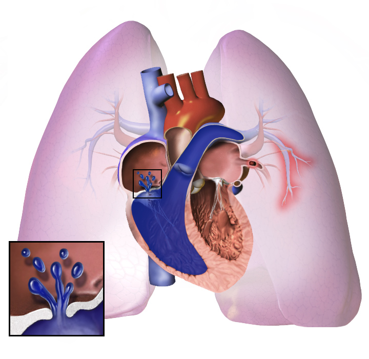
Pulmonary hypertension (PH) is a hemodynamic syndrome defined by a mean pulmonary artery pressure (mPAP) > 20 mmHg at rest measured by right heart catheterization, a threshold lowered in 2018 from the historical >25 mmHg to reflect that a normal mPAP is 14 ± 3 mmHg and values between 20-25 mmHg carry increased risk [11]D5. Also called pulmonary arterial hypertension (PAH, specifically for WHO Group 1), this is distinct from pulmonary venous hypertension (PVH, Groups 2-5). The condition encompasses all five WHO clinical groups despite their divergent etiologies: group 1 (PAH), group 2 (PH due to left heart disease), group 3 (PH due to lung diseases/hypoxia), group 4 (chronic thromboembolic PH), and group 5 (PH with unclear multifactorial mechanisms).
The pulmonary circulation is unique in accommodating the entire cardiac output while maintaining low pressure (normal mPAP 14 ± 3 mmHg) and low resistance (normal pulmonary vascular resistance, PVR, <2 Wood units) [2]D5. Pulmonary arteries have thinner walls, less vascular smooth muscle, and lower basal tone than systemic arteries, a design that favors efficient gas exchange over high-pressure perfusion [2]D5. The homeostatic hierarchy places PH as a derangement of pulmonary vascular resistance control, not merely a pressure number; it is the chronic elevation of afterload on the right ventricle that determines morbidity and mortality [7]D5.
Synonyms and Historical Terms
- WHO Group 1: idiopathic PAH (IPAH), heritable PAH (gene mutations in BMPR2, KCNK3), drug/toxin-induced PAH, PAH associated with connective tissue disease, HIV, , congenital heart disease [17]D5
- WHO Group 2: pulmonary venous hypertension, post-capillary PH (wedge pressure >15 mmHg)
- WHO Group 3: hypoxic PH, PH due to , , sleep-disordered breathing [1]D5
- WHO Group 4: chronic thromboembolic pulmonary hypertension (CTEPH)
- WHO Group 5: PH due to sarcoidosis, metabolic disorders, hematologic conditions
- Historical term: "Ayerza's disease" (old term for IPAH with severe hypoxia)
Clinical Significance
PH is the third leading cause of cardiovascular death after heart failure and stroke, with a 5-year survival of only 28% in advanced (WHO functional class IV) patients, even in the modern therapy era [11]D5. The syndrome affects approximately 1% of the global population, rising to >10% in those over age 65, driven largely by Group 2 PH from the epidemic of heart failure with preserved ejection fraction [5]D5.
Boundaries of This Article
This article covers the full spectrum of PH, from its molecular underpinnings (endothelial dysfunction, metabolic switching in vascular smooth muscle cells, potassium channel dysregulation [17]D5[6]D5) through hemodynamic classification, diagnostic algorithm, and group-specific medical, interventional, and surgical . It does not address pulmonary hypertension in neonates or pediatric-specific etiologies, which have distinct definitions and treatment algorithms [16]D5.
Pearl: The single most important diagnostic step is to perform a right heart catheterization to confirm an mPAP >20 mmHg and to determine whether the PH is pre-capillary (PVR >2 WU, wedge pressure ≤15 mmHg) or post-capillary (wedge >15 mmHg), as this distinction entirely alters the treatment pathway [2]D5.
| Group | Name | Key Feature | Hemodynamic Pattern |
|---|---|---|---|
| 1 | PAH | Remodeling of distal pulmonary arterioles | Pre-capillary (PCWP ≤15) |
| 2 | PH due to left heart disease | Passive backward transmission of elevated left-sided filling pressures | Post-capillary (PCWP >15) |
| 3 | PH due to lung disease/hypoxia | Hypoxic vasoconstriction and parenchymal destruction | Pre-capillary (PCWP ≤15) |
| 4 | CTEPH | Organized thrombus obstructing proximal or distal pulmonary arteries | Pre-capillary (PCWP ≤15) |
| 5 | PH with unclear mechanisms | Multifactorial (sarcoidosis, metabolic, hematologic) | Variable |
Normal Process & Mechanism (First Principles)
- ▸Hypoxic pulmonary vasoconstriction is initiated by mitochondrial complex III-derived ROS, not by cellular hypoxia per se, and chronic HIF-1α stabilization drives fixed remodeling [35, 36].
- ▸Pulmonary artery smooth muscle cells in PH exhibit a Warburg-like metabolic shift (glycolysis over oxidative phosphorylation), suppressed Kv1.5 channels, and mitochondrial fission, collectively conferring apoptosis resistance [26, 38, 39].
- ▸The adventitia becomes a proinflammatory niche driven by alternative complement pathway activation and ROCK1-mediated leukocyte recruitment, creating a self-sustaining cycle of remodeling [24, 25].
The pulmonary circulation is a low-pressure, high-flow system designed for gas exchange, not systemic perfusion. Normal mean pulmonary artery pressure (mPAP) is 14 ± 3 mm Hg at rest, with pulmonary vascular resistance (PVR) typically < 3 Wood units. Understanding how this homeostasis is maintained, and where it breaks, requires tracing the pathway from oxygen sensing through vascular tone regulation to structural remodeling.
The Oxygen-Sensing Machinery
The pulmonary circulation uniquely constricts in response to hypoxia rather than dilating. This phenomenon, hypoxic pulmonary vasoconstriction (HPV), optimizes ventilation-perfusion matching by diverting blood away from poorly ventilated alveoli [35]D5. The oxygen sensor resides at complex III of the mitochondrial electron transport chain, where hypoxia increases reactive oxygen species (ROS) released into the intermembrane space [35]D5. These ROS activate downstream targets, including Rho-kinase (ROCK) and hypoxia-inducible factor 1α (HIF-1α), that ultimately raise intracellular calcium and trigger smooth muscle contraction [35]D5[36]D5.
In the acute setting, HPV is reversible. In chronic global hypoxia (e.g., high altitude), sustained HIF-1α stabilization upregulates vasoconstrictor and proliferative genes, driving fixed remodeling [36]D5. The von Hippel-Lindau (VHL) ubiquitin ligase normally degrades HIF-1α under normoxia; loss of this regulation produces a PH phenotype [36]D5.
The Vasodilator-Vasoconstrictor Balance
Endothelial homeostasis depends on three major pathways:
| Pathway | Normal Effect | Key Mediator | Derangement in PH |
|---|---|---|---|
| NO-sGC-cGMP | Vasodilation, antiproliferation | Endothelial NOS → NO → sGC → cGMP | Impaired NO production; reduced sGC activity [29]A1a |
| Prostacyclin pathway | Vasodilation, antiplatelet | Prostacyclin → IP receptor → cAMP | Decreased prostacyclin synthase expression [25]D5 |
| Endothelin system | Vasoconstriction, proliferation | Endothelin-1 → ETA/ETB receptors | Upregulated ET-1; increased ETA signaling [40]D5 |
Endothelin-1 (ET-1) exhibits circadian variation, with peak plasma levels in the early morning, a pattern that may contribute to diurnal variation in pulmonary pressures and raise questions about chronotherapy for ET receptor antagonists [40]D5.
In health, NO-mediated vasodilation predominates. In PH, endothelial dysfunction shifts the balance toward vasoconstriction, with reduced NO bioavailability and upregulated ET-1 [25]D5[29]A1a.
Right Ventricular Adaptation and Failure
The right ventricle (RV) is geometrically and functionally distinct from the left ventricle (LV). It is a thin-walled, crescent-shaped chamber optimized for volume displacement against low afterload. When PVR rises, the RV hypertrophies to preserve cardiac output, an initially adaptive response mediated by sarcomere addition in series (eccentric hypertrophy) [7]D5. However, the RV has limited reserve. Afterload-sensitive, it fails when mPAP exceeds roughly 40 mm Hg, transitioning from hypertrophy to dilatation and reduced contractility [7]D5.
RV failure in PH is not simply a consequence of pressure overload. Maladaptive molecular pathways, including Rho-kinase activation in the RV myocardium, impair relaxation and promote fibrosis [25]D5. Unlike the LV, the RV shows greater dependence on glucose metabolism and less on fatty acid oxidation; this metabolic inflexibility may contribute to energy deficit under stress [7]D5.
The Cell Proliferation-Apoptosis Imbalance
At the cellular level, pulmonary artery smooth muscle cells (PASMCs) exhibit a cancer-like phenotype in PAH: proliferative, apoptosis-resistant, and metabolically reprogrammed [26]D5[37]D5. Key features include:
- Suppressed Kv1.5 potassium channels, which normally promote apoptosis by maintaining mitochondrial membrane potential. Loss of Kv1.5 depolarizes the cell, inhibits apoptosis, and triggers contraction [26]D5.
- Mitochondrial fragmentation (fission) driven by dynamin-related protein 1 (Drp1), shifting metabolism toward glycolysis (the Warburg effect) [38]D5[39]D5.
- Activation of HIF-1α under normoxia (pseudohypoxia), which upregulates glycolytic enzymes and suppresses oxidative phosphorylation [26]D5.
- Serotonin-mediated remodeling: 5-HT, through transglutaminase 2 (TG2)-dependent serotonylation of fibronectin and Rho, promotes PASMC proliferation and contraction [27]D5.
This metabolic switch from oxidative phosphorylation to glycolysis generates less ATP per glucose molecule but supports rapid cell division and confers resistance to apoptotic signals, a key therapeutic challenge [37]D5[39]D5.
The Adventitial Niche and Inflammation
The adventitia is not a passive scaffold. In PH, it becomes a proinflammatory niche populated by complement-rich fibroblasts, granzyme K+ CD8 T cells, and activated macrophages [24]D5. The alternative complement pathway is a key driver: complement activation within the pulmonary vasculature amplifies chemotaxis, fibroblast activation, and extracellular matrix deposition [24]D5. Rho-kinase in circulating inflammatory cells (ROCK1) promotes leukocyte adhesion and migration into the vessel wall, further fueling the inflammatory cycle [25]D5.
Animal Models and Experimental Systems
No single model recapitulates all features of human PH. Models are grouped by mechanism [23]D5:
| Model Type | Examples | Relevant Human Group |
|---|---|---|
| Tone-related | Chronic hypoxia; Sugen-hypoxia (SU5416 + hypoxia) | Group 3 (hypoxia-driven) |
| Inflammation-driven | Monocrotaline (rat); Schistosoma-induced | Group 1 (PAH with connective tissue disease) |
| Genetic | BMPR2 mutant mice; Hph-1 mice (HIF hydroxylase deficient) | Heritable PAH |
The Sugen-hypoxia model uniquely produces obliterative plexiform lesions, mimicking severe human PAH. Genetic models (e.g., BMPR2 mutations) reveal that a second hit, inflammation, shear stress, is required for overt disease [23]D5.
Pearl: Pulmonary hypertension arises from a convergence of impaired oxygen sensing (HIF-ROS-Kv1.5 axis), endothelial vasodilator loss (NO/cGMP, prostacyclin), and a proliferative, apoptosis-resistant PASMC phenotype driven by mitochondrial reprogramming, an understanding that directly explains current therapeutic targets (NO pathway augmentation, prostacyclin analogs, endothelin blockade) and points toward metabolic and anti-inflammatory strategies still under investigation [25]D5[26]D5[37]D5.
Regulation & Feedback Control
- ▸Pulmonary vascular resistance is regulated by integrated feedback loops involving PASMC K+ channels, endothelial vasoactive mediators, oxygen sensing via HIF, and BMP signaling.
- ▸Disruption of any component (e.g., KV channel downregulation, endothelial dysfunction, BMPR2 mutation, metabolic reprogramming) shifts the set point toward sustained vasoconstriction and vascular remodeling.
- ▸Circadian, epigenetic, and sex-specific factors (e.g., mineralocorticoid receptor, microRNAs) modulate these feedback loops and contribute to disease heterogeneity.
Pulmonary vascular resistance (PVR) is held near its set point by a network of sensor-controller-effector loops that integrate ion channel activity, endothelial mediators, oxygen sensing, and metabolic feedback [41]D5[42]D5. Disruption of any component shifts the set point toward vasoconstriction and remodeling, the hallmark of pulmonary hypertension.
Ion Channels as the Effector Arm
Pulmonary artery smooth muscle cells (PASMCs) express five classes of K+ channels: large-conductance Ca2+-activated (BK), voltage-dependent (KV), inward-rectifier (Kir), ATP-sensitive (KATP), and two-pore domain (K2P) [42]D5. These channels set the resting membrane potential. When KV channels are inhibited, by hypoxia, inflammatory mediators, or genetic downregulation, membrane depolarization opens voltage-gated Ca2+ channels, raising cytosolic Ca2+ and triggering vasoconstriction [41]D5[44]D5. In PAH, KV channel expression is reduced, shifting the balance toward sustained contraction [41]D5. Conversely, KATP channel activation (e.g., by hypoxia) can promote vasodilation, but this compensatory mechanism is often overwhelmed [42]D5.
Endothelial Feedback Loops
The endothelium releases vasodilators (nitric oxide [NO], prostacyclin) and vasoconstrictors (endothelin-1). NO diffuses to PASMCs, activates soluble guanylyl cyclase, and reduces Ca2+ sensitivity via cGMP [22]D5. Endothelin-1 acts through ETA receptors on PASMCs (vasoconstriction) and ETB receptors on endothelial cells (clearance and NO release) [40]D5. In PH, endothelial dysfunction reduces NO bioavailability and increases endothelin-1, shifting the loop toward vasoconstriction [41]D5. The circadian clock modulates endothelin-1 expression; clock disruption can further dysregulate vascular tone [40]D5.
Oxygen Sensing and the HIF Pathway
Hypoxia is a primary driver of pulmonary vasoconstriction. PASMCs sense low O2 via mitochondrial electron transport chain complexes and NADPH oxidases, leading to inhibition of KV channels and Ca2+ influx [55]D5. Hypoxia-inducible factors (HIF-1α, HIF-2α) upregulate genes that promote proliferation, metabolic reprogramming, and extracellular matrix remodeling [55]D5. Chronic HIF activation, as in high-altitude exposure or sleep apnea, contributes to irreversible vascular remodeling [51]D5[55]D5.
BMP Signaling and Genetic Set Point
Bone morphogenetic protein (BMP) signaling through BMPR2 maintains PASMC quiescence and promotes contractile differentiation [57]D5. Loss-of-function mutations in BMPR2 (found in ~70% of heritable PAH) disrupt this feedback, leading to unchecked proliferation and apoptosis resistance [57]D5. BMP signaling also cross-talks with TGF-β and Wnt pathways, and its dysregulation is a key determinant of the disease set point [57]D5.
Metabolic and Mitochondrial Feedback
Mitochondria regulate PASMC phenotype via reactive oxygen species (mtROS) and metabolic intermediates. In PH, a shift from oxidative phosphorylation to glycolysis (Warburg-like) increases lactate and promotes proliferation [50]D5[38]D5. Mitochondrial fission and mitophagy drive a synthetic, pro-proliferative phenotype [38]D5. Polyamine metabolism (putrescine, spermidine, spermine) is upregulated, supporting cell growth and redox imbalance [20]D5. These metabolic changes create a positive feedback loop: sustained proliferation further alters metabolism, reinforcing the pathological state.
Epigenetic and Sex-Specific Modulation
Epigenetic mechanisms, DNA methylation, histone modification, and noncoding RNAs, fine-tune gene expression in response to environmental cues [21]D5. MicroRNAs such as miR-21 and miR-145 are dysregulated in PH and target pathways including BMP signaling and ion channels [56]D5. The mineralocorticoid receptor (MR) in endothelial and smooth muscle cells has sex-specific roles: in females, MR activation promotes endothelial dysfunction and vascular inflammation, contributing to PH [59]D5. Thyroid hormone also modulates angiogenesis via integrin αvβ3, with implications for pulmonary vascular remodeling [45]D5.
Pearl: The pulmonary circulation's low-resistance state is maintained by a delicate balance of K+ channel activity, endothelial NO/endothelin-1, and BMP signaling; disruption of any of these feedback loops, through genetic mutation, hypoxia, or metabolic reprogramming, shifts the set point toward vasoconstriction and remodeling, the hallmark of pulmonary hypertension [41]D5[42]D5[55]D5[57]D5.
| Component | Sensor | Controller | Effector | Dysregulation in PH |
|---|---|---|---|---|
| Ion channels | Membrane potential | K+ channel activity (KV, BK, KATP) | Ca2+ influx → contraction | KV downregulation → depolarization → vasoconstriction [41]D5[42]D5 |
| Endothelial mediators | Shear stress, O2 | NO, prostacyclin, endothelin-1 | PASMC relaxation/contraction | Reduced NO, increased ET-1 → vasoconstriction [22]D5[40]D5 |
| Oxygen sensing | Mitochondria, NADPH oxidase | HIF-1α/2α | Gene expression (proliferation, metabolism) | Chronic HIF activation → remodeling [55]D5 |
| BMP signaling | Ligand binding | BMPR2, Smad1/5/8 | PASMC quiescence | BMPR2 loss → proliferation [57]D5 |
| Metabolic feedback | Substrate availability, ROS | Glycolysis, OXPHOS, polyamines | Phenotype switch | Glycolytic shift → proliferation [50]D5[38]D5 |
Integration with Other Systems
- ▸Right ventricular-pulmonary artery uncoupling (Ees/Ea <0.8) is the central hemodynamic event driving deterioration in PH and can be assessed by RV strain [11, 65]
- ▸Mitochondrial metabolic reprogramming (glycolysis switch) and perivascular inflammation are unifying cellular mechanisms across WHO groups 1, 3, and 4 [11, 63, 64]
- ▸PH associated with HFpEF (Group 2), portopulmonary hypertension, and SLE-PAH require system-specific management, not generic PAH therapy [5, 68, 69]
Pulmonary hypertension is not a disease of the pulmonary vasculature in isolation; it disrupts and is disrupted by virtually every organ system. Understanding these couplings is essential for recognizing the full clinical syndrome and avoiding pitfalls.
Right Ventricular-Pulmonary Artery Uncoupling: The Central Hemodynamic Axis
The right ventricle (RV) is a flow generator designed to eject against low afterload. When pulmonary vascular resistance rises, the RV adapts by increasing contractility, a compensatory mechanism termed RV-pulmonary artery (PA) coupling. As PH progresses, this relationship fails: the RV cannot generate sufficient pressure to maintain forward flow, leading to RV-PA uncoupling [11]D5. The gold-standard measure is the ratio of end-systolic elastance (Ees) to arterial elastance (Ea), with a normal Ees/Ea ratio >1.0. A ratio <0.8 identifies uncoupling and predicts clinical worsening and mortality [11]D5[65]B2a. RV free-wall longitudinal strain (RV-FWS) and global longitudinal strain (RV-GLS) detected by speckle-tracking echocardiography correlate closely with invasive Ees/Ea and provide a non-invasive surrogate [65]B2a.
Heart-Lung Interactions: Diastolic Cross-Talk and Venous Congestion
In PH due to left heart disease (WHO Group 2), particularly heart failure with preserved ejection fraction (HFpEF), chronic pulmonary venous congestion drives pulmonary arterial remodeling through a venous-to-arterial pressure transmission [5]D5. A key concept is the pulmonary artery wedge pressure (PAWP) threshold: a PAWP ≤15 mmHg at rest defines pre-capillary PH, while >15 mmHg suggests post-capillary disease requiring different management [5]D5. During exercise, even patients with normal resting PAWP may exhibit latent pulmonary venous hypertension (exercise PAWP >25 mmHg), identified by exercise stress echocardiography; this pattern carries prognostic value in and [66]B2a[67]B2a. The absence of effective PAH-specific therapies for Group 2 PH reflects this fundamental different pathobiology: vasodilators may reduce preload but do not address upstream left ventricular stiffness [5]D5.
Systemic Hemodynamics and the Lung-Kidney-Vessel Axis
PH alters systemic afterload, particularly in pediatric chronic lung disease (bronchopulmonary dysplasia). Infants with severe bronchopulmonary dysplasia exhibit increased carotid artery intima-media thickness and central aortic stiffness, a sign of systemic vascular injury driven by chronic hypoxemia, inflammation, and altered autonomic tone [61]D5. This systemic afterload elevation contributes to left ventricular diastolic dysfunction, which in turn worsens pulmonary venous congestion. In these infants, angiotensin-converting enzyme inhibitors have been used to reduce systemic afterload, though data are limited [61]D5. In adults, systemic sclerosis (SSc) represents a paradigmatic multiorgan disease: microvascular disease, myocardial fibrosis, and converge to produce progressive RV dysfunction, and RV-FWS is a strong independent predictor of mortality in SSc [65]B2a.
Hepato-Pulmonary and Porto-Pulmonary Crosstalk
(often from cirrhosis or MASLD) can produce two distinct PH syndromes. (PoPH), medial hypertrophy and endothelial dysfunction in the pulmonary arterioles, responds to PAH-specific therapy but is frequently under-recognized [69]B2a. Hepatopulmonary syndrome, conversely, involves intrapulmonary vascular dilatation causing hypoxemia, not pulmonary vasoconstriction. The distinction is critical: vasodilators worsen hepatopulmonary syndrome, while they are first-line in PoPH. MASLD, through its associated systemic inflammation and adipose tissue dysfunction (adipose tissue hypoxia, macrophage infiltration), may also promote pulmonary vascular remodeling via a liver-lung inflammatory axis [69]B2a.
Mitochondrial Dysfunction: A Unifying Cellular Mechanism
Across PH groups, a shared molecular signature is mitochondrial metabolic reprogramming in pulmonary vascular cells. Pulmonary endothelial cells and smooth muscle cells (VSMCs) shift from oxidative phosphorylation to glycolysis (the Warburg effect), driven by mitochondrial reactive oxygen species (ROS) production and Kv channel dysfunction [11]D5[63]D5. This bioenergetic switch enables proliferation and apoptosis resistance in VSMCs, fueling medial hypertrophy [42]D5[6]D5. In skeletal muscle, similar mitochondrial dysfunction contributes to exercise limitation and systemic fatigue, independent of cardiac function [63]D5. Mitochondria also serve as oxygen sensors: in hypoxia, mitochondrial ROS stabilize hypoxia-inducible factor (HIF)-1α, which upregulates vasoconstrictors and growth factors that remodel the pulmonary vasculature [71]D5[64]D5.
Inflammatory Pathways in Hypoxic PH
Chronic hypoxia, whether from sleep apnea [1]D5, [64]D5, or high-altitude exposure, activates a perivascular inflammatory response involving macrophages, dendritic cells, and T lymphocytes that secrete endothelin-1, interleukin-6, and platelet-derived growth factor [64]D5. This inflammation amplifies VSMC proliferation and promotes irreversible remodeling in a subset of patients with "out-of-proportion" hypoxic PH, who develop a histology resembling WHO Group 1 disease [64]D5. The link between intermittent hypoxia in sleep apnea and pulmonary hypertension is mediated partly by sympathetic nervous system activation, which increases pulmonary vascular tone [1]D5.
and Autoimmune Connections
Systemic lupus erythematosus (SLE) associated with pulmonary arterial hypertension (PAH) may present with (IPO), a rare but life-threatening complication [68]C4. Among 43 reported SLE-IPO cases, concurrent PAH was present in a subset and predicted worse outcomes; immunosuppressive therapy (corticosteroids, ) can reverse IPO if recognized early [68]C4. The pathophysiological link likely involves vasculitic damage to the enteric nervous system and pulmonary microvasculature from the same autoimmune process.
The Red Blood Cell as a Vasoregulatory Effector
Erythrocytes are not passive oxygen carriers. Through S-nitrosylated hemoglobin (SNO-Hb), red blood cells couples oxygen release to nitric oxide bioactivity: at low oxygen tension, SNO-Hb releases vasodilating S-nitrosothiols that relax downstream resistance vessels, matching local perfusion to metabolic demand [22]D5. In PH, impaired SNO-Hb formation or accelerated NO scavenging by free hemoglobin contributes to loss of hypoxic vasodilation and exacerbates pulmonary vascular resistance [22]D5.
Pearl: Pulmonary hypertension must be evaluated as a systemic syndrome: RV-PA uncoupling (Ees/Ea <0.8) defines hemodynamic progression, mitochondrial metabolic reprogramming drives cellular remodeling across groups, and associated conditions, HFpEF, portal hypertension, sleep apnea, MASLD, SLE, require simultaneous management to improve outcomes [5]D5[11]D5[61]D5[64]D5[68]C4[69]B2a.
| System | Interaction | Clinical Consequence | Reference |
|---|---|---|---|
| Cardiovascular | RV-PA uncoupling | RV failure, ↓ cardiac output, death | [11]D5[65]B2a |
| Pulmonary (left heart) | Venous-to-arterial pressure transmission; exercise PAWP rise | Latent PH-HFpEF; no benefit from PAH drugs | [5]D5[66]B2a[67]B2a |
| Systemic vasculature | Arterial stiffness, ↑ afterload | LV diastolic dysfunction in pediatric CLD | [61]D5 |
| Liver | Portal hypertension → PoPH vs HPS | Differentiate: vasodilators for PoPH, contraindicated in HPS | [69]B2a |
| Skeletal muscle | Mitochondrial dysfunction, glycolysis | Exercise limitation, fatigue | [63]D5 |
| Immune/Inflammatory | Perivascular macrophages, cytokines | Irreversible remodeling in a subset of hypoxic PH | [64]D5 |
| Autoimmune | SLE → PAH + intestinal pseudo-obstruction | Immunosuppression may reverse IPO | [68]C4 |
| Erythrocyte | Impaired SNO-Hb formation | Loss of hypoxic vasodilation, ↑ PVR | [22]D5 |
Quantitative Parameters, Equations & Curves
The pulmonary circulation is a low-resistance, low-pressure, and high-compliance system designed to accommodate the entire cardiac output with minimal energy expenditure [41]D5. Quantitative relationships govern how pressure, flow, resistance, and vessel geometry interact, and deviations from these norms define the hemodynamic state of pulmonary hypertension (PH).
The Ohm's Law Analogy: Pressure = Flow × Resistance
The fundamental equation of pulmonary hemodynamics is the analog of Ohm's law:
Mean pulmonary artery pressure (mPAP) = Cardiac output (CO) × Pulmonary vascular resistance (PVR) + Pulmonary artery wedge pressure (PAWP)
More precisely, PVR is calculated as:
PVR = (mPAP - PAWP) / CO
where PVR is expressed in Wood units (WU). One Wood unit equals 1 mmHg·min/L. A normal PVR is < 3.0 WU; values ≥ 3.0 WU are considered elevated and are a key diagnostic criterion for precapillary PH [41]D5.
The Vessel-Radius Relationship: The Fourth Power
PVR is not simply a measure of resistance; it is inversely proportional to the fourth power of the intraluminal radius of the pulmonary artery (as described by Poiseuille's law):
PVR ∝ 1 / r⁴
This means that a very small decrease in the pulmonary vascular lumen diameter results in a significant increase in PVR and, consequently, mPAP [41]D5. For example, a 10% reduction in radius produces a 46% increase in resistance (1/0.9⁴ ≈ 1.46). This relationship underpins the devastating effect of even mild vascular remodeling in PAH.
Wave Mechanics: Beyond Steady Flow
While PVR captures the non-oscillatory component of right ventricular (RV) hydraulic load, it neglects the dynamic compliance of the pulmonary arteries and the contribution of wave transmission [81]D5. The pulmonary vasculature is not a rigid tube; it is a compliant system that generates and reflects pressure waves. Key parameters include:
- Pulse wave velocity (PWV): A measure of arterial stiffness. Higher PWV indicates stiffer vessels and is associated with increased RV afterload. Formula: PWV = √(E·h / ρ·D), where E is elastic modulus, h is wall thickness, ρ is blood density, and D is vessel diameter.
- Wave reflection: Occurs at impedance mismatches (e.g., bifurcations, high-resistance distal vessels). The magnitude and timing of reflected waves provide information on the degree of mismatch between proximal and distal circulation [81]D5.
- Wave intensity analysis (WIA): Separates forward-traveling and backward-traveling (reflected) waves. In pulmonary hypertension, there is early wave reflection from stiffer distal vessels, which augments RV afterload [81]D5.
RV-Pulmonary Artery Coupling: The Force-Velocity Relationship
RV function must be matched to its afterload. The optimal clinical index of this coupling is the ratio of RV end-systolic elastance (Ees, a load-independent measure of contractility) to arterial elastance (Ea, a measure of net afterload).
Normal RV-PA coupling: Ees/Ea ≈ 1.0 to 2.0
In compensated PH, the RV increases contractility (Ees) to match the increased Ea, maintaining the ratio near normal. As PH progresses, RV contractility fails to keep pace, and the Ees/Ea ratio falls below 0.8, indicating uncoupling and imminent RV failure [65]B2a. This ratio can be approximated from routine right heart catheterization data using formulas involving stroke volume, end-systolic volume, and mPAP.
Pressure-Volume Loop Parameters
The relationship between pressure and volume is also critical. During right heart catheterization, key measurements include:
- mPAP: Normal ≤ 20 mmHg; PH is defined as > 20 mmHg.
- PAWP: Normal ≤ 15 mmHg; elevated in postcapillary PH.
- Transpulmonary gradient (TPG): mPAP - PAWP. Normal < 12 mmHg. Elevated TPG indicates a component of precapillary disease.
- Diastolic pressure gradient (DPG): Pulmonary artery diastolic pressure - PAWP. Normal < 7 mmHg. A higher DPG is more specific for precapillary involvement.
Table: Key Hemodynamic Equations
| Parameter | Equation | Normal Value | Clinical Significance |
|---|---|---|---|
| PVR | (mPAP - PAWP) / CO | < 3.0 WU | Index of precapillary resistance [41]D5 |
| TPG | mPAP - PAWP | < 12 mmHg | Suggests precapillary component if elevated |
| DPG | PA diastolic - PAWP | < 7 mmHg | Specific marker of pulmonary vascular disease |
| Ees | End-systolic pressure / end-systolic volume | ~0.5-1.0 mmHg/mL | RV contractility index |
| Ea | End-systolic pressure / stroke volume | ~0.5-1.0 mmHg/mL | Arterial afterload index |
Flow Regimes: Laminar vs. Turbulent
In health, pulmonary blood flow is predominantly laminar. However, with vessel obstruction (e.g., in CTEPH) or high-flow states (e.g., congenital heart disease), flow becomes turbulent. The Reynolds number (Re) predicts the transition:
Re = (ρ · V · D) / μ
where ρ = density, V = velocity, D = vessel diameter, μ = viscosity. Turbulence occurs when Re > 2000. Turbulent flow dramatically increases the afterload beyond that predicted by PVR alone, as the loss of energy to vortices reduces net forward flow.
Coupling with the Systemic Circulation
The pulmonary circulation interacts with the systemic side through the bronchial circulation and interatrial shunting (e.g., through a patent foramen ovale). In severe PH with right atrial pressure > left atrial pressure, a right-to-left shunt can occur, leading to systemic desaturation but also acting as a "pop-off" valve that reduces RV preload and may transiently improve forward flow at the cost of hypoxemia.
Stress Testing: Flow vs. Pressure Reserve
Normal pulmonary vasculature can accommodate a three- to fourfold increase in cardiac output during exercise with only a modest rise in mPAP (typically to < 30 mmHg). In PH, even mild exertion leads to a steep rise in mPAP, reflecting reduced vasodilator reserve and increased stiffness. Exercise hemodynamics (measured by exercise right heart catheterization) can unmask early disease when resting values are borderline.
Pearl: The fourth-power relationship between vessel radius and PVR means that even a 10% reduction in radius increases resistance by 46% [41]D5; this extreme sensitivity to luminal narrowing is why early vascular remodeling is so dangerous in pulmonary hypertension, amplifying the load on the right ventricle long before symptoms appear.
Measurement & Assessment Methods
- ▸Right heart catheterization with mPAP ≥25 mmHg is essential for diagnosis and classification, with PVR and PCWP differentiating WHO groups.
- ▸Echocardiographic RV-PA coupling indices (RV-FWS/sPAP) and wave intensity analysis detect early RV dysfunction and dynamic afterload not captured by PVR alone.
- ▸NT-proBNP and CMR provide complementary prognostic information beyond hemodynamics.
Right Heart Catheterization: The Gold Standard
Right heart catheterization (RHC) is the essential diagnostic test for pulmonary hypertension (PH). A mean pulmonary artery pressure (mPAP) ≥25 mmHg at rest remains the hemodynamic threshold that defines PH [81]D5. Beyond confirming the diagnosis, RHC measures pulmonary capillary wedge pressure (PCWP), cardiac output (by thermodilution or Fick method), and pulmonary vascular resistance (PVR = (mPAP - PCWP) / cardiac output). These parameters are critical for differentiating WHO Group 1 (pre-capillary PH: PCWP ≤15 mmHg, PVR >3 Wood units) from Group 2 (post-capillary PH: PCWP >15 mmHg) [81]D5[86]D5. PVR, however, only captures the non-oscillatory component of right ventricular (RV) afterload. Wave intensity analysis, an emerging technique, reveals that wave speed (a measure of arterial stiffness) and the timing and magnitude of wave reflections differ substantially between healthy subjects and PH patients, information not provided by PVR alone [81]D5.
Echocardiography: The Preferred Screening Tool
Transthoracic echocardiography is the first-line imaging modality for PH suspicion. The estimated systolic pulmonary artery pressure (sPAP) derived from the tricuspid regurgitation jet velocity (using the simplified Bernoulli equation: 4v² + right atrial pressure) correlates well with invasively measured sPAP when the signal is adequate. Advanced echocardiographic parameters have become essential for detecting early RV dysfunction. RV free-wall longitudinal strain (RV-FWS) and RV global longitudinal strain (RV-GLS) identify subclinical RV impairment before conventional measures decline, and the RV-PA coupling ratio (RV-FWS / sPAP or tricuspid annular plane systolic excursion [TAPSE] / sPAP) predicts adverse outcomes in systemic sclerosis-associated PH [65]B2a. AI-based analysis of echocardiographic images for tricuspid regurgitation, frequently secondary to PH, demonstrates high sensitivity (meta-analytic sensitivity >90%) for automated detection, potentially reducing operator-dependent variability in screening [85]A1a. Exercise stress echocardiography (ESE) unmasks latent hemodynamic burden: in patients with and , ESE-derived changes in sPAP and RV function provide incremental prognostic information beyond resting measures, a principle directly applicable to PH risk stratification [66]B2a[67]B2a.
Biomarkers and Imaging
N-terminal pro-B-type natriuretic peptide (NT-proBNP) is a validated biomarker of RV wall stress and correlates with PH severity and prognosis. Levels guide risk stratification: NT-proBNP <300 ng/L is low-risk, ≥1400 ng/L is high-risk. Cardiac magnetic resonance imaging (CMR) offers gold-standard RV volumetric and functional assessment (ejection fraction, end-systolic volume index) without ionizing radiation, and CMR-derived RV mass is an independent predictor of mortality.
Controversies and Guideline Disagreement
| Question | Position A | Position B | Strength | Implication |
|---|---|---|---|---|
| Should RHC be performed before initiating PAH therapy? | AHA/ACC: Yes, mandatory for Group 1 diagnosis and classification [86]D5 | ESC/ERS: Yes, mandatory, with acute vasoreactivity testing in selected patients [81]D5 | Consensus | RHC is non-negotiable for Group 1 diagnosis |
| Is PVR alone sufficient to guide ? | Traditional teaching: PVR is the key hemodynamic target | Emerging evidence: wave reflection and RV-PA coupling metrics provide incremental value [65]B2a[81]D5 | Low | PVR remains primary, but coupling indices are gaining acceptance |
Pearl: Hemodynamic assessment by right heart catheterization with mPAP ≥25 mmHg confirms PH, but integrating echocardiographic RV-PA coupling (RV-FWS/sPAP) and wave reflection analysis captures dynamic load not reflected in PVR alone, improving risk stratification [65]B2a[81]D5.
| Parameter | Normal | PH (Group 1 Pre-capillary) | PH (Group 2 Post-capillary) |
|---|---|---|---|
| Mean PAP (mPAP) | <25 mmHg | ≥25 mmHg | ≥25 mmHg |
| PCWP | ≤15 mmHg | ≤15 mmHg | >15 mmHg |
| PVR | ≤3 Wood units | >3 Wood units | Variable |
| Cardiac output | 4-8 L/min | May be reduced | Reduced or normal |
Physiological Variation: The Normal Envelope of the Pulmonary Circulation
- ▸During exercise, mPAP rises linearly with cardiac output but should remain below 40-50 mmHg and capillary wedge pressure below 25 mmHg to avoid interstitial edema [88].
- ▸Female sex is paradoxically associated with both higher incidence of PAH and better survival, mediated by estrogen's vasodilatory and proliferative effects [90].
The pulmonary circulation is not a static resistor; its pressures, resistance, and recruitment change dynamically across physiological states. Understanding this normal envelope is essential to distinguish healthy adaptation from the beginnings of pathology.
Exercise: High Flow Without High Resistance
During exercise, the pulmonary circulation recruits and distends capillaries, keeping the rise in mean pulmonary artery pressure (mPAP) proportionate to the increase in cardiac output. In young adults, the pulmonary pressure-flow relationship is almost linear, with a slope of roughly 1-2 mmHg·min/L [88]D5. The average pulmonary vascular resistance (PVR) at rest is 1 Wood unit; this falls to 0.5-0.8 Wood units during exercise as vessels distend [88]D5. The key safety limit: exercise-induced mPAP should not exceed 40-50 mmHg, and capillary wedge pressure must remain below 20-25 mmHg. Above those thresholds, interstitial lung edema and ventilation-perfusion mismatch occur, marking the upper boundary of the tolerable right ventricular afterload [88]D5.
Age: Progressive Stiffening
Healthy aging stiffens the pulmonary vasculature. PVR rises from ~1 Wood unit at age 20 to ~2.5 Wood units by age 70, driven by increased collagen and reduced elastin in the pulmonary artery wall [88]D5. This age-related increase is independent of left heart disease and must be factored into interpretation of hemodynamic measurements, particularly when diagnosing pulmonary .
Altitude: Hypoxic Pulmonary Vasoconstriction and Adaptation
Acute ascent to high altitude triggers hypoxic pulmonary vasoconstriction (HPV), a conserved reflex that diverts blood from poorly ventilated alveoli [[87]D5, [36]D5]. Under global hypoxia (e.g., high altitude), generalized HPV elevates mPAP. At 3,800 meters, the average mPAP in healthy lowland natives rises from 14 mmHg at sea level to 24 mmHg [98]D5. Chronic hypoxia induces pulmonary vascular remodeling, smooth muscle hypertrophy and adventitial thickening, that sustains the pressure elevation [[102]D5, [36]D5].
Some populations display genetic adaptation. Tibetans and Ethiopians carry variants in the HIF-2α (EPAS1) and PPARA genes that blunt HPV, giving them near-normal mPAP at altitude (and low rates of chronic mountain sickness) [101]D5. Andean highlanders, by contrast, have higher mPAP and are susceptible to chronic mountain sickness, characterized by excessive polycythemia and pulmonary hypertension [101]D5.
Pregnancy: Volume Overload with Vasodilation
Pregnancy is a high-flow state with a 30-50% increase in cardiac output by the third trimester. Normally, mPAP does not rise because pulmonary vascular resistance falls by roughly 30-40%, mediated by estrogen-induced nitric oxide production and prostacyclin [90]D5. However, the right ventricle dilates and increases stroke volume; any superimposed vasoconstrictive stimulus, such as preeclampsia, can rapidly unmask a limited reserve [70]B2a.
Posture: Gravity Gradients
In the upright position, gravity creates a vertical gradient in pulmonary blood flow: the lung apices receive minimal flow (West's zone 1) while the bases are well perfused (zone 3). This redistribution does not change mPAP but does lower the effective pulmonary capillary volume available for gas exchange. Supine posture abolishes the gradient, increasing capillary recruitment and slightly lowering PVR [89]A1c.
Sex: Baseline Differences and Hormonal Modulation
At rest, women have a slightly lower mPAP (by 1-2 mmHg) compared with men of the same age, and their PVR is also lower [90]D5. Mechanistically, estradiol upregulates endothelial nitric oxide synthase and K⁺ channels (especially BK channels), promoting vasodilation [42]D5. Paradoxically, the incidence of pulmonary arterial hypertension (Group 1) is 3-4 times higher in women, yet female patients have better survival than males, the so-called "estrogen paradox" [90]D5. This may reflect the dual role of estrogen: protective on the pulmonary vasculature (promoting NO production) but permissive for maladaptive proliferation in predisposed individuals.
Pearl: The healthy pulmonary circulation accommodates a 4- to 5-fold increase in cardiac output with only a modest rise in mPAP (<50 mmHg), thanks to recruitment, distension, and fall in PVR [88]D5; deviation from this pattern, a steep slope or plateau beyond the normal threshold, signals early disease.
| Condition | mPAP (mmHg) | PVR (Wood units) | Cardiac Output (L/min) | Key Mechanism |
|---|---|---|---|---|
| Rest, supine, sea level | 12-16 | 1.0-1.5 | 4-6 | Baseline |
| Exercise (maximal) | 30-50 | 0.5-0.8 | 15-25 | Recruitment and distension |
| High altitude (3,800 m) | 20-30 | 1.5-2.5 | 5-8 | Hypoxic pulmonary vasoconstriction |
| Pregnancy (3rd trimester) | 14-18 | 0.7-1.0 | 7-9 | Hormonal vasodilation |
| Upright posture | 12-16 | 1.0-1.5 | 4-6 | Gravity redistribution (no net PVR change) |
| Age 70 (rest) | 15-20 | 2.0-2.5 | 4-5 | Arterial stiffening |
Clinical Correlation: From Derangement to Disease
- ▸Dyspnea on exertion is the earliest symptom; syncope indicates advanced disease with cardiac index often <2.0 L/min/m².
- ▸Physical exam findings reflect right heart strain: elevated JVP, RV heave, loud P2, and tricuspid regurgitation murmur.
- ▸WHO group classification (1-5) guides management and prognosis; group 2 (left heart disease) is the most common cause.
The transition from normal pulmonary vascular physiology to disease begins with endothelial dysfunction, which disrupts the balance between vasodilators (NO, prostacyclin) and vasoconstrictors (endothelin-1, serotonin) [41]D5[109]D5. This imbalance triggers sustained vasoconstriction, smooth muscle cell proliferation, and extracellular matrix remodeling, collectively increasing pulmonary vascular resistance (PVR) [74]D5[107]D5. The clinical consequence is a progressive rise in mean pulmonary artery pressure (mPAP), which imposes a chronic afterload on the right ventricle.
Presenting Symptoms
Dyspnea on exertion is the earliest and most common symptom, reflecting the inability to augment cardiac output during exercise due to fixed PVR [91]D5. As disease advances, patients report fatigue, presyncope, and exertional chest pain. Syncope is a late and ominous sign, indicating severely limited cardiac reserve (cardiac index often <2.0 L/min/m²). Peripheral edema and abdominal distension signal right ventricular (RV) failure. The timeline is typically insidious: symptoms progress over months to years, with a nadir in functional capacity at 2-4 weeks after decompensation. Acute worsening may be triggered by infection, nonadherence to therapy, or pulmonary embolism.
Physical Examination Findings
The examination targets the right heart and pulmonary circulation. Key findings include:
- Elevated jugular venous pressure (JVP) with prominent a and v waves.
- Right ventricular heave at the left parasternal border.
- Loud P2 (pulmonary component of S2) due to high pulmonary artery pressure.
- Tricuspid regurgitation murmur (holosystolic, increases with inspiration).
- Hepatomegaly, , and peripheral edema in RV failure.
- Signs of underlying cause: crackles in group 2 (left heart disease), digital clubbing in group 3 (lung disease), or evidence of connective tissue disease in group 1.
Phenotypic Variants
The WHO classification groups 1-5 capture distinct etiologies and pathophysiologies [91]D5. The table below summarizes key features.
| WHO Group | Etiology | Key Pathophysiology | Frequency |
|---|---|---|---|
| 1 (PAH) | Idiopathic, heritable, drug-induced, CTD, HIV, | Precapillary PH; plexiform lesions; endothelial proliferation | ~1-2 per million (rare) |
| 2 (PH-LHD) | Left heart failure (HFrEF, HFpEF), valvular disease | Postcapillary PH; elevated PAWP; passive congestion | Most common (50-70% of PH) |
| 3 (PH-lung disease) | , ILD, sleep apnea, high altitude | Hypoxic vasoconstriction; parenchymal destruction | Second most common |
| 4 (CTEPH) | Chronic thromboemboli | Organized thrombi; vascular remodeling; precapillary PH | ~3-5% after acute PE |
| 5 (Miscellaneous) | Sarcoidosis, vasculitis, metabolic disorders | Multifactorial; variable mechanisms | Rare |
Group 2 PH is the most prevalent, driven by left heart disease [113]D5. In HFpEF, metabolic syndrome and inflammation contribute to pulmonary vascular remodeling, leading to combined pre- and postcapillary PH (cpc-PH) [110]D5. Group 3 PH arises from chronic hypoxemia, which triggers HIF-mediated vasoconstriction and remodeling [36]D5[55]D5. Group 4 CTEPH is potentially curable with pulmonary endarterectomy [120]D5.
Red Flags
Certain symptoms and signs demand urgent action:
- Syncope - indicates critically low cardiac output; risk of sudden death is high.
- Hemoptysis - may signal pulmonary artery rupture or bronchial artery hypertrophy.
- Rapid progression of dyspnea over days - consider acute pulmonary embolism or decompensated RV failure.
- Signs of RV failure (JVP >10 cm H₂O, massive edema, ascites) - require diuresis and consideration of inotropes or mechanical support.
- Hypoxemia at rest (SpO₂ <90%) - suggests advanced disease or shunt physiology.
Atypical Presentations
PH can present in unusual contexts that may be missed:
- Sickle cell disease: -driven PH with high cardiac output and low PVR; often detected on screening echocardiography [112]D5.
- Metabolic syndrome: mild PH that exacerbates HFpEF; often overlooked [121]D5.
- Preterm infants: bronchopulmonary dysplasia (BPD) leads to PH due to impaired alveolarization and vascular growth [103]D5[122]D5.
- Alveolar capillary dysplasia with misaligned pulmonary veins (ACDMPV): neonatal PH with refractory hypoxemia; diagnosis requires lung biopsy [118]C4.
- High altitude: chronic mountain sickness can cause PH with polycythemia; risk increases with altitude >2500 m [99]D5[101]D5.
Pearl: The clinical presentation of PH is nonspecific, but the combination of exertional dyspnea, elevated JVP, and a loud P2 should prompt echocardiographic screening; right heart catheterization remains essential for confirmation and classification [91]D5[113]D5.
Key Pearls, Common Misconceptions & Self-Test
- ▸Right heart catheterization is mandatory for PH diagnosis and classification; echocardiography alone is insufficient.
- ▸PAH-specific therapies are only approved for WHO Group 1 and may be harmful in Group 2 PH.
- ▸The estrogen paradox (higher incidence in women but better survival) is a critical sex-specific feature of PAH.
The five WHO groups of pulmonary hypertension demand fundamentally different treatment strategies; misclassifying a patient can lead to harm [91]D5. This section consolidates the essential clinical takeaways, corrects persistent errors, and provides retrieval-practice anchors for durable learning.
Key Pearls
- Hemodynamic diagnosis requires right heart catheterization (RHC). Pre-capillary PH is defined by mean pulmonary artery pressure >20 mmHg, pulmonary vascular resistance >2 Wood units, and pulmonary artery wedge pressure ≤15 mmHg [91]D5. Echocardiography estimates probability but cannot confirm the diagnosis or group.
- The "estrogen paradox" governs sex differences in PAH. Women have a 2-4 fold higher incidence of PAH, yet they exhibit better right ventricular function and survival compared to men [106]D5[90]D5. This paradox is attributed to sex hormone effects on pulmonary vascular remodeling and RV adaptation.
- Sotatercept, an activin signaling inhibitor, improves clinical outcomes in PAH. In a meta-analysis of randomized trials, sotatercept reduced the risk of clinical worsening (HR 0.52, 95% CI 0.38-0.71) and improved 6-minute walk distance (mean difference +33 m) compared to placebo [96]A1a. It targets vascular remodeling beyond vasodilation.
- Hypoxic pulmonary vasoconstriction (HPV) is a double-edged sword. In focal hypoxia (e.g., pneumonia), HPV optimizes ventilation-perfusion matching. In global hypoxia (e.g., high altitude), generalized HPV raises pulmonary pressures and can precipitate PH [87]D5[102]D5.
Common Misconceptions
| Misconception | Correct Understanding |
|---|---|
| PH is a single disease. | PH comprises five WHO groups with distinct etiologies, pathobiology, and treatments [91]D5. |
| Echocardiogram alone can diagnose PH. | RHC is mandatory for definitive diagnosis and classification [91]D5. |
| PAH-specific drugs (e.g., bosentan, sildenafil) work for all PH groups. | These therapies are approved only for Group 1 PAH; they can worsen hemodynamics in Group 2 PH (left heart disease) [91]D5[86]D5. |
| Most PH is due to pulmonary arterial hypertension. | Group 2 (left heart disease) and Group 3 (lung disease) account for the majority of PH cases worldwide [110]D5[86]D5. |
Self-Test Questions
-
A 65-year-old woman with dyspnea has an echocardiogram showing estimated RVSP 55 mmHg. What is the next diagnostic step? Answer: Right heart catheterization to confirm PH and determine the WHO group.
-
Which WHO group of PH is most commonly associated with heart failure with preserved ejection fraction? Answer: Group 2 (post-capillary PH) [110]D5.
-
True or false: Women with PAH have worse survival than men. Answer: False. Despite higher incidence, women have better RV function and survival (estrogen paradox) [106]D5.
-
Name one novel therapy that targets activin signaling in PAH. Answer: Sotatercept [96]A1a.
Pearl: The cornerstone of PH is accurate classification via RHC; misclassification leads to inappropriate therapy and potential harm. The estrogen paradox and the role of HPV illustrate key pathophysiologic nuances that inform prognosis and treatment [91]D5[106]D5[87]D5.
References
- [1]
Dempsey JA, Veasey SC, Morgan BJ et al.. “Pathophysiology of sleep apnea.” Physiological reviews (2010). PMID: 20086074 ↗
L5REVIEW_NARRATIVECited in: Definition & Physiological Scope, Regulation & Feedback Control, Integration with Other Systems, Clinical Correlation: From Derangement to Disease, Key Pearls, Common Misconceptions & Self-Test - [2]
Suresh K, Shimoda LA. “Lung Circulation.” Comprehensive Physiology (2016). PMID: 27065170 ↗
L5REVIEW_NARRATIVECited in: Definition & Physiological Scope, Integration with Other Systems - [3]
Bravo-Sagua R, Parra V, López-Crisosto C et al.. “Calcium Transport and Signaling in Mitochondria.” Comprehensive Physiology (2017). PMID: 28333383 ↗
L5REVIEW_NARRATIVECited in: Definition & Physiological Scope, Regulation & Feedback Control, Clinical Correlation: From Derangement to Disease, Key Pearls, Common Misconceptions & Self-Test - [4]
Schmidt EP, Kuebler WM, Lee WL et al.. “Adhesion Molecules: Master Controllers of the Circulatory System.” Comprehensive Physiology (2016). PMID: 27065171 ↗
L5REVIEW_NARRATIVECited in: Definition & Physiological Scope, Clinical Correlation: From Derangement to Disease - [5]
Prabala P, Raza F, Chesler NC. “Heart-Lung Interactions in Pulmonary Hypertension due to Heart Failure With Preserved Ejection Fraction.” Comprehensive Physiology (2026). PMID: 42138604 ↗
L5REVIEW_NARRATIVECited in: Definition & Physiological Scope, Integration with Other Systems, Quantitative Parameters, Equations & Curves, Physiological Variation - [6]
Shi J, Yang Y, Cheng A et al.. “Metabolism of vascular smooth muscle cells in vascular diseases.” American journal of physiology. Heart and circulatory physiology (2020). PMID: 32762559 ↗
L5REVIEW_NARRATIVECited in: Definition & Physiological Scope, Integration with Other Systems, Physiological Variation, Clinical Correlation: From Derangement to Disease, Key Pearls, Common Misconceptions & Self-Test - [7]
Poels EM, da Costa Martins PA, van Empel VP. “Adaptive capacity of the right ventricle: why does it fail?” American journal of physiology. Heart and circulatory physiology (2015). PMID: 25681425 ↗
L5REVIEW_NARRATIVECited in: Definition & Physiological Scope, Normal Process & Mechanism (First Principles), Physiological Variation - [8]
Lennon FE, Singleton PA. “Role of hyaluronan and hyaluronan-binding proteins in lung pathobiology.” American journal of physiology. Lung cellular and molecular physiology (2011). PMID: 21571904 ↗
L5REVIEW_NARRATIVECited in: Definition & Physiological Scope, Regulation & Feedback Control - [9]
Lin Q, Johns RA. “Resistin family proteins in pulmonary diseases.” American journal of physiology. Lung cellular and molecular physiology (2020). PMID: 32692581 ↗
L5REVIEW_NARRATIVECited in: Definition & Physiological Scope, Clinical Correlation: From Derangement to Disease - [10]
Mora Massad K, Dai Z, Petrache I et al.. “Lung endothelial cell heterogeneity in health and pulmonary vascular disease.” American journal of physiology. Lung cellular and molecular physiology (2025). PMID: 39772753 ↗
L5REVIEW_NARRATIVECited in: Definition & Physiological Scope, Clinical Correlation: From Derangement to Disease, Key Pearls, Common Misconceptions & Self-Test - [11]
Marshall JD, Bazan I, Zhang Y et al.. “Mitochondrial dysfunction and pulmonary hypertension: cause, effect, or both.” American journal of physiology. Lung cellular and molecular physiology (2018). PMID: 29345195 ↗
L5REVIEW_NARRATIVECited in: Definition & Physiological Scope, Integration with Other Systems, Clinical Correlation: From Derangement to Disease, Key Pearls, Common Misconceptions & Self-Test - [12]
Lasa JJ, Dhillon GS, Duff JP et al.. “Part 8: Pediatric Advanced Life Support: 2025 American Heart Association and American Academy of Pediatrics Guidelines for Cardiopulmonary Resuscitation and Emergency Cardiovascular Care.” Circulation (2025). PMID: 41122885 ↗
L1GUIDELINECited in: Definition & Physiological Scope - [13]
Lasa JJ, Dhillon GS, Duff JP et al.. “Part 8: Pediatric Advanced Life Support: 2025 American Heart Association and American Academy of Pediatrics Guidelines for Cardiopulmonary Resuscitation and Emergency Cardiovascular Care.” Pediatrics (2026). PMID: 41122862 ↗
L1GUIDELINECited in: Definition & Physiological Scope - [14]
Marozkina NV, Gaston B. “Nitrogen chemistry and lung physiology.” Annual review of physiology (2015). PMID: 25668023 ↗
L5REVIEW_NARRATIVECited in: Definition & Physiological Scope, Clinical Correlation: From Derangement to Disease, Key Pearls, Common Misconceptions & Self-Test - [15]
Chaurembo AI, Xing N, Huang ZF et al.. “Echoes in the powerhouse: mito-lncRNAs contribution to cardiac function and disease.” Acta pharmacologica Sinica (2026). PMID: 42236984 ↗
L5REVIEW_NARRATIVECited in: Definition & Physiological Scope, Regulation & Feedback Control - [16]
Hari Gopal S, Parmekar S, Dempsey E et al.. “Caring for the smallest hearts: cardiovascular phenotypes and assessment in tiny babies.” Pediatric research (2026). PMID: 42236923 ↗
L5REVIEW_NARRATIVECited in: Definition & Physiological Scope - [17]
Saint-Martin Willer A, Santos-Gomes J, Adão R et al.. “Physiological and pathophysiological roles of the KCNK3 potassium channel in the pulmonary circulation and the heart.” The Journal of physiology (2023). PMID: 37477289 ↗
L5REVIEW_NARRATIVECited in: Definition & Physiological Scope, Clinical Correlation: From Derangement to Disease - [18]
Feliciangeli S, Chatelain FC, Bichet D et al.. “The family of K2P channels: salient structural and functional properties.” The Journal of physiology (2015). PMID: 25530075 ↗
L5REVIEW_NARRATIVECited in: Definition & Physiological Scope - [19]
Duke JW, Lewandowski AJ, Abman SH et al.. “Physiological aspects of cardiopulmonary dysanapsis on exercise in adults born preterm.” The Journal of physiology (2022). PMID: 34961925 ↗
L5REVIEW_NARRATIVECited in: Definition & Physiological Scope, Physiological Variation, Clinical Correlation: From Derangement to Disease - [20]
Schibalski RS, Shulha AS, Tsao BP et al.. “The role of polyamine metabolism in cellular function and physiology.” American journal of physiology. Cell physiology (2024). PMID: 38881422 ↗
L5REVIEW_NARRATIVECited in: Definition & Physiological Scope, Regulation & Feedback Control, Physiological Variation, Clinical Correlation: From Derangement to Disease - [21]
Hagood JS. “Beyond the genome: epigenetic mechanisms in lung remodeling.” Physiology (Bethesda, Md.) (2014). PMID: 24789982 ↗
L5REVIEW_NARRATIVECited in: Definition & Physiological Scope, Regulation & Feedback Control, Physiological Variation, Clinical Correlation: From Derangement to Disease - [22]
Sonveaux P, Lobysheva II, Feron O et al.. “Transport and peripheral bioactivities of nitrogen oxides carried by red blood cell hemoglobin: role in oxygen delivery.” Physiology (Bethesda, Md.) (2007). PMID: 17420301 ↗
L5REVIEW_NARRATIVECited in: Definition & Physiological Scope, Regulation & Feedback Control, Integration with Other Systems - [23]
West J, Hemnes A. “Experimental and transgenic models of pulmonary hypertension.” Comprehensive Physiology (2011). PMID: 23737202 ↗
L5REVIEW_NARRATIVECited in: Normal Process & Mechanism (First Principles), Integration with Other Systems, Quantitative Parameters, Equations & Curves, Clinical Correlation: From Derangement to Disease - [24]
Zhang H, Prasad RR, Kumar S et al.. “Adventitial Niches, Complement and Inflammation in Pulmonary Vascular Disease: Current Status and Future Directions.” Comprehensive Physiology (2026). PMID: 41889113 ↗
L5REVIEW_NARRATIVECited in: Normal Process & Mechanism (First Principles) - [25]
Satoh K, Fukumoto Y, Shimokawa H. “Rho-kinase: important new therapeutic target in cardiovascular diseases.” American journal of physiology. Heart and circulatory physiology (2011). PMID: 21622831 ↗
L5REVIEW_NARRATIVECited in: Normal Process & Mechanism (First Principles) - [26]
Archer SL, Gomberg-Maitland M, Maitland ML et al.. “Mitochondrial metabolism, redox signaling, and fusion: a mitochondria-ROS-HIF-1alpha-Kv1.5 O2-sensing pathway at the intersection of pulmonary hypertension and cancer.” American journal of physiology. Heart and circulatory physiology (2007). PMID: 18083891 ↗
L5REVIEW_NARRATIVECited in: Normal Process & Mechanism (First Principles) - [27]
Penumatsa KC, Fanburg BL. “Transglutaminase 2-mediated serotonylation in pulmonary hypertension.” American journal of physiology. Lung cellular and molecular physiology (2013). PMID: 24375797 ↗
L5REVIEW_NARRATIVECited in: Normal Process & Mechanism (First Principles), Clinical Correlation: From Derangement to Disease - [28]
Liu H, Song T, Huang Y. “The mechanism of m6A modification in cardiovascular diseases: A systematic review.” Genes & diseases (2025). PMID: 41112525 ↗
L5SR_OBSCited in: Normal Process & Mechanism (First Principles) - [29]
Alqarni AA, Alghamdi SA, Aldhahir AM et al.. “Drugs targeting the NO-sGC-cGMP pathway in the treatment of patients with COPD-associated pulmonary hypertension: a systematic review.” Frontiers in pharmacology (2025). PMID: 40978475 ↗
L1SR_OBSCited in: Normal Process & Mechanism (First Principles) - [30]
Epelde F. “Heterogeneity in Heart Failure with Preserved Ejection Fraction: A Systematic Review of Phenotypic Classifications and Clinical Implications.” Journal of clinical medicine (2025). PMID: 40725513 ↗
L5SR_OBSCited in: Normal Process & Mechanism (First Principles) - [31]
Amirsardari Z, Khalili A, Sharafi M et al.. “Metabolomics profiling in venous thromboembolism and its chronic sequelae - A systematic review.” Thrombosis research (2025). PMID: 40121920 ↗
L1SR_OBSCited in: Normal Process & Mechanism (First Principles) - [32]
Pang L, Du S, Wu S et al.. “Genetic assessment of the causal relationship between obstructive sleep apnea and pulmonary hypertension: Mendelian randomization and meta-analysis.” Respiratory medicine (2026). PMID: 41812932 ↗
L2SR_OBSCited in: Normal Process & Mechanism (First Principles) - [33]
Zhang J, Chen J, Li SG et al.. “Case Report: A multidisciplinary, protocol-driven pathway from recurrent pregnancy loss to live birth in an anti-Ro/SSA-positive primary Sjögren's syndrome pregnancy with literature review.” Frontiers in immunology (2026). PMID: 41624857 ↗
L4CASE_REPORTCited in: Normal Process & Mechanism (First Principles) - [34]
Huang AP, Voskamp S, Ebadi AA et al.. “Meta-analysis of differential gene expression in idiopathic pulmonary arterial hypertension.” Cardiovascular pathology : the official journal of the Society for Cardiovascular Pathology (2025). PMID: 41352442 ↗
L1SR_OBSCited in: Normal Process & Mechanism (First Principles) - [35]
Smith KA, Schumacker PT. “Sensors and signals: the role of reactive oxygen species in hypoxic pulmonary vasoconstriction.” The Journal of physiology (2018). PMID: 30091476 ↗
L5REVIEW_NARRATIVECited in: Normal Process & Mechanism (First Principles) - [36]
Slingo ME. “Oxygen-sensing pathways and the pulmonary circulation.” The Journal of physiology (2023). PMID: 37843154 ↗
L5REVIEW_NARRATIVECited in: Normal Process & Mechanism (First Principles), Physiological Variation, Clinical Correlation: From Derangement to Disease, Key Pearls, Common Misconceptions & Self-Test - [37]
Hu CJ, Zhang H, Laux A et al.. “Mechanisms contributing to persistently activated cell phenotypes in pulmonary hypertension.” The Journal of physiology (2018). PMID: 29920674 ↗
L5REVIEW_NARRATIVECited in: Normal Process & Mechanism (First Principles), Clinical Correlation: From Derangement to Disease - [38]
Pearce WJ. “Mitochondrial influences on smooth muscle phenotype.” American journal of physiology. Cell physiology (2023). PMID: 38009196 ↗
L5REVIEW_NARRATIVECited in: Normal Process & Mechanism (First Principles), Regulation & Feedback Control - [39]
Qin HL, Bao JH, Tang JJ et al.. “Arterial remodeling: the role of mitochondrial metabolism in vascular smooth muscle cells.” American journal of physiology. Cell physiology (2022). PMID: 36468843 ↗
L5REVIEW_NARRATIVECited in: Normal Process & Mechanism (First Principles), Quantitative Parameters, Equations & Curves, Clinical Correlation: From Derangement to Disease, Key Pearls, Common Misconceptions & Self-Test - [40]
Douma LG, Barral D, Gumz ML. “Interplay of the Circadian Clock and Endothelin System.” Physiology (Bethesda, Md.) (2021). PMID: 33325818 ↗
L5REVIEW_NARRATIVECited in: Normal Process & Mechanism (First Principles), Regulation & Feedback Control, Clinical Correlation: From Derangement to Disease, Key Pearls, Common Misconceptions & Self-Test - [41]
Balistrieri A, Makino A, Yuan JX. “Pathophysiology and pathogenic mechanisms of pulmonary hypertension: role of membrane receptors, ion channels, and Ca2+ signaling.” Physiological reviews (2022). PMID: 36422993 ↗
L5REVIEW_NARRATIVECited in: Regulation & Feedback Control, Quantitative Parameters, Equations & Curves, Clinical Correlation: From Derangement to Disease, Key Pearls, Common Misconceptions & Self-Test - [42]
Garrud TAC, Collier DM, Jaggar JH. “Potassium channels in vascular smooth muscle cells.” Physiological reviews (2026). PMID: 41526008 ↗
L5REVIEW_NARRATIVECited in: Regulation & Feedback Control, Integration with Other Systems, Physiological Variation - [43]
Gao Y, Raj JU. “Regulation of the pulmonary circulation in the fetus and newborn.” Physiological reviews (2010). PMID: 20959617 ↗
L5REVIEW_NARRATIVECited in: Regulation & Feedback Control - [44]
Makino A, Firth AL, Yuan JX. “Endothelial and smooth muscle cell ion channels in pulmonary vasoconstriction and vascular remodeling.” Comprehensive Physiology (2011). PMID: 23733654 ↗
L5REVIEW_NARRATIVECited in: Regulation & Feedback Control, Quantitative Parameters, Equations & Curves, Clinical Correlation: From Derangement to Disease - [45]
Davis PJ, Sudha T, Lin HY et al.. “Thyroid Hormone, Hormone Analogs, and Angiogenesis.” Comprehensive Physiology (2015). PMID: 26756636 ↗
L5REVIEW_NARRATIVECited in: Regulation & Feedback Control - [46]
Xu W, Erzurum SC. “Endothelial cell energy metabolism, proliferation, and apoptosis in pulmonary hypertension.” Comprehensive Physiology (2011). PMID: 23737177 ↗
L5REVIEW_NARRATIVECited in: Regulation & Feedback Control, Clinical Correlation: From Derangement to Disease, Key Pearls, Common Misconceptions & Self-Test - [47]
Gilfillan M, Bhandari V. “Moving bronchopulmonary dysplasia research from the bedside to the bench.” American journal of physiology. Lung cellular and molecular physiology (2022). PMID: 35437999 ↗
L5REVIEW_NARRATIVECited in: Regulation & Feedback Control - [48]
Chen Q, Ren C, Shu C et al.. “Hypertension-gut microbiota research trends: a bibliometric and visualization analysis (2000-2025).” Frontiers in microbiology (2025). PMID: 40873701 ↗
L2SR_OBSCited in: Regulation & Feedback Control - [49]
Freire TC, Ferreira MS, De Angelis K et al.. “Respiratory, cardiovascular and musculoskeletal mechanisms involved in the pathophysiology of pulmonary hypertension: An updated systematic review of preclinical and clinical studies.” Heart & lung : the journal of critical care (2024). PMID: 38941771 ↗
L1SR_OBSCited in: Regulation & Feedback Control - [50]
Xu W, Janocha AJ, Erzurum SC. “Metabolism in Pulmonary Hypertension.” Annual review of physiology (2021). PMID: 33566674 ↗
L5REVIEW_NARRATIVECited in: Regulation & Feedback Control, Clinical Correlation: From Derangement to Disease, Key Pearls, Common Misconceptions & Self-Test - [51]
Song J, Zhang X, Mu H et al.. “Research progress on high-altitude hypoxia pulmonary injury: pathogenesis and Chinese herbal medicine for prevention and treatment.” Frontiers in pharmacology (2026). PMID: 42131810 ↗
L5REVIEW_NARRATIVECited in: Regulation & Feedback Control - [52]
Leong XF. “Pterostilbene in Cardiovascular Diseases: From Molecular Mechanisms to Therapeutic Potential.” Biomedicines (2026). PMID: 42072399 ↗
L5REVIEW_NARRATIVECited in: Regulation & Feedback Control - [53]
Zhu Y, Yang X, Cao X et al.. “From nanotechnology to immunomodulation: emerging strategies targeting macrophages in high altitude pulmonary hypertension.” Journal of nanobiotechnology (2026). PMID: 41933337 ↗
L5REVIEW_NARRATIVECited in: Regulation & Feedback Control - [54]
Wang WT, Li X, Wang T et al.. “Pulmonary Vascular Signaling in Pulmonary Hypertension: Potential Role of Endothelial Ca2+ Signaling in Cellular Senescence and Inflammasome Activation.” Arteriosclerosis, thrombosis, and vascular biology (2026). PMID: 41815086 ↗
L5REVIEW_NARRATIVECited in: Regulation & Feedback Control - [55]
Veith C, Schermuly RT, Brandes RP et al.. “Molecular mechanisms of hypoxia-inducible factor-induced pulmonary arterial smooth muscle cell alterations in pulmonary hypertension.” The Journal of physiology (2015). PMID: 26228924 ↗
L5REVIEW_NARRATIVECited in: Regulation & Feedback Control, Quantitative Parameters, Equations & Curves, Clinical Correlation: From Derangement to Disease, Key Pearls, Common Misconceptions & Self-Test - [56]
Harvey LD, Chan SY. “Evolving systems biology approaches to understanding non-coding RNAs in pulmonary hypertension.” The Journal of physiology (2018). PMID: 30113078 ↗
L5REVIEW_NARRATIVECited in: Regulation & Feedback Control - [57]
Luo JY, Zhang Y, Wang L et al.. “Regulators and effectors of bone morphogenetic protein signalling in the cardiovascular system.” The Journal of physiology (2015). PMID: 25952563 ↗
L5REVIEW_NARRATIVECited in: Regulation & Feedback Control - [58]
MacKay CE, Knock GA. “Control of vascular smooth muscle function by Src-family kinases and reactive oxygen species in health and disease.” The Journal of physiology (2014). PMID: 25384773 ↗
L5REVIEW_NARRATIVECited in: Regulation & Feedback Control - [59]
Wolter NL, Jaffe IZ. “Emerging vascular cell-specific roles for mineralocorticoid receptor: implications for understanding sex differences in cardiovascular disease.” American journal of physiology. Cell physiology (2022). PMID: 36440858 ↗
L5REVIEW_NARRATIVECited in: Regulation & Feedback Control, Clinical Correlation: From Derangement to Disease, Key Pearls, Common Misconceptions & Self-Test - [60]
Fuenderich MT, Yogeswaran A, Janetzko P et al.. “PVRI-GoDeep-A Global Meta-Registry at the Crossroads of Heart and Lung.” Comprehensive Physiology (2026). PMID: 41876955 ↗
L2REVIEW_NARRATIVECited in: Integration with Other Systems - [61]
Sehgal A, South AM, Menahem S. “Systemic hemodynamics and pediatric lung disease: mechanistic links and therapeutic relevance.” American journal of physiology. Heart and circulatory physiology (2024). PMID: 38968163 ↗
L5REVIEW_NARRATIVECited in: Integration with Other Systems, Clinical Correlation: From Derangement to Disease - [62]
Rodrigues PG, Leite-Moreira AF, Falcão-Pires I. “Myocardial reverse remodeling: how far can we rewind?” American journal of physiology. Heart and circulatory physiology (2016). PMID: 26993225 ↗
L5REVIEW_NARRATIVECited in: Integration with Other Systems, Clinical Correlation: From Derangement to Disease - [63]
Agrawal A, Mabalirajan U. “Rejuvenating cellular respiration for optimizing respiratory function: targeting mitochondria.” American journal of physiology. Lung cellular and molecular physiology (2015). PMID: 26566906 ↗
L5REVIEW_NARRATIVECited in: Integration with Other Systems - [64]
Pugliese SC, Poth JM, Fini MA et al.. “The role of inflammation in hypoxic pulmonary hypertension: from cellular mechanisms to clinical phenotypes.” American journal of physiology. Lung cellular and molecular physiology (2014). PMID: 25416383 ↗
L5REVIEW_NARRATIVECited in: Integration with Other Systems - [65]
Cealera E, Gurzun MM, Gache AC et al.. “Right Ventricular Strain and RV-Pulmonary Artery Coupling in Systemic Sclerosis: A Systematic Review.” Journal of clinical medicine (2026). PMID: 42123103 ↗
L2SR_OBSCited in: Integration with Other Systems, Quantitative Parameters, Equations & Curves, Measurement & Assessment Methods, Key Pearls, Common Misconceptions & Self-Test - [66]
Sonaglioni A, Baravelli M, Gramaglia GF et al.. “Unmasking Risk in Mitral Regurgitation: Prognostic Value of Exercise Stress Echocardiography-A Systematic Review.” Journal of clinical medicine (2026). PMID: 42122985 ↗
L2SR_OBSCited in: Integration with Other Systems, Measurement & Assessment Methods, Physiological Variation, Key Pearls, Common Misconceptions & Self-Test - [67]
Sonaglioni A, Lombardo M, Gramaglia GF et al.. “The Prognostic Value of Exercise Stress Echocardiography in Asymptomatic Moderate and Severe Aortic Stenosis: A Systematic Review of Stress-Derived Hemodynamic and Functional Markers.” Journal of clinical medicine (2026). PMID: 42122979 ↗
L2SR_OBSCited in: Integration with Other Systems, Measurement & Assessment Methods, Physiological Variation - [68]
Long T, Li J, Li SG et al.. “SLE-associated intestinal pseudo-obstruction: a case report and analysis of 43 cases.” Frontiers in immunology (2026). PMID: 42088489 ↗
L4SR_OBSCited in: Integration with Other Systems - [69]
Georgakopoulou VE, Steiropoulos P, Androutsakos T et al.. “Metabolic Dysfunction-Associated Steatotic Liver Disease and Respiratory Disorders: A Systematic Review of Clinical and Pathophysiological Associations.” Current obesity reports (2026). PMID: 41995935 ↗
L2SR_OBSCited in: Integration with Other Systems - [70]
Luo Z, Li J, Liu X et al.. “Prediction Models for Severe Maternal Outcomes in Non-ICU Settings: Systematic Review.” BJOG : an international journal of obstetrics and gynaecology (2026). PMID: 41487074 ↗
L2SR_OBSCited in: Integration with Other Systems, Physiological Variation - [71]
Dromparis P, Michelakis ED. “Mitochondria in vascular health and disease.” Annual review of physiology (2012). PMID: 23157555 ↗
L5REVIEW_NARRATIVECited in: Integration with Other Systems, Clinical Correlation: From Derangement to Disease, Key Pearls, Common Misconceptions & Self-Test - [72]
Chan SY, Loscalzo J. “Pulmonary vascular disease related to hemodynamic stress in the pulmonary circulation.” Comprehensive Physiology (2011). PMID: 23737167 ↗
L5REVIEW_NARRATIVECited in: Quantitative Parameters, Equations & Curves - [73]
Auger WR, Krasuski RA, Mahmud E et al.. “Balloon Pulmonary Angioplasty for Treatment of Chronic Thromboembolic Pulmonary Hypertension: Statement From the BPA-CTEPH Alliance.” JACC. Cardiovascular interventions (2026). PMID: 42055655 ↗
L1GUIDELINECited in: Quantitative Parameters, Equations & Curves - [74]
Thenappan T, Chan SY, Weir EK. “Role of extracellular matrix in the pathogenesis of pulmonary arterial hypertension.” American journal of physiology. Heart and circulatory physiology (2018). PMID: 30141981 ↗
L5REVIEW_NARRATIVECited in: Quantitative Parameters, Equations & Curves, Clinical Correlation: From Derangement to Disease - [75]
Price LC, McAuley DF, Marino PS et al.. “Pathophysiology of pulmonary hypertension in acute lung injury.” American journal of physiology. Lung cellular and molecular physiology (2012). PMID: 22246001 ↗
L5REVIEW_NARRATIVECited in: Quantitative Parameters, Equations & Curves - [76]
Hundscheid TM, Amodeo I, Cavallaro G et al.. “Antenatal Sildenafil for Congenital Diaphragmatic Hernia: A Systematic Review and Bayesian Meta-Analysis of Preclinical Studies.” Biomedicines (2025). PMID: 41007834 ↗
L1SR_OBSCited in: Quantitative Parameters, Equations & Curves - [77]
Richardson NT, Jahnel RE, Makanjuola BO et al.. “Meta-analysis of poultry organ weights and their relationship with meat yield.” Poultry science (2025). PMID: 40912146 ↗
L5SR_OBSCited in: Quantitative Parameters, Equations & Curves - [78]
Alghamdi KS, Maiter A, Hyde GA et al.. “Pulmonary hypertension and the role of MRI flow assessment: a systematic review.” The British journal of radiology (2025). PMID: 40711861 ↗
L2SR_OBSCited in: Quantitative Parameters, Equations & Curves - [79]
Kramer M, Rosenkranz S, Kramer T. “Depression in Pulmonary Hypertension: A Systematic Review of Clinical Outcomes, Treatment Interactions, and Emerging Technologies.” Journal of clinical medicine (2025). PMID: 39941652 ↗
L2SR_OBSCited in: Quantitative Parameters, Equations & Curves - [80]
Heuts S, Kawczynski MJ, Leus A et al.. “The volume-outcome relationship for pulmonary endarterectomy in chronic thromboembolic pulmonary hypertension.” The European respiratory journal (2025). PMID: 39572220 ↗
L1SR_OBSCited in: Quantitative Parameters, Equations & Curves - [81]
Su J, Hilberg O, Howard L et al.. “A review of wave mechanics in the pulmonary artery with an emphasis on wave intensity analysis.” Acta physiologica (Oxford, England) (2016). PMID: 27636734 ↗
L5REVIEW_NARRATIVECited in: Quantitative Parameters, Equations & Curves, Measurement & Assessment Methods, Clinical Correlation: From Derangement to Disease, Key Pearls, Common Misconceptions & Self-Test - [82]
Creager MA, Barnes GD, Giri J et al.. “2026 AHA/ACC/ACCP/ACEP/CHEST/SCAI/SHM/SIR/SVM/SVN Guideline for the Evaluation and Management of Acute Pulmonary Embolism in Adults: A Report of the American College of Cardiology/American Heart Association Joint Committee on Clinical Practice Guidelines.” Circulation (2026). PMID: 41712677 ↗
L1GUIDELINECited in: Measurement & Assessment Methods - [83]
Gilbert RM, Gleghorn JP. “Connecting clinical, environmental, and genetic factors point to an essential role for vitamin A signaling in the pathogenesis of congenital diaphragmatic hernia.” American journal of physiology. Lung cellular and molecular physiology (2023). PMID: 36749917 ↗
L5REVIEW_NARRATIVECited in: Measurement & Assessment Methods, Physiological Variation - [84]
Creager MA, Barnes GD, Giri J et al.. “2026 AHA/ACC/ACCP/ACEP/CHEST/SCAI/SHM/SIR/SVM/SVN Guideline for the Evaluation and Management of Acute Pulmonary Embolism in Adults: A Report of the American College of Cardiology/American Heart Association Joint Committee on Clinical Practice Guidelines.” Journal of the American College of Cardiology (2026). PMID: 41712898 ↗
L1GUIDELINECited in: Measurement & Assessment Methods - [85]
Eini P, Serpoush H, Rezayee M et al.. “"Diagnostic Performance of Artificial Intelligence in Evaluating Tricuspid Regurgitation: A Systematic Review and Meta-Analysis".” Clinical cardiology (2026). PMID: 42244453 ↗
L1SR_OBSCited in: Measurement & Assessment Methods, Clinical Correlation: From Derangement to Disease - [86]
Lundgren J, Rådegran G. “Pathophysiology and potential treatments of pulmonary hypertension due to systolic left heart failure.” Acta physiologica (Oxford, England) (2014). PMID: 24703457 ↗
L5REVIEW_NARRATIVECited in: Measurement & Assessment Methods, Clinical Correlation: From Derangement to Disease, Key Pearls, Common Misconceptions & Self-Test - [87]
Sylvester JT, Shimoda LA, Aaronson PI et al.. “Hypoxic pulmonary vasoconstriction.” Physiological reviews (2012). PMID: 22298659 ↗
L5REVIEW_NARRATIVECited in: Physiological Variation, Key Pearls, Common Misconceptions & Self-Test - [88]
Naeije R, Chesler N. “Pulmonary circulation at exercise.” Comprehensive Physiology (2012). PMID: 23105961 ↗
L5REVIEW_NARRATIVECited in: Physiological Variation, Clinical Correlation: From Derangement to Disease - [89]
Belkin MN, Fudim M, Baratto C et al.. “Standardization of Baseline and Provocative Invasive Hemodynamic Protocols for the Evaluation of Heart Failure and Pulmonary Hypertension: A Scientific Statement From the American Heart Association.” Circulation. Heart failure (2026). PMID: 41493051 ↗
L1GUIDELINECited in: Physiological Variation - [90]
Martin YN, Pabelick CM. “Sex differences in the pulmonary circulation: implications for pulmonary hypertension.” American journal of physiology. Heart and circulatory physiology (2014). PMID: 24610923 ↗
L5REVIEW_NARRATIVECited in: Physiological Variation, Clinical Correlation: From Derangement to Disease, Key Pearls, Common Misconceptions & Self-Test - [91]
Hensley MK, Levine A, Gladwin MT et al.. “Emerging therapeutics in pulmonary hypertension.” American journal of physiology. Lung cellular and molecular physiology (2018). PMID: 29388467 ↗
L5REVIEW_NARRATIVECited in: Physiological Variation, Clinical Correlation: From Derangement to Disease, Key Pearls, Common Misconceptions & Self-Test - [92]
Gomez-Arroyo J, Saleem SJ, Mizuno S et al.. “A brief overview of mouse models of pulmonary arterial hypertension: problems and prospects.” American journal of physiology. Lung cellular and molecular physiology (2012). PMID: 22307907 ↗
L5REVIEW_NARRATIVECited in: Physiological Variation - [93]
Karim L, Kosmider B, Bahmed K. “Mitochondrial ribosomal stress in lung diseases.” American journal of physiology. Lung cellular and molecular physiology (2021). PMID: 34873929 ↗
L5REVIEW_NARRATIVECited in: Physiological Variation, Clinical Correlation: From Derangement to Disease, Key Pearls, Common Misconceptions & Self-Test - [94]
Said SI. “Mediators and modulators of pulmonary arterial hypertension.” American journal of physiology. Lung cellular and molecular physiology (2006). PMID: 16698850 ↗
L5REVIEW_NARRATIVECited in: Physiological Variation, Clinical Correlation: From Derangement to Disease - [95]
Frump AL, Bonnet S, de Jesus Perez VA et al.. “Emerging role of angiogenesis in adaptive and maladaptive right ventricular remodeling in pulmonary hypertension.” American journal of physiology. Lung cellular and molecular physiology (2017). PMID: 29097426 ↗
L5REVIEW_NARRATIVECited in: Physiological Variation - [96]
R Murad M, Awashra A, S Elgendy M et al.. “Clinical, hemodynamic, and safety outcomes of sotatercept in pulmonary arterial hypertension: a meta-analysis of randomized trials with time-to-event data.” Therapeutic advances in respiratory disease (2026). PMID: 42312805 ↗
L1SR_OBSCited in: Physiological Variation, Clinical Correlation: From Derangement to Disease, Key Pearls, Common Misconceptions & Self-Test - [97]
Cekirdekci EI, Onar LC. “Clinical and Hemodynamic Effects of Pulmonary Artery Denervation in Pulmonary Hypertension Despite Optimized Pharmacotherapy: An Updated Systematic Review and Meta-Analysis.” Journal of clinical medicine (2026). PMID: 41976921 ↗
L1SR_OBSCited in: Physiological Variation - [98]
Heath-Freudenthal A, Estrada A, von Alvensleben I et al.. “Surviving birth at high altitude.” The Journal of physiology (2024). PMID: 38520695 ↗
L5REVIEW_NARRATIVECited in: Physiological Variation, Clinical Correlation: From Derangement to Disease, Key Pearls, Common Misconceptions & Self-Test - [99]
Titz A, Schneider S, Mueller J et al.. “Symposium review: high altitude travel with pulmonary vascular disease.” The Journal of physiology (2024). PMID: 38780974 ↗
L5REVIEW_NARRATIVECited in: Physiological Variation, Clinical Correlation: From Derangement to Disease - [100]
Lai YC, Wang L, Gladwin MT. “Insights into the pulmonary vascular complications of heart failure with preserved ejection fraction.” The Journal of physiology (2018). PMID: 30549058 ↗
L5REVIEW_NARRATIVECited in: Physiological Variation - [101]
Ronen R, Zhou D, Bafna V et al.. “The genetic basis of chronic mountain sickness.” Physiology (Bethesda, Md.) (2014). PMID: 25362634 ↗
L5REVIEW_NARRATIVECited in: Physiological Variation, Clinical Correlation: From Derangement to Disease, Key Pearls, Common Misconceptions & Self-Test - [102]
Kylhammar D, Rådegran G. “The principal pathways involved in the in vivo modulation of hypoxic pulmonary vasoconstriction, pulmonary arterial remodelling and pulmonary hypertension.” Acta physiologica (Oxford, England) (2016). PMID: 27381367 ↗
L5REVIEW_NARRATIVECited in: Physiological Variation, Key Pearls, Common Misconceptions & Self-Test - [103]
Ambalavanan N, Deutsch G, Pryhuber G et al.. “The evolving pathophysiology of bronchopulmonary dysplasia.” Physiological reviews (2025). PMID: 40880213 ↗
L5REVIEW_NARRATIVECited in: Clinical Correlation: From Derangement to Disease, Key Pearls, Common Misconceptions & Self-Test - [104]
Francis SH, Blount MA, Corbin JD. “Mammalian cyclic nucleotide phosphodiesterases: molecular mechanisms and physiological functions.” Physiological reviews (2011). PMID: 21527734 ↗
L5REVIEW_NARRATIVECited in: Clinical Correlation: From Derangement to Disease - [105]
Navarro-Llinares L, García-León B, de la Bastida-Casero L et al.. “The Vascular-Immune Cells Crosstalk and Microenvironment in Pulmonary Hypertension.” Comprehensive Physiology (2026). PMID: 41687017 ↗
L5REVIEW_NARRATIVECited in: Clinical Correlation: From Derangement to Disease - [106]
Hester J, Ventetuolo C, Lahm T. “Sex, Gender, and Sex Hormones in Pulmonary Hypertension and Right Ventricular Failure.” Comprehensive Physiology (2019). PMID: 31853950 ↗
L5REVIEW_NARRATIVECited in: Clinical Correlation: From Derangement to Disease, Key Pearls, Common Misconceptions & Self-Test - [107]
Tajsic T, Morrell NW. “Smooth muscle cell hypertrophy, proliferation, migration and apoptosis in pulmonary hypertension.” Comprehensive Physiology (2011). PMID: 23737174 ↗
L5REVIEW_NARRATIVECited in: Clinical Correlation: From Derangement to Disease, Key Pearls, Common Misconceptions & Self-Test - [108]
Kang Y, Wang S, Huang J et al.. “Right ventricular dysfunction and remodeling in diabetic cardiomyopathy.” American journal of physiology. Heart and circulatory physiology (2018). PMID: 30412438 ↗
L5REVIEW_NARRATIVECited in: Clinical Correlation: From Derangement to Disease, Key Pearls, Common Misconceptions & Self-Test - [109]
Christou H, Khalil RA. “Mechanisms of pulmonary vascular dysfunction in pulmonary hypertension and implications for novel therapies.” American journal of physiology. Heart and circulatory physiology (2022). PMID: 35213243 ↗
L5REVIEW_NARRATIVECited in: Clinical Correlation: From Derangement to Disease - [110]
Aradhyula V, Vyas R, Dube P et al.. “Novel insights into the pathobiology of pulmonary hypertension in heart failure with preserved ejection fraction.” American journal of physiology. Heart and circulatory physiology (2024). PMID: 38639739 ↗
L5REVIEW_NARRATIVECited in: Clinical Correlation: From Derangement to Disease, Key Pearls, Common Misconceptions & Self-Test - [111]
Mathew R. “Pathogenesis of pulmonary hypertension: a case for caveolin-1 and cell membrane integrity.” American journal of physiology. Heart and circulatory physiology (2013). PMID: 24163076 ↗
L5REVIEW_NARRATIVECited in: Clinical Correlation: From Derangement to Disease - [112]
Potoka KP, Gladwin MT. “Vasculopathy and pulmonary hypertension in sickle cell disease.” American journal of physiology. Lung cellular and molecular physiology (2014). PMID: 25398989 ↗
L5REVIEW_NARRATIVECited in: Clinical Correlation: From Derangement to Disease - [113]
Breitling S, Ravindran K, Goldenberg NM et al.. “The pathophysiology of pulmonary hypertension in left heart disease.” American journal of physiology. Lung cellular and molecular physiology (2015). PMID: 26502478 ↗
L5REVIEW_NARRATIVECited in: Clinical Correlation: From Derangement to Disease - [114]
Gibson W, Wunderlich NC, Alranini I et al.. “Predictors of Procedural and Clinical Outcomes Following Transcatheter Tricuspid Edge-to-Edge Repair: An Expert Overview.” JACC. Cardiovascular interventions (2026). PMID: 42191312 ↗
L1SR_OBSCited in: Clinical Correlation: From Derangement to Disease, Key Pearls, Common Misconceptions & Self-Test - [115]
Li G, Du X, Luo W et al.. “Efficacy and safety of bosentan-related therapy in neonates with persistent pulmonary hypertension of the newborn: a systematic review and meta-analysis.” Frontiers in pediatrics (2026). PMID: 42317665 ↗
L1SR_OBSCited in: Clinical Correlation: From Derangement to Disease - [116]
Nakahara S, Hirao Y, Fujiuchi B et al.. “Inhaled nitric oxide in fibrotic and advanced interstitial lung disease: A systematic review and meta-analysis of randomized controlled trials.” PloS one (2026). PMID: 42329957 ↗
L1SR_MA_RCTCited in: Clinical Correlation: From Derangement to Disease - [117]
An JE, Cho J, Kim MJ et al.. “Indirect Treatment Comparison of Riociguat Replacement Therapy and Selexipag Add-on Therapy in Patients With Pulmonary Arterial Hypertension: Results From a Systematic Review.” Reviews in cardiovascular medicine (2026). PMID: 42238623 ↗
L2SR_OBSCited in: Clinical Correlation: From Derangement to Disease - [118]
Guasti E, Tardini G, Buratti S et al.. “Alveolar Capillary Dysplasia With Misaligned Pulmonary Veins (ACDMPV): Description of Two Cases and Literature Review.” Pediatric pulmonology (2026). PMID: 42324883 ↗
L4CASE_REPORTCited in: Clinical Correlation: From Derangement to Disease - [119]
Wszelaki P, Karczmarska A, Szymoniuk S et al.. “The Role of Platelets in Pulmonary Hypertension: From Activation to Pulmonary Vascular Remodeling-A Review Article.” Biomedicines (2026). PMID: 42351631 ↗
L5REVIEW_NARRATIVECited in: Clinical Correlation: From Derangement to Disease - [120]
Mohammed A, Hussein S, Mahdi G et al.. “The Utility of Extracorporeal Membrane Oxygenation in the Setting of Chronic Thromboembolic Pulmonary Hypertension.” Medical sciences (Basel, Switzerland) (2026). PMID: 42346812 ↗
L5REVIEW_NARRATIVECited in: Clinical Correlation: From Derangement to Disease - [121]
Willson C, Watanabe M, Tsuji-Hosokawa A et al.. “Pulmonary vascular dysfunction in metabolic syndrome.” The Journal of physiology (2018). PMID: 30125956 ↗
L5REVIEW_NARRATIVECited in: Clinical Correlation: From Derangement to Disease, Key Pearls, Common Misconceptions & Self-Test - [122]
Goss K. “Long-term pulmonary vascular consequences of perinatal insults.” The Journal of physiology (2018). PMID: 30067297 ↗
L5REVIEW_NARRATIVECited in: Clinical Correlation: From Derangement to Disease - [123]
Saco TV, Parthasarathy PT, Cho Y et al.. “Role of epigenetics in pulmonary hypertension.” American journal of physiology. Cell physiology (2014). PMID: 24717578 ↗
L5REVIEW_NARRATIVECited in: Clinical Correlation: From Derangement to Disease, Key Pearls, Common Misconceptions & Self-Test - [124]
Rode B, Bailey MA, Marthan R et al.. “ORAI Channels as Potential Therapeutic Targets in Pulmonary Hypertension.” Physiology (Bethesda, Md.) (2018). PMID: 29897302 ↗
L5REVIEW_NARRATIVECited in: Clinical Correlation: From Derangement to Disease, Key Pearls, Common Misconceptions & Self-Test