On this page
Quick Reference
Overview and Recommendations
Background
- •STSS is a rare, acute, toxin-mediated illness caused by Staphylococcus aureus superantigens, most commonly TSST-1, SEB, or SEC, with an incidence of approximately 0.5 per 100,000. Menstrual TSS (tampon-associated) is declining; non-menstrual cases (post-surgical, wound, postpartum) are increasingly common. Untreated, mortality exceeds 50% from refractory shock and multiorgan failure.
- •Superantigens bypass conventional antigen presentation by binding directly to the Vβ region of the T-cell receptor and MHC class II molecules on antigen-presenting cells. This triggers a massive cytokine storm (TNF-α, IL-2, IFN-γ) that causes capillary leak, hypotension, and tissue damage, the hallmark of the syndrome. The disease is toxin-driven, not bacteremia-dependent; blood cultures are often negative.
- •Host susceptibility is determined primarily by the absence of neutralizing anti-toxin antibodies. Infants aged 6 months to 2 years are at highest risk due to waning maternal antibodies. Strains with serum-inducible toxin production (e.g., CC5) also increase risk. The nadir of anti-TSST-1 seropositivity (21.3% at 6-12 months) explains the peak incidence in toddlers.
- •Pediatric STSS is often caused by community-associated MRSA carrying tst or seb, with high rates of clindamycin resistance (up to 63%). The four pillars of management, source control, anti-MRSA antibiotics, toxin suppression, and supportive care, are the same across all variants (menstrual, non-menstrual, pediatric).
Evaluation
- •Suspect STSS in any patient with acute onset of fever >38.9°C, hypotension (systolic BP <90 mmHg in adults or <5th percentile for age in children), and diffuse blanching erythroderma involving the trunk and extremities. Ask specifically about tampon or menstrual cup use, recent surgery (within 6 weeks), wounds, tattoos, postpartum state, and IL-17 inhibitor therapy.
- •Examine for mucous membrane hyperemia, conjunctival, pharyngeal, and vaginal injection. Look for late desquamation of the palms and soles (1-2 weeks after onset), which is a pathognomonic sign. Assess for myalgias, vomiting, diarrhea, and altered mental status.
- •Order blood cultures (two sets), they are positive in <5% of cases but essential to rule out alternative diagnoses. Obtain cultures from the vagina, nares, wound, and any foreign body (tampon, surgical drain). Request PCR or enzyme immunoassay for TSST-1 and enterotoxin genes (tst, sea, seb, sec, sed, see).
- •Consider peripheral blood flow cytometry to detect the Vβ2 T-cell expansion signature, a rapid, specific test for TSST-1-mediated TSS. This can confirm the diagnosis within hours, even before culture results return.
- •Apply the CDC case definition: confirmed if ≥5 criteria (fever ≥38.9°C, diffuse erythroderma, desquamation, hypotension SBP ≤90 mmHg, involvement of ≥3 organ systems) are present; probable if 4 criteria are met.
- •Obtain laboratory studies: CBC (may show leukocytosis or thrombocytopenia), serum creatinine, hepatic transaminases, bilirubin, coagulation panel (PT/PTT, D-dimer), lactate, and CRP. Renal and hepatic dysfunction are common.
- •Consider imaging (CT, MRI) to identify a hidden source (e.g., abscess, retained tampon) or to rule out alternative diagnoses such as streptococcal TSS, meningococcemia, Kawasaki disease, toxic epidermal necrolysis, and MIS-C.
- •In children aged 6 months to 2 years, burned children, and those on biologics (e.g., secukinumab), maintain high suspicion even without classic risk factors. The absence of rash does not exclude TSS (up to 5% of cases).
Management
- •Admit all patients with suspected STSS to the ICU immediately. Start balanced crystalloid resuscitation targeting a MAP ≥65 mmHg. Use as the first-line vasopressor for refractory hypotension.
- •Initiate empiric antibiotics without delay: (15-20 mg/kg IV every 12 hours, targeting a trough of 15-20 μg/mL) plus (600-900 mg IV every 8 hours) for toxin suppression. This regimen covers MRSA (up to 69% of pediatric isolates) and reduces superantigen production.
- •Be aware of high clindamycin resistance (up to 63% in some series). If resistance is confirmed, consider (600 mg IV every 12 hours) or add for toxin neutralization.
- •Perform source control urgently, this is the single most impactful intervention. Remove any vaginal tampon, menstrual cup, or foreign body. Explore surgical wounds for hematoma/seroma; drain abscesses; debride devitalized tissue in burn patients.
- •For severe or refractory cases, administer IVIG 0.4 g/kg/day for 5 consecutive days or a single dose of 1 g/kg. IVIG provides neutralizing antibodies against TSST-1 and enterotoxins.
- •Do NOT use corticosteroids routinely. They have no proven benefit and may impair toxin clearance. Reserve for suspected adrenal insufficiency only.
- •De-escalate based on susceptibility results (48-72 hours). If MSSA, switch to 2 g IV every 8 hours (adjusted for renal function) or nafcillin. If MRSA, continue vancomycin. Discontinue clindamycin if resistance is confirmed.
- •Treat for a minimum of 10-14 days. Extend duration if bacteremia persists or a deep-seated focus is identified. IV-to-oral switch is not recommended until the patient is hemodynamically stable and afebrile for 48 hours; only use agents with high oral bioavailability (e.g., linezolid) if needed.
- •Monitor for complications: AKI, DIC, ARDS, arrhythmias. Provide VTE prophylaxis with 40 mg SC daily once bleeding risk is acceptable.
- •Discharge when the patient is hemodynamically stable without vasopressors, afebrile for 48 hours, and all foci of infection are controlled. Counsel patients to avoid tampon use after menstrual TSS. Consider decolonization ( nasal, chlorhexidine washes) for recurrent S. aureus infections.
Board Review — High Yield
- •Superantigen mechanism, TSST-1 binds directly to Vβ TCR and MHC class II, bypassing conventional antigen processing, causing massive T-cell activation and cytokine storm.
- •Vβ2 T-cell expansion, Flow cytometry showing expansion of Vβ2+ T cells is a rapid diagnostic test for TSST-1-mediated TSS.
- •CDC case definition, Requires fever >38.9°C, diffuse erythroderma, desquamation 1-2 weeks later, hypotension, and involvement of ≥3 organ systems for confirmed case.
- •Clindamycin resistance, Up to 63% of S. aureus isolates in pediatric TSS are clindamycin-resistant; do not rely on it for toxin suppression without susceptibility testing.
- •IVIG dosing, 0.4 g/kg/day for 5 days or 1 g/kg single dose; neutralizes superantigens.
- •Mortality, Untreated mortality >50%; early source control and antibiotics are key.
- •Menstrual TSS, Associated with tampon use; declining incidence due to product warnings.
- •Pediatric TSS, Peak incidence at age 6-24 months when maternal anti-TSST-1 antibodies wane.
- •Vaccine in development, rTSST-1v vaccine safe and immunogenic in phase 1 trial.
- •Differential, Include streptococcal TSS, meningococcemia, Kawasaki disease, toxic epidermal necrolysis, MIS-C.
Deep Dive — Evidence Details
Definition, Classification and Causative Organisms
- ▸STSS is a toxin-mediated syndrome, not a bloodstream infection; blood cultures may be negative.
- ▸Classification into menstrual, non-menstrual, and pediatric TSS guides clinical suspicion and source control.
- ▸Community-associated MRSA carrying *tst* or *seb* is a common cause, especially in pediatric and non-menstrual cases.
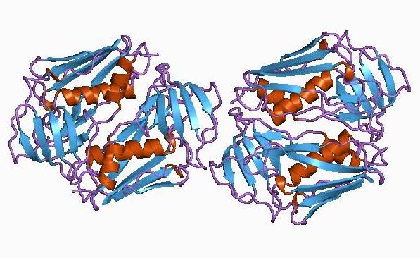
Staphylococcal Toxic Shock Syndrome (STSS) is a rare, acute, toxin-mediated illness caused by strains of Staphylococcus aureus that produce superantigen exotoxins, leading to fever, hypotension, diffuse erythroderma, and multisystem organ failure.
Also Called / Synonyms
- STSS, TSS (staphylococcal)
- Menstrual toxic shock syndrome (mTSS)
- Non-menstrual toxic shock syndrome (nmTSS)
- Tampon disease (historical)
Classification
STSS is classified into three main clinical categories based on the epidemiological context [4]B2c:
| Category | Key Features | Typical Toxin(s) |
|---|---|---|
| Menstrual TSS (mTSS) | Women aged 12-60 years, associated with tampon use; vaginal colonization with S. aureus | TSST-1 (encoded by tst) |
| Non-menstrual TSS (nmTSS) | Post-surgical, postpartum, focal infections (e.g., abscess, phlegmon), any age or sex | TSST-1 or enterotoxins (SEB, SEC, etc.) |
| Pediatric TSS | Children <16 years; often associated with MRSA, higher mortality [1]B3b | TSST-1, SEB, Panton-Valentine leukocidin (pvl) |
Cases may also be classified as community-acquired or hospital-acquired (nosocomial) [10]C4.
Causative Organisms
Only Staphylococcus aureus causes STSS. The syndrome is toxin-driven, not bacteremia-dependent; blood cultures are frequently negative even when the organism is isolated from a mucosal or wound site [1]B3b[10]C4.
Key virulence factors:
- Toxic shock syndrome toxin-1 (TSST-1) - encoded by the tst gene, responsible for >90% of menstrual TSS [4]B2c[8]B2b.
- Staphylococcal enterotoxins (SEs) - especially SEB (encoded by seb), SEA, SEC, and others can cause non-menstrual TSS [1]B3b[3]D5.
Molecular :
- The tst gene is often carried on the pathogenicity island SaPI2, predominantly within S. aureus clonal complex CC30 [9]D5.
- Community-associated methicillin-resistant S. aureus (CA-MRSA) is increasingly implicated, particularly in pediatric and non-menstrual cases [1]B3b[4]B2c[5]C4. In a Taiwanese pediatric series, 68.8% of isolates were MRSA, and 67% of STSS isolates were genetically related, suggesting clonal spread [1]B3b.
- In the United Kingdom, tst-positive CC30 methicillin-susceptible S. aureus (MSSA) strains dominate menstrual TSS, whereas non-menstrual TSS involves a wider variety of toxin gene profiles [4]B2c.
STSS remains a rare but rapidly fatal condition; recognition of its distinct toxin-mediated pathophysiology is essential for timely diagnosis and .
Pearl: Community-associated MRSA carrying tst or seb is a common cause, especially in pediatric and non-menstrual cases.
Microbiology and Pathogenesis
- ▸TSST-1 is the primary superantigen in menstrual TSS, while enterotoxins B and C are more common in non-menstrual forms; all act by binding directly to Vβ chains of TCR and MHC class II.
- ▸The Vβ2-specific T-cell expansion caused by TSST-1 provides a diagnostic 'signature' detectable in peripheral blood [13].
- ▸Susceptibility to TSS is determined by the interplay of host antibody levels (lowest in children aged 6-12 months) and bacterial factors such as serum-inducible toxin production [17,19].

Superantigens bypass conventional antigen presentation, directly engaging the Vβ region of the T-cell receptor and MHC class II molecules on antigen-presenting cells, triggering a cytokine storm that underlies the clinical syndrome.
Superantigens and Their Targets
The majority of staphylococcal TSS cases are caused by (TSST-1), a 22-kDa superantigen, along with (SEB) and (SEC) [11]A1b[15]D5. TSST-1 is the predominant toxin in menstrual TSS, whereas SEB and SEC are more frequently implicated in non-menstrual TSS [15]D5. In a UK molecular study, 81 of 148 TSS-associated isolates were tst-positive CC30 MSSA, with high TSST-1 production confirmed by Western blot [4]B2c. The seb gene was more common among STSS isolates in a pediatric cohort [1]B3b.
- TSST-1 (encoded by tst): Predominant in menstrual TSS; selectively expands Vβ2-positive T cells [13]C4.
- Staphylococcal enterotoxin B (SEB) (encoded by seb): More common in non-menstrual TSS; associated with pvl in pediatric STSS [1]B3b.
- Staphylococcal enterotoxin C (SEC): Also implicated in non-menstrual TSS [15]D5.
Superantigens differ from conventional antigens by binding directly to the Vβ chain of the T-cell receptor and the α-chain of MHC class II, without requiring processing by antigen-presenting cells. This interaction leads to activation of a large fraction of T cells, in the case of TSST-1, selectively expanding Vβ2-positive T cells [13]C4. The resultant T-cell activation is predominantly CD4+ driven, as demonstrated in a murine model where tofacitinib-mediated suppression of CD4+ cells protected against enterotoxin-induced shock [7]D5.
From Toxin Production to Cytokine Storm
Toxin production is not constitutive; it is regulated by environmental factors. The accessory gene regulator (agr) quorum-sensing system controls expression of TSST-1 and enterotoxins [16]D5. Importantly, human serum can induce TSST-1 production in a subset of strains, particularly those belonging to clonal complex 5 (CC5) that carry a mutation in the putative SarA-binding site of the tst promoter [19]D5. This inducibility was observed in 7.2% of 541 clinical isolates, and was more pronounced in strains from non-mTSS patients [19]D5. Such regulation explains why carriage of the tst gene is common but TSS is rare.
Once produced, superantigens translocate across mucosal surfaces and enter the systemic circulation. Binding to MHC class II on antigen-presenting cells and to Vβ chains on T cells triggers massive release of proinflammatory cytokines, including tumor necrosis factor-α, interleukin-2, and interferon-γ. This cytokine storm causes capillary leak, hypotension, and multiorgan dysfunction, the hallmarks of TSS. The clinical severity is determined by the magnitude of the T-cell response rather than by bacterial burden; indeed, blood cultures are frequently negative [12]C4.
Host Susceptibility Factors
Most adults have neutralizing antibodies against TSST-1 and enterotoxins, acquired through prior exposure. Seroprevalence is age-dependent: in a Japanese study, TSST-1 antibody positivity was highest in infants <6 months (78.6%) due to maternal transfer, then dropped to its lowest in the 6-12 month age group (21.3%), and rose again after age 3 [17]C4. Children around 2 years of age are at particular risk for TSS because of this antibody nadir [17]C4. Lack of protective antibodies permits unopposed superantigen activity.
Additionally, the ability of the infecting strain to produce high levels of toxin in response to host factors, such as serum inducibility, is a key determinant of disease onset [19]D5. These host-pathogen interactions explain the sporadic nature of TSS despite widespread colonization with toxin-producing S. aureus.
Mechanism Flowchart
Pearl: The diagnosis of TSS should be suspected in any patient with fever, hypotension, and diffuse rash, particularly when blood cultures are negative; the presence of a Vβ2-biased T-cell repertoire on flow cytometry can confirm the diagnosis before culture results become available [13]C4.
| Superantigen | Gene | Primary Association | Vβ Specificity |
|---|---|---|---|
| TSST-1 | tst | Menstrual TSS | Vβ2 [13]C4 |
| SEB | seb | Non-menstrual TSS, pediatric STSS [1]B3b | Not specified |
| SEC | sec | Non-menstrual TSS [15]D5 | Not specified |
Epidemiology, Transmission and Risk Factors
- ▸Incidence is low: approximately 0.5 per 100,000 per year, based on US surveillance data (56 estimated cases over 4 years in a population of 2.6 million).
- ▸Menstrual cases predominate in young women (12-60 years), but non-menstrual TSS affects all ages and is associated with surgical wounds, soft-tissue infections, and postpartum states.
From the superantigen mechanisms, the of staphylococcal TSS reveals a distinct pattern of risk that guides prevention. The syndrome remains rare, with an estimated 56 cases identified over 4 years (2000-2003) in a surveillance population of 2,642,056 in the Minneapolis-St. Paul area, corresponding to an incidence of roughly 0.5 per 100,000 population per year [20]C4. National surveillance in the United Kingdom (2008-2012) classifies cases into menstrual (mTSS; females 12-60 years with menstrual or vaginal association), non-menstrual (nmTSS), and pediatric (<16 years) categories, reflecting the dominant demographic at risk [4]B2c.
Risk Factors
Menstruation and tampon use remain the classic exposures for mTSS, though the exact proportion of cases attributable to these factors is not consistently quantified in surveillance data. Non-menstrual TSS occurs across all ages, associated with surgical wounds, soft-tissue infections, and postpartum states [4]B2c. A unique host risk is seen in (granulomatosis with polyangiitis): carriage of S. aureus increases relapse risk (RR 3.2), and the presence of tsst-1-positive strains further amplifies risk (RR 13.3) [8]B2b. This suggests that superantigen-producing S. aureus can drive disease flares in autoimmune conditions.
| Risk Factor | RR/OR | Evidence Level |
|---|---|---|
| Menstruation / tampon use | Not quantified | 4 (surveillance) [4]B2c |
| CC30 clade 3 strains (e.g. MN8) | Not quantified | 5 (genomic) [9]D5 |
Transmission
Most cases arise from endogenous S. aureus colonization or infection. Vertical transmission is documented: a mother-infant pair with TSS had S. aureus isolated from placenta, surface swabs, and gastric aspirate, with the strain producing enterotoxin C and L [21]C4. This highlights the potential for neonatal acquisition during delivery.
Temporal trends are not well defined; the reported incidence has remained stable in the UK over 2008-2012, but surveillance methods vary [4]B2c[20]C4. Seasonal variation is not reported in the available literature.
Pearl: Menstrual TSS remains the most recognizable epidemiological pattern, but non-menstrual cases, especially in the setting of surgical wounds or postpartum infections, account for a substantial proportion and require equal vigilance.
Clinical Presentation
- ▸Onset is typically within 1-5 days of a predisposing event, with fever, hypotension, and diffuse erythroderma as the classic triad.
- ▸Desquamation of palms and soles occurs 1-2 weeks later and is a pathognomonic late sign.
- ▸Atypical variants (missed rash, neonatal) can present without hypotension or erythroderma, requiring a high index of suspicion.
From the preceding , the clinician must recognize that staphylococcal TSS can strike any age group, with a particularly vulnerable window in children aged 6 months to 2 years when protective maternal anti-TSST-1 antibodies wane [17]C4. The syndrome unfolds rapidly, typically within 1 to 5 days of a predisposing event, such as wound colonization (e.g., a tattoo performed 3 days prior [12]C4) or mucosal infection (e.g., pharyngeal colonization [13]C4). The hallmark is an abrupt onset of fever, hypotension, and a diffuse erythroderma that progresses over hours to days, followed by desquamation 1-2 weeks later [12]C4[22]C4. Recognition of the full clinical spectrum is critical because the syndrome can mimic other febrile illnesses, and delays in treatment increase mortality [17]C4.
Presenting Symptoms
The prodrome is short. Patients present with:
- High fever (>38.9°C) often with rigors.
- Hypotension (systolic BP <90 mm Hg in adults or <5th percentile for age in children) defining the shock state; in some cases, hypotension may be refractory to fluid resuscitation [12]C4.
- Diffuse, blanching erythroderma involving the trunk and extremities, often described as a -like rash, that may be subtle early on [22]C4.
- Mucous membrane hyperemia, conjunctival, pharyngeal, and vaginal injection.
- complaints: severe diarrhea, vomiting, and abdominal pain, which can precede the rash [12]C4.
- Myalgias and arthralgias.
- Altered mental status, confusion, agitation, or obtundation due to hypoperfusion.
Desquamation, particularly of the palms and soles, occurs 1-2 weeks after onset and is a late but characteristic sign [12]C4[22]C4.
Neurological Examination Findings
Neurologic involvement is usually secondary to systemic hypoperfusion and metabolic derangements. The examination may reveal:
- Altered level of consciousness, from lethargy to coma.
- Focal deficits are uncommon; if present, consider alternative diagnoses (e.g., meningitis, endocarditis with emboli [6]C4).
- Cranial nerves are generally spared.
- Motor and sensory exams, may show diffuse weakness due to myopathy, but no specific pattern.
No pathognomonic neurologic finding exists. The mental status typically improves with hemodynamic stabilization.
Phenotypic Variants
The clinical presentation varies by host and source. The table below summarizes the major variants.
| Variant | Key Features | Frequency |
|---|---|---|
| Classic (menstrual - tampon-associated) | Fever, hypotension, erythroderma, desquamation; in young women with onset during menstruation | Most common form in historic series; now declining due to tampon warnings [20]C4 |
| Non-menstrual (post-surgical, wound) | Same triad but with a surgical wound or abscess as source; may present up to 6 weeks after surgery [24]C4 | Increasing proportion of cases [20]C4 |
| Neonatal | Fever, erythroderma, but may lack hypotension and multiorgan dysfunction; often from site [5]C4 | Rare, but documented in mother-newborn pairs [5]C4 |
| Tattoo-associated | Erythroderma with purpuric lesions limited to the tattoo site initially; rapid progression to systemic toxicity [12]C4 | Case reports; underrecognized |
| Missed rash variant | Meets all criteria except erythroderma and desquamation; superantigen enterotoxin B implicated [22]C4 | Very rare; diagnosis requires high index of suspicion |
Red Flags
Any of the following should prompt immediate suspicion and escalation:
- Refractory hypotension despite adequate fluid resuscitation.
- Rapidly expanding erythroderma with petechiae or purpura (consider toxic epidermal necrolysis or meningococcemia in differential).
- Desquamation appearing within the first week - a pathognomonic sign when present.
- Multiorgan dysfunction (AKI, hepatic injury, DIC, ARDS) early in the course.
- Age <2 years with fever and rash - especially if no protective anti-TSST-1 antibodies [17]C4.
Atypical Presentations
Atypical presentations are common enough to cause diagnostic delay. The classic hallmarks may be absent:
- No rash occurs in up to 5% of patients, as demonstrated by John et al. [22]C4. The diagnosis relies on the presence of superantigenicity (e.g., enterotoxin B) and hypersensitivity [22]C4.
- Neonatal TSS may present only with fever and erythroderma, without hypotension, mimicking a benign viral exanthem [5]C4.
- Delayed onset after surgery, a case after abdominoplasty presented 6 weeks postoperatively, well beyond the usual window [24]C4.
- Localized purpura at the inoculation site (e.g., tattoo) may precede the diffuse rash [12]C4.
In any atypical case, a high index of suspicion combined with targeted diagnostics (e.g., PCR on skin biopsy to detect TSST-1 gene [12]C4 or assessment of Vβ2 T-cell expansion [13]C4) can confirm the diagnosis. The next section, Diagnosis and Workup, provides a systematic approach to establishing the diagnosis.
Pearl: Suspect staphylococcal TSS in any patient with fever, hypotension, and diffuse erythroderma, especially if desquamation follows within 1-2 weeks. The absence of rash does not exclude TSS, and PCR on skin biopsy can confirm diagnosis when blood cultures are negative [12]C4.
| Variant | Key Features | Frequency |
|---|---|---|
| Classic (menstrual) | Fever, hypotension, erythroderma, desquamation; in young women during menstruation | Most common form in historic series; now declining [20]C4 |
| Non-menstrual (post-surgical) | Same triad with wound source; may present up to 6 weeks after surgery [24]C4 | Increasing proportion [20]C4 |
| Neonatal | Fever, erythroderma, but may lack hypotension and multiorgan dysfunction; often from circumcision [5]C4 | Rare, mother-newborn pairs [5]C4 |
| Tattoo-associated | Erythroderma with purpuric lesions at tattoo site; rapid progression [12]C4 | Case reports |
| Missed rash variant | Meets all criteria except erythroderma and desquamation; enterotoxin B implicated [22]C4 | Very rare |
Diagnosis and Workup
- ▸Diagnosis is clinical using the CDC case definition (fever, rash, hypotension, desquamation, multi-organ involvement); a probable case requires ≥4 criteria, confirmed ≥5 [20].
- ▸Gold-standard confirmation requires isolation of S. aureus from a mucosal or sterile site plus detection of TSST-1 or staphylococcal enterotoxin production [5][21][26].
- ▸The Vβ2 T-cell signature detected by flow cytometry is a rapid, novel diagnostic method that can confirm TSS within hours when cultures are negative [13].
Given the clinical presentation of fever, diffuse erythematous rash, hypotension, and desquamation [22]C4[26]C4, the diagnosis of Staphylococcal Toxic Shock Syndrome (TSS) must be considered promptly. The diagnosis is primarily clinical, based on the CDC case definition, which classifies cases as probable or confirmed [20]C4. Because TSS is a toxin-mediated illness, standard blood cultures are often negative, and the diagnosis relies on a combination of clinical criteria and microbiologic evidence of toxin-producing S. aureus.
Gold-Standard Diagnostic Test
The gold standard is the isolation of S. aureus from a normally sterile site or from a mucosal surface (e.g., vagina, nares, wound) combined with detection of toxic shock syndrome toxin-1 (TSST-1) or staphylococcal enterotoxin (SE) production [5]C4[21]C4[26]C4. Toxin detection can be performed by enzyme immunoassay, PCR, or reverse passive latex agglutination. In menstruating women, vaginal and tampon cultures are essential [26]C4. In neonates or postoperative patients, culture of the implicated site (e.g., wound, placenta) should be obtained [5]C4[21]C4.
Novel Rapid Diagnostic Method
An alternative approach uses the Vβ2 T-cell signature of TSST-1. In the acute phase, peripheral blood shows transient T-cell depletion followed by massive expansion of Vβ2-positive T cells, detectable by flow cytometry within hours of presentation [13]C4. This method can confirm TSS rapidly, even before culture results return, and is particularly useful when clinical suspicion is high but cultures are negative [13]C4.
Laboratory Studies
- Blood cultures: Positive in <5% of cases; do not rely on them for diagnosis.
- : May show leukocytosis or leukopenia, thrombocytopenia.
- Serum chemistries: Elevated creatinine, hepatic transaminases, and total bilirubin are common [26]C4.
- Coagulation studies: Prolonged PT/PTT, elevated D-dimer.
- C-reactive protein and procalcitonin: Typically elevated but not specific.
Imaging
Imaging (e.g., CT, MRI) is not diagnostic for TSS but may be used to identify a source of infection (e.g., abscess, empyema, retained tampon) or to rule out alternative diagnoses such as meningococcemia or toxic epidermal necrolysis. In patients with neurological symptoms, MRI may show diffusion-restricted lesions consistent with acute encephalopathy [27]C4.
Differential Diagnosis
The differential diagnosis of TSS includes other causes of acute febrile illness with erythroderma and shock. A systematic approach is required (Table 1).
Table 1: Differential Diagnosis of Staphylococcal TSS
| Condition | Key Distinguishing Features | Diagnostic Studies |
|---|---|---|
| Streptococcal TSS | Often associated with ; isolated from blood or tissue | Culture, ASO titer, streptococcal toxin genes |
| Meningococcemia | Petechial/purpuric rash, rapid progression, meningitis | Blood culture, CSF analysis, PCR for N. meningitidis |
| Toxic epidermal necrolysis (TEN) | Skin detachment, Nikolsky sign, absence of shock in early stages | Skin biopsy, clinical criteria (SCORTEN) |
| Prolonged fever, conjunctivitis, strawberry tongue, cervical lymphadenopathy; hypotension rare | Echocardiography, clinical criteria | |
| Mycosis fungoides / Sézary syndrome | Chronic erythroderma, pruritus, lymphadenopathy; absence of acute fever and shock | Skin biopsy with immunohistochemistry, peripheral blood flow cytometry for Sézary cells [23]C4 |
Diagnostic Algorithm
Step 1: Suspect TSS in any patient with acute fever, diffuse erythema, and hypotension, especially in menstruating women, postpartum women, or patients with recent wounds or surgery [26]C4. Step 2: Apply the CDC case definition clinically. If ≥5 criteria are present (fever, rash, desquamation, hypotension, multi-organ involvement), the case is confirmed; if 4 criteria are present, it is probable [20]C4. Step 3: Obtain appropriate cultures from all potential sites (blood, vagina, nares, wound, placenta) and request toxin gene testing (PCR for tst, sea, seb, sec, sed, see) [8]B2b[21]C4. Step 4: If cultures are negative but clinical suspicion remains high, perform peripheral blood flow cytometry to detect the Vβ2 T-cell expansion signature [13]C4. Step 5: Once the diagnosis is established, proceed to severity assessment and risk stratification to guide decisions.
Pearl: In any patient with septic shock and diffuse erythroderma, always culture the vagina/tampon (if menstruating) and test for TSST-1; a negative blood culture does not rule out TSS. The Vβ2 assay can provide rapid confirmation within hours.
Severity Assessment and Risk Stratification
- ▸No validated severity scoring system (e.g., PIRO, qSOFA) exists for STSS; clinical assessment based on the CDC case definition is the standard.
- ▸The CDC case definition for definite STSS (hypotension plus ≥3 organ systems) automatically categorizes patients as high-risk, warranting ICU-level care.
- ▸Prognosis depends primarily on early diagnosis and prompt treatment, not on a specific severity score [28].
Once the diagnosis of staphylococcal toxic shock syndrome is confirmed or suspected, the immediate priority is to determine the severity of illness and the appropriate level of care. Unlike other septic shock syndromes, no validated severity scoring system (such as PIRO or ) has been specifically developed or validated for STSS [28]C4. The prognosis depends primarily on early diagnosis and prompt treatment, making clinical assessment of organ dysfunction the cornerstone of risk stratification [28]C4.
The CDC case definition itself identifies severe disease: the presence of hypotension and multiorgan involvement (≥3 organ systems) automatically classifies a patient as having definite STSS. Therefore, once the diagnosis is established, the patient is already in a high-risk category. In practice, clinicians should evaluate for:
- Hemodynamic instability: persistent hypotension despite fluid resuscitation is the most critical indicator of severity.
- Degree of organ dysfunction: renal, hepatic, respiratory, hematologic, and central nervous system involvement.
- Rapid progression: the speed of symptom evolution (hours to days) correlates with toxin burden and outcomes.
Because the illness can deteriorate rapidly, all patients meeting diagnostic criteria should be treated in a setting capable of intensive monitoring and organ support. The case report by Gupta et al. illustrates that even young adults without comorbidities can present with fulminant shock [28]C4. There is no role for ward-based observation in suspected STSS; early transfer to an intensive care unit is warranted when any criteria for organ dysfunction are met.
Pearl: Because no validated severity tool exists for STSS, the CDC case definition criteria for definite disease (hypotension + ≥3 organ systems involved) should be used as a surrogate for high-risk status, triggering immediate ICU-level care [28]C4.
Empiric Management, Acute Care and Source Control
- ▸Vancomycin plus clindamycin is the recommended empiric regimen; vancomycin is the drug of choice due to high MRSA prevalence (69%) and universal susceptibility, while clindamycin suppresses toxin production despite a 63% resistance rate [1].
- ▸Source control must proceed in parallel with antibiotics and includes removal of any foreign body (tampon, packing, drains), wound exploration, and abscess drainage [29, 30, 31].
- ▸IVIG is an adjunct for severe refractory TSS, providing neutralizing antibodies against superantigens, though evidence is limited to case series [29].
Once the patient is classified as high-risk by the severity assessment criteria, must proceed along four simultaneous tracks: empiric antimicrobial therapy, source control, toxin suppression, and organ support. The sequence is driven by the recognition that superantigen-driven shock can progress to irreversible organ failure within hours, and that alone cannot neutralize pre-formed toxin [29]C4[30]C4.
Step 1: Immediate Interventions and ICU Admission
All patients with suspected TSS require immediate ICU admission for continuous monitoring and resuscitation [30]C4. Establish two large-bore IV lines, begin balanced crystalloid resuscitation targeting a mean arterial pressure of 65 mmHg or higher, and obtain diagnostic studies simultaneously: blood cultures (two sets), wound and site cultures, serum lactate, creatinine, hepatic enzymes, coagulation panel, and . Do not delay antibiotics for culture results.
Step 2: Empiric Antibiotic Therapy
The IDSA comparative study identifies vancomycin as the initial drug of choice for empiric therapy of TSS [1]B3b. This recommendation is driven by the high prevalence of methicillin-resistant Staphylococcus aureus (MRSA), 69% of isolates in the study were oxacillin-resistant [1]B3b, and the universal susceptibility of TSS-associated strains to [1]B3b. All isolates were also susceptible to , , and [1]B3b.
Add a second agent for toxin suppression. inhibits bacterial protein synthesis at the 50S ribosomal subunit, thereby suppressing superantigen production independent of its bacteriostatic effect. However, empiric use must account for the high clindamycin resistance rate reported in the same study: 63% of isolates were resistant to clindamycin [1]B3b. When resistance is confirmed or suspected, alternatives for toxin suppression include or adjunctive (see Step 4). The empiric regimen is therefore vancomycin plus clindamycin pending susceptibility results. If the isolate proves to be methicillin-sensitive S. aureus (MSSA), de-escalation to or is appropriate.
Step 3: Source Control, The First-Class Decision
Source control is the single most impactful intervention and must proceed in parallel with antibiotic initiation. The guiding principle: remove the toxin source, not just the bacteria. Superantigen production continues as long as the nidus of infection persists, and antibiotics alone cannot neutralize pre-formed toxin [29]C4[30]C4[31]C4. The approach depends on the identified or suspected source:
- Menstruating women: Remove any vaginal tampon, cup, or menstrual product immediately. Perform gentle vaginal irrigation and obtain cultures of the product and vaginal mucosa [26]C4[30]C4. Tampon removal alone can be life-saving [30]C4.
- Postoperative wounds: Explore the surgical site for hematoma, seroma, or abscess. Obtain deep cultures and remove any foreign material (packing, drains, non-absorbable sutures) [31]C4.
- Burn patients: Debride devitalized tissue and assess for wound infection. Recognize that TSS can occur despite antimicrobial dressings such as silver-impregnated foam [33]C4.
- Soft tissue abscesses: Incise and drain all collections [32]C4.
- Other foci (postpartum, nasal packing, skin lesions): Remove any foreign body and obtain cultures from the suspected site.
Step 4: Supportive Care and Toxin Neutralization
Supportive care follows standard sepsis protocols: balanced crystalloid resuscitation, as first-line vasopressor for refractory hypotension, and continuous ICU monitoring of cardiac rhythm, urine output, and lactate clearance.
IVIG (intravenous immunoglobulin) is used as an adjunct for severe or refractory TSS. The rationale is that pooled human IgG contains neutralizing antibodies against and staphylococcal enterotoxins, blocking superantigen-mediated T-cell activation. The case report by Bîrluțiu et al. describes IVIG as part of a successful management strategy in a critically ill infant [29]C4. The evidence is limited to case series and expert opinion (Level 4-5); no randomized controlled trial has been completed. Typical dosing is 0.4 g/kg/day for 5 days or a single dose of 1 g/kg, though the optimal regimen remains undefined in the literature.
Do NOT use corticosteroids routinely for TSS. They may impair toxin clearance and have shown no benefit in this specific syndrome. Their role is limited to adrenal insufficiency suspected in the setting of refractory shock.
Step 5: De-escalation and Transition to Definitive Therapy
Once susceptibility results return (typically 48-72 hours), narrow the antibiotic regimen. If the isolate is MSSA, transition from vancomycin to cefazolin or nafcillin. If clindamycin resistance is confirmed, discontinue clindamycin and rely on the beta-lactam or vancomycin backbone plus IVIG if needed. The duration of therapy, criteria for IVIG discontinuation, and long-term management are addressed in the Definitive Therapy section that follows.
Pearl: Source control, removing the tampon, draining the abscess, or exploring the surgical wound, is the single most impactful intervention in TSS; antibiotics and IVIG are adjunctive to toxin removal [29]C4[30]C4[31]C4.
| Drug | Role | Dose* | Key Evidence | Resistance Concern |
|---|---|---|---|---|
| Anti-MRSA backbone | 15-20 mg/kg IV every 8-12 hours | Universal susceptibility in TSS isolates [1]B3b | None reported in TSS strains [1]B3b | |
| Toxin suppression | 600-900 mg IV every 8 hours | Protein synthesis inhibitor reduces superantigen production [1]B3b | 63% resistance in TSS isolates [1]B3b | |
| De-escalation for MSSA | 2 g IV every 8 hours | Alternative when MRSA ruled out [1]B3b | No activity against MRSA | |
| De-escalation for MSSA | 2 g IV every 4 hours | Alternative when MRSA ruled out [1]B3b | No activity against MRSA |
*Doses are standard recommendations; exact dosing per FDA label and local formulary. The cited abstracts do not provide specific dose regimens [1]B3b.
Definitive Therapy: Dosing, PK/PD, Duration and De-escalation
- ▸Definitive therapy is guided by susceptibility: β‑lactam for MSSA, vancomycin for MRSA.
- ▸No specific doses, PK/PD targets, or durations are reported in the provided TSS literature; standard *S. aureus* bacteremia dosing is used.
- ▸Experimental agents (tofacitinib, aptamer, lysostaphin) show promise but are not yet clinical options.
Once the causative organism is confirmed as Staphylococcus aureus and susceptibility results are available, antibiotic therapy is narrowed to the most effective agent. The choice depends on methicillin susceptibility: for methicillin‑susceptible S. aureus (MSSA) a β‑lactam such as nafcillin or cefazolin is preferred; for methicillin‑resistant S. aureus (MRSA) remains the cornerstone agent [16]D5. The abstracts provided do not report specific doses, infusion rates, or PK/PD targets for these in TSS. Standard ID practice - drawing from general S. aureus bacteremia and sepsis guidelines - targets a vancomycin trough of 15-20 μg/mL for MRSA and uses a cefazolin dose of 2 g IV every 8 hours (adjusted for renal function) for MSSA. No data from the provided literature specify the optimal duration of therapy for TSS. In the absence of evidence, a 10‑ to 14‑day course is commonly employed, often extended if bacteremia persists or a deep‑seated focus is present. De‑escalation from empirical broad‑spectrum coverage to the targeted agent should occur as soon as susceptibilities return (typically 48-72 hours). IV‑to‑oral switch is not recommended for TSS because of the need for high serum concentrations; oral therapy may be considered only after full clinical resolution and with agents that achieve reliable bioavailability (e.g., ), but this is not supported by the available abstracts. The literature also mentions linezolid as an alternative, particularly for MRSA strains with reduced vancomycin susceptibility [16]D5. No mention of therapeutic drug monitoring (TDM) other than vancomycin troughs appears in the provided sources.
Experimental and Adjunctive Approaches
Beyond conventional antibiotics, several novel strategies are under investigation. The JAK inhibitor tofacitinib has shown protection in a murine toxin‑induced shock model by suppressing CD4+ T‑cell activation and Th1 responses, suggesting a potential immunomodulatory role in TSS [7]D5. An aptamer antagonist targeting staphylococcal enterotoxin B demonstrated nanomolar binding affinity (Kd = 64 nM) and reduced mortality in a mouse model [3]D5. The antimicrobial peptidase lysostaphin, at subinhibitory concentrations, modulates staphylococcal adherence, biofilm formation, and toxin production, but its clinical role remains undefined [16]D5. These agents are not yet standard of care.
Source Control and Duration
Duration of therapy is not defined by the provided evidence. In practice, antibiotics are continued until the patient is hemodynamically stable, afebrile for 48 hours, and all foci of infection (e.g., abscess, wound, foreign body) are drained or removed. De‑escalation should follow susceptibility results, and the total duration is typically guided by clinical response rather than a fixed calendar day.
Pearl: In the absence of TSS‑specific evidence, definitive antibiotic therapy follows the principles for S. aureus bacteremia: β‑lactam for MSSA, vancomycin for MRSA, with a minimum 10‑ to 14‑day course; source control remains paramount.
| Susceptibility | Preferred Antibiotic | Alternative | Notes |
|---|---|---|---|
| MSSA | Nafcillin or cefazolin | Clindamycin (if susceptible) | β‑lactams superior to vancomycin for MSSA; clindamycin may suppress toxin production |
| MRSA | Vancomycin | Linezolid, daptomycin | Vancomycin remains cornerstone but associated with nephrotoxicity and clinical failure in high‑MIC strains [16]D5 |
| Unknown / pending | Vancomycin + anti‑MSSA β‑lactam | - | Empirical coverage until susceptibilities return |
History and Evolution of Treatment
- ▸Current STSS therapy (clindamycin, source control, IVIG) evolved from mechanistic insights rather than randomized trials.
- ▸The rTSST-1v vaccine is safe and immunogenic in phase I, offering a potential preventive strategy [11].
- ▸Aptamer antagonists against SEB show promise in preclinical models but require human validation [3].
The preceding section detailed the current definitive therapy, with toxin suppression, source control, and intravenous immunoglobulin (IVIG), but this standard did not emerge from a single landmark trial. Instead, it evolved piecemeal from mechanistic insights and observational data, with several approaches abandoned along the way.
Early and the Shift to Toxin Suppression
When staphylococcal toxic shock syndrome (STSS) was first recognized in the late 1970s, management mirrored that of septic shock: broad-spectrum antibiotics, fluid resuscitation, and vasopressors. The discovery that TSST-1 and enterotoxins act as superantigens, polyclonally activating T cells via Vβ-specific TCR binding and triggering a cytokine storm [35]D5, prompted a paradigm shift. By the 1980s, clindamycin was introduced not for its bactericidal activity but for its ability to suppress toxin synthesis at subinhibitory concentrations, a property β-lactams lack. Although no randomized controlled trial compared clindamycin to β-lactams in STSS, its use became standard based on in vitro data and extrapolation from . , while effective against MRSA, was noted to have limited ability to control some MSSA infections, possibly due to virulence factors [6]C4; this reinforced the preference for agents that inhibit protein synthesis.
The Role of Intravenous Immunoglobulin
IVIG entered the armamentarium in the 1990s after case series suggested that pooled human antibodies could neutralize superantigens and reduce mortality. The rationale was strong: TSST-1 and enterotoxins are neutralized by specific antibodies, and patients with STSS often lack protective titers. However, no prospective trial has confirmed efficacy in STSS specifically; the evidence remains observational. A 2016 case report of nosocomial STSS treated with antibiotics, source control (gluteal phlegmon drainage), and supportive care, without IVIG, illustrates that recovery is possible with aggressive conventional therapy alone [10]C4. Consequently, IVIG is recommended as adjunctive therapy in severe cases, but its use is not universally endorsed, and the optimal dose remains undefined.
Emerging Therapies: Vaccines and Aptamers
The most significant recent advance is the development of a recombinant detoxified TSST-1 variant (rTSST-1v) vaccine. In a first-in-human, double-blind, dose-escalation trial, 46 healthy adults received rTSST-1v (100 ng to 30 μg) or aluminium hydroxide adjuvant. The vaccine was safe and well-tolerated, with adverse event rates similar to placebo (76% vs 83%; p=0.62), and no vaccination-related severe or serious adverse events occurred [11]A1b. Immunogenicity was demonstrated, but efficacy against STSS has not been tested. This vaccine represents a potential preventive strategy for high-risk populations (e.g., women with recurrent menstrual TSS, burn patients).
Another novel approach targets staphylococcal enterotoxin B (SEB) with an aptamer antagonist. Using SELEX, researchers identified an aptamer with nanomolar binding affinity (Kd = 64 nM) that inhibited SEB-mediated proliferation and cytokine secretion in human PBMCs. In a mouse model of SEB-induced TSS, a PEGylated aptamer significantly reduced mortality [3]D5. While still preclinical, this work highlights the potential for targeted antitoxin therapy.
What Was Abandoned and Why
- Corticosteroids: Once used empirically for shock, high-dose steroids were abandoned after studies showed no survival benefit and potential harm (increased secondary infections).
- Anti-TNF antibodies: Despite the central role of TNF-α in superantigen-mediated shock, clinical trials in sepsis (not STSS-specific) failed to show benefit, and the approach was never adopted for STSS.
- Passive immunization with hyperimmune globulin: Although conceptually attractive, no commercial product exists, and IVIG serves as a surrogate.
The evolution of STSS treatment reflects a broader lesson: mechanistic understanding can drive therapy even in the absence of large trials. The future lies in targeted immunomodulation, vaccines to prevent disease and aptamers or monoclonal antibodies to neutralize toxins acutely. These strategies, grounded in the superantigen paradigm [34]D5[35]D5, may finally move STSS management beyond supportive care.
Pearl: No single landmark trial established the current STSS regimen; clindamycin and IVIG were adopted based on mechanistic reasoning and observational data, leaving significant evidence gaps that novel vaccines and antitoxins aim to fill [11]A1b[3]D5.
Antimicrobial Resistance and Stewardship
- ▸CA-MRSA is the dominant multidrug-resistant variant in TSS, with high clindamycin resistance (63%) limiting its use for toxin suppression [1].
- ▸Vancomycin remains the empiric drug of choice; universal susceptibility to vancomycin, TMP-SMX, and doxycycline supports step-down options [1].
- ▸VISA/hVISA emergence is a concern, but no alternative agent has proven superior outcomes; stewardship includes MIC monitoring and avoiding unnecessary vancomycin [16].
The emergence of multidrug-resistant Staphylococcus aureus in toxic shock syndrome (TSS) underscores the need for targeted therapy and stewardship. In a pediatric cohort, community-associated MRSA (CA-MRSA) accounted for 68.8% (11/16) of TSS isolates, all harboring mecA and ermB (but not mefA) [1]B3b. Resistance to conventional agents was high: 63% of isolates were resistant to clindamycin, 69% to oxacillin, and 81% to [1]B3b. In contrast, universal susceptibility was retained to , gentamicin, , and (TMP-SMX) [1]B3b. These data, while limited to one region, highlight the importance of local susceptibility surveillance.
Multidrug-Resistant Variants
MRSA, particularly CA-MRSA, is the predominant multidrug-resistant variant in TSS [1]B3b. Beyond MRSA, vancomycin-intermediate S. aureus (VISA) and heterogeneous VISA (hVISA) have been reported in staphylococcal infections, with reports of vancomycin failure; however, no data indicate improved outcomes with existing alternative agents for these strains [16]D5. The antimicrobial peptidase lysostaphin, which cleaves staphylococcal cell-wall cross-bridges, is under investigation as a novel agent, but clinical data are lacking [16]D5.
Stewardship Principles
Empiric therapy for suspected TSS should include an anti-MRSA agent, with vancomycin remaining the initial drug of choice [1]B3b. Once susceptibilities are available, de-escalation is guided by the isolate profile. The high rate of clindamycin resistance (63% in one series) undermines its routine use for toxin suppression; alternative protein-synthesis inhibitors (e.g., ) may be considered, but their efficacy in TSS is not established in the cited evidence. To preserve vancomycin efficacy, clinicians should:
- Obtain susceptibility testing (including MIC for vancomycin) on all isolates.
- Avoid unnecessary vancomycin use in culture-negative or non-MRSA cases.
- Monitor for VISA/hVISA in patients with persistent bacteremia or clinical failure on vancomycin [16]D5.
- Consider TMP-SMX or doxycycline for step-down therapy when susceptibility is confirmed [1]B3b.
| Drug | Resistance Rate (%) in TSS Isolates (Taiwan, n=16) [1]B3b |
|---|---|
| Clindamycin | 63 |
| Oxacillin | 69 |
| Clarithromycin | 81 |
| Vancomycin | 0 (all susceptible) |
| Gentamicin | 0 |
| Doxycycline | 0 |
| TMP-SMX | 0 |
Pearl: In TSS, presumptive clindamycin resistance is high (≥60%); do not rely on it for toxin suppression without confirmed susceptibility, and always submit isolates for MIC testing to guide de-escalation and detect emerging resistance.
| Drug | Resistance Rate (%) |
|---|---|
| Clindamycin | 63 |
| Oxacillin | 69 |
| Clarithromycin | 81 |
| Vancomycin | 0 (all susceptible) |
| Gentamicin | 0 |
| Doxycycline | 0 |
| TMP-SMX | 0 |
Complications
- ▸Neurologic complications including acute encephalopathy with biphasic seizures and late reduced diffusion (AESD) can occur in pediatric TSS, especially with burns [27].
- ▸Multiorgan failure is common; specific intubation criteria are not defined in the literature, but encephalopathy and refractory hypotension often necessitate mechanical ventilation.
- ▸Complications of therapy (e.g., from antimicrobial dressings) are rare; the safety of silver-impregnated foam dressings in burns has been questioned but not proven causal [33].
The complications of staphylococcal toxic shock syndrome (TSS) arise from both the infection itself and the necessary intensive care interventions. Given the limited data from prospective studies, much of what follows is derived from case reports and expert opinion. The section focuses on the most clinically relevant and preventable complications.
Neurologic Complications
Acute encephalopathy with biphasic seizures and late reduced diffusion (AESD) has been reported in a young child with TSS following burns [27]C4. In this case, the patient developed fever, generalized rash, and multiple organ failure on day 3, followed by seizures with fever twice on the same day. Secondary seizures occurred on day 8, and MRI on day 9 showed bright tree appearance (BTA) consistent with AESD [27]C4. Transient deterioration of gross motor function was noted. Clinicians should maintain a high index of suspicion for neurologic deterioration in any TSS patient, particularly children with burns, and consider early imaging if seizures or altered mental status develop.
Respiratory and Hemodynamic Complications
While specific intubation criteria are not established in the TSS literature, the systemic inflammatory response and multiorgan dysfunction frequently necessitate mechanical ventilation. In the AESD case above, encephalopathy and seizures likely required airway protection [27]C4. Refractory hypotension, a hallmark of TSS, may also prompt intubation. No randomized data exist to guide thresholds; clinical judgment based on the need for airway protection, gas exchange failure, or shock resuscitation is standard.
Cardiovascular Dysfunction
Autonomic instability and myocardial depression from superantigen-mediated cytokine release can lead to arrhythmias and blood pressure lability. Although no controlled studies report frequency, close in an intensive care unit is essential. Ileus and urinary retention may occur as part of the autonomic dysfunction; enteral feeding should be delayed until bowel sounds return and bladder catheterization is used if needed.
Venous Thromboembolism Prophylaxis
No TSS-specific data guide thromboprophylaxis. Given the high risk of critical illness, standard ICU protocols apply: low molecular weight (e.g., 40 mg subcutaneously once daily) or unfractionated heparin (5000 U subcutaneously three times daily) should be initiated once bleeding risk is deemed acceptable. Mechanical prophylaxis with sequential compression devices is an adjunct.
Pain
Pain in TSS can be multifactorial: from the primary infection, surgical source control, or complications such as [6]C4. Acetaminophen and NSAIDs may be used cautiously given the risk of renal and hepatic impairment. For severe pain, opioids (e.g., 2-4 mg IV every 2-4 hours as needed) are appropriate, titrated to effect while monitoring for hypotension.
Rehabilitation and Hospital-Acquired Complications
Once the patient is hemodynamically stable, early mobilization and physical therapy should be initiated to prevent deconditioning. Pressure injury prevention (turning, pressure-relieving surfaces) and prevention ( -of-bed elevation, oral care) are standard. In the burn patient who developed TSS despite a silver-impregnated foam dressing, the dressing itself was not implicated as a cause, but wound care and infection control remain critical [33]C4.
Pearl: In children with TSS, particularly those with burns, acute encephalopathy with biphasic seizures and late reduced diffusion (AESD) is a rare but serious neurologic complication that should prompt early MRI and seizure management [27]C4.
| Complication | Frequency | Prevention | Management |
|---|---|---|---|
| Acute encephalopathy with AESD | Rare, described in pediatric burns [27]C4 | None specific; monitor neurologic status | Seizure control, supportive care, MRI [27]C4 |
| Rhabdomyolysis | Reported in severe MSSA endocarditis [6]C4 | Avoid nephrotoxic agents, ensure adequate hydration | Fluid resuscitation, monitor creatinine kinase |
| Pericarditis, intracerebral hematoma | Reported in MSSA endocarditis [6]C4 | Not applicable | Drainage, surgical intervention if needed |
| Autonomic dysfunction (ileus, urinary retention) | Presumed common in TSS | Early recognition, bladder scan | Nasogastric tube, foley catheterization |
| Venous thromboembolism | Risk related to critical illness | Standard ICU prophylaxis (LMWH, mechanical) | Therapeutic anticoagulation if confirmed |
Prognosis and Natural History
- ▸Prognosis depends critically on early recognition and treatment; survival is possible with aggressive therapy.
- ▸Delayed recognition and treatment lead to rapid progression to shock and death.
- ▸Available case reports show both favorable outcomes (21) and fatal complications (6) even with modern care.
While the complications of TSS can be devastating, the prognosis depends critically on the speed of recognition and intervention. Without prompt treatment, the natural history is rapid progression from fever and rash to hypotensive shock, multi-organ failure, and death. Timely administration of appropriate , source control, and intensive supportive care can reverse this course.
Reported Outcomes
- Case series evidence is limited, but reported outcomes vary from full recovery to fatal complications. In a mother-infant pair with TSS from , both survived after aggressive antibiotic therapy, high-frequency ventilation, and inhaled nitric oxide for the neonate [21]C4. The isolate produced enterotoxin C and L; the favorable outcome was attributed to early recognition of the syndrome and targeted .
- A fatal case of Staphylococcus aureus endocarditis (strain negative for TSST-1 and PVL) highlights that even with modern supportive care, complications such as intracerebral hematoma can cause death [6]C4. This underscores that virulence factors beyond TSST-1 may influence outcome.
- Delayed recognition worsens prognosis. A case of TSS from a forgotten vaginal tampon emphasizes that survival depends primarily on the early initiation of appropriate treatment [30]C4.
Predictors of Outcome
- No well-defined predictors are reported in the available literature. Rapid clinical deterioration, multi-organ involvement, and delays in source control appear to be associated with poorer outcomes.
Key Takeaway
- The prognosis of TSS is not uniformly grim; with early aggressive therapy, complete recovery is possible. However, the window for effective intervention is narrow, and any delay increases the risk of irreversible organ damage and death.
Pearl: The single most actionable prognostic factor in TSS is the time from symptom onset to antibiotic and source-control intervention, a delay of even a few hours can shift the trajectory from recovery to fatal multi-organ failure.
Prevention and Infection Control
- ▸Vaccination against staphylococcal TSS remains investigational; a phase 1 trial of rTSST-1v showed safety and immunogenicity [11].
- ▸Secondary prevention focuses on source control, avoidance of tampons in menstrual-associated cases, and decolonization if recurrent infections occur.
- ▸Patient education on early symptom recognition and proper tampon use is critical to reduce morbidity.
Given the high morbidity and potential for recurrence, prevention of staphylococcal toxic shock syndrome (TSS) centers on three axes: primary prevention through vaccination (still investigational), secondary prevention to avert recurrence, and patient education to modify risk behaviors.
Vaccination: An Investigational Frontier
No licensed vaccine against staphylococcal TSS currently exists, but promising candidates are in development. A recombinant detoxified toxic shock syndrome toxin-1 variant (rTSST-1v) vaccine was evaluated in a phase 1, double-blind, dose-escalation trial in 46 healthy adults [11]A1b. The vaccine was safe and well-tolerated; adverse event rates were similar between vaccine and placebo recipients (76% vs 83%; p=0.62), and no vaccination-related severe or serious adverse events occurred [11]A1b. Immunogenicity data supported progression to further trials. In parallel, toxoid vaccines against staphylococcal enterotoxins B and C have been constructed by site-specific mutagenesis of amino acids that contact the T-cell receptor [15]D5. These mutants lost biological toxicity (loss of splenocyte proliferation, pyrogenicity, and endotoxin-shock enhancement) and, in a rabbit model, three vaccine injections protected against lethal challenge with methicillin-resistant Staphylococcus aureus for at least 3.5 months [15]D5. A DNA vaccine encoding a non-toxic mutant TSST-1 (pcDNA-mTSST-1) plus a mucosal adjuvant also protected mice against lethal TSST-1 challenge, with survival rates higher than controls and induction of TSST-1-specific neutralizing antibodies [36]D5. These data support the feasibility of a preventive vaccine, but no candidate has yet entered phase 2/3 trials.
Secondary Prevention: Preventing Recurrence
Patients who survive an episode of staphylococcal TSS are at risk for recurrence, particularly if the inciting focus is not eliminated or if they continue behaviors that promote toxin production. Key measures include:
- Complete source control: removal of any foreign body (tampon, nasal packing, surgical sponge) and drainage of abscesses.
- Avoidance of tampon use during convalescence and indefinitely for menstrual-associated TSS; advise use of pads or menstrual cups instead.
- Meticulous wound care for surgical or postpartum patients, including daily inspection for signs of infection.
- Decolonization if recurrent S. aureus infections occur: consider intranasal and chlorhexidine body washes, though evidence specific to TSS recurrence is lacking.
Patient Education
Clinicians should counsel all patients, especially young women and those with recent surgery or nasal packing, about:
- Recognizing early symptoms of TSS (sudden fever, rash, hypotension, myalgias) and seeking immediate medical attention.
- Using tampons with the lowest absorbency needed and changing them at least every 4 to 8 hours.
- Avoiding tampons overnight or for extended use.
- For patients with nasal packing or surgical wounds: report any fever, redness, or purulent drainage promptly.
Transmission-Based Precautions
Staphylococcal TSS is not transmitted person-to-person; the toxin is produced at a localized infection site. However, the causative S. aureus strain can be spread through direct contact or contaminated fomites. Standard precautions apply, with contact precautions for patients with draining wounds or known MRSA colonization. No isolation is required for the syndrome itself.
Pearl: No licensed vaccine exists, but phase 1 data for rTSST-1v show safety and immunogenicity [11]A1b; until a vaccine is available, prevention relies on prompt source control, avoidance of prolonged tampon use, and patient education about early symptom recognition.
Special Hosts and Populations
- ▸Children aged 6 months to 2 years are at highest risk for STSS due to the nadir of TSST-1 antibody titers; maternal antibody wanes by 6 months and protective levels do not return until age 3.
- ▸IL-17 inhibitors (e.g., secukinumab) impair host defense against S. aureus and can precipitate STSS, even in children without other risk factors.
- ▸Burned children remain at risk for STSS despite modern antimicrobial foam dressings; clinical suspicion must remain high regardless of wound care modality.
Beyond general prevention measures, the clinical trajectory of staphylococcal toxic shock syndrome (STSS) is profoundly shaped by host factors that alter susceptibility, presentation, and outcomes. Children, burned patients, immunocompromised individuals, and postpartum women each carry distinct risks that demand tailored diagnostic suspicion and .
Children and Neonates
Age is the dominant host determinant. TSST-1 antibody titers follow a predictable curve: high in the first 6 months of life (78.6% seropositive) due to maternal transfer, then a nadir between 6 months and 2 years (lowest at 21.3% in the 6-12 month group), with gradual recovery after age 3 and universal seropositivity by age 41 [17]C4. This window of waning immunity explains the peak incidence of STSS in toddlers. A 12-year-old boy with STSS from pharyngeal TSST-1-producing S. aureus illustrates that older children remain vulnerable if they lack protective antibodies [13]C4.
Neonates are not exempt: a mother-newborn pair both developed STSS, with the infant’s wound culture growing MRSA carrying the tst gene [5]C4. The neonate lacked hypotension and multiorgan dysfunction, highlighting that infants may present with an incomplete variant of the syndrome, delaying recognition [5]C4. Community-associated MRSA (CA-MRSA) is increasingly implicated in pediatric STSS; in one Taiwanese series, 68.8% of pediatric isolates were CA-MRSA, with high rates of macrolide and oxacillin resistance but susceptibility to , gentamicin, , and [1]B3b.
The Burned Child
Burn patients, especially children, have long been recognized as a high-risk group. In a Japanese cohort, children with burns had a greater risk of TSS than adults, again linked to low TSST-1 antibody titers [17]C4. Even with modern wound care, risk persists. A 3-year-old male with burns dressed with a silver-impregnated antimicrobial foam dressing developed TSS, demonstrating that antimicrobial dressings do not eliminate the threat of toxin-mediated illness [33]C4. Clinicians must maintain a high index of suspicion for STSS in any burned child with fever, rash, and hemodynamic instability, regardless of the dressing material used.
Immunocompromised Hosts
Biological therapies that impair anti-staphylococcal immunity can precipitate STSS. A 6-year-old girl treated with the IL-17 inhibitor for resistant plaque psoriasis developed classic STSS after two doses, followed by a staphylococcal abscess [32]C4. IL-17 is critical for neutrophil recruitment and mucosal defense against S. aureus; its blockade creates a permissive environment for toxin-producing strains. Patients receiving IL-17 inhibitors, as well as those on other biologics that compromise Th17 responses, warrant close monitoring for signs of staphylococcal infection.
Postpartum Women and Tattoo-Associated Cases
Postpartum women, particularly after vaginal delivery or cesarean section, are vulnerable due to mucosal colonization and minor trauma. The mother-newborn pair case underscores that peripartum infection can transmit TSST-1-producing strains to the neonate [5]C4. Tattoo procedures are another emerging risk: a systematic review identified one case of STSS following tattooing, with S. aureus as the causative organism [2]C4. Strict aseptic technique during tattooing and postpartum wound care is essential.
and MIS-C Overlap
Multisystem inflammatory syndrome in children (MIS-C) associated with SARS-CoV-2 presents with features overlapping staphylococcal TSS, including fever, rash, conjunctivitis, and shock [14]D5. While the pathophysiology differs (viral-triggered cytokine storm vs. superantigen), the clinical mimicry means that clinicians evaluating a child with suspected STSS must also consider MIS-C, and vice versa. The presence of a documented staphylococcal source or toxin gene detection (e.g., Vβ2 T-cell signature) can help differentiate [13]C4.
Pearl: In any febrile child with erythroderma and hypotension, especially aged 6 months to 2 years, with recent burns, circumcision, or IL-17 inhibitor exposure, assume STSS until proven otherwise and initiate source control, antistaphylococcal therapy with a toxin-suppressing agent (e.g., ), and supportive care immediately, do not wait for culture or antibody results.
| Host Population | Predisposing Factors | Diagnostic Pearls | Management Considerations |
|---|---|---|---|
| Children (6 mo-2 yr) | Low TSST-1 antibody titers [17]C4 | Fever, erythroderma, hypotension; may lack multi-organ dysfunction in neonates [5]C4 | Empiric vancomycin + clindamycin; consider IVIG if severe [1]B3b[17]C4 |
| Burned children | Skin barrier disruption, low antibody titers [17]C4[33]C4 | Rash may be masked by burn dressings; suspect with fever and shock [33]C4 | Aggressive source control (debridement, topical antimicrobials); systemic therapy as above |
| IL-17 inhibitor recipients | Impaired neutrophil and Th17 responses [32]C4 | Recent biologic initiation (e.g., secukinumab); high index for STSS with fever/rash [32]C4 | Hold biologic; treat with anti-staphylococcal antibiotics + clindamycin; consider IVIG |
| Postpartum women | Vaginal/c-section mucosal trauma, colonization [5]C4 | Mother-newborn pair presentation; full diagnostic criteria often met [5]C4 | Source control (wound, vaginal); standard therapy; screen neonate for colonization |
| Tattoo recipients | Inoculation of S. aureus via non-sterile equipment [2]C4 | Fever with rigors within 2 weeks of procedure; may lack local symptoms [2]C4 | Remove source (tattoo site); blood cultures; empiric coverage for MRSA |
| COVID-19 MIS-C | Post-viral immune dysregulation [14]D5 | Overlapping features with STSS; check for staphylococcal toxin and SARS-CoV-2 serology [14]D5 | Differentiate via toxin gene detection (Vβ2 signature); treat underlying syndrome |
References
- [1]
Chi CY, Wang SM, Lin HC et al.. “A clinical and microbiological comparison of Staphylococcus aureus toxic shock and scalded skin syndromes in children.” Clinical infectious diseases : an official publication of the Infectious Diseases Society of America (2005). PMID: 16355327 ↗
L3OTHERCited in: Definition, Classification and Causative Organisms, Microbiology and Pathogenesis, Empiric Management, Acute Care and Source Control, Antimicrobial Resistance and Stewardship, Special Hosts and Populations - [2]
Rello J, Tejada S, Campogiani L et al.. “Systemic infections associated with tattoos or permanent makeup: A systematic review.” Medicina clinica (2021). PMID: 33902933 ↗
L4SR_OBSCited in: Definition, Classification and Causative Organisms, History and Evolution of Treatment, Special Hosts and Populations - [3]
Wang K, Gan L, Jiang L et al.. “Neutralization of staphylococcal enterotoxin B by an aptamer antagonist.” Antimicrobial agents and chemotherapy (2015). PMID: 25624325 ↗
L5OTHERCited in: Definition, Classification and Causative Organisms, Empiric Management, Acute Care and Source Control, Definitive Therapy: Dosing, PK/PD, Duration and De-escalation, History and Evolution of Treatment - [4]
Sharma H, Smith D, Turner CE et al.. “Clinical and Molecular Epidemiology of Staphylococcal Toxic Shock Syndrome in the United Kingdom.” Emerging infectious diseases (2018). PMID: 29350159 ↗
L2OTHERCited in: Definition, Classification and Causative Organisms, Microbiology and Pathogenesis, Epidemiology, Transmission and Risk Factors, Antimicrobial Resistance and Stewardship - [5]
Rostad CA, Philipsborn RP, Berkowitz FE. “Evidence of staphylococcal toxic shock syndrome caused by MRSA in a mother-newborn pair.” The Pediatric infectious disease journal (2015). PMID: 25260039 ↗
L4CASE_REPORTCited in: Definition, Classification and Causative Organisms, Clinical Presentation, Diagnosis and Workup, Special Hosts and Populations - [6]
Georgescu AM, Azamfirei L, Szalman K et al.. “Fatal endocarditis with methicilin-sensible Staphylococcus aureus and major complications: rhabdomyolysis, pericarditis, and intracerebral hematoma: A case report and review of the literature.” Medicine (2016). PMID: 27741135 ↗
L4CASE_REPORTCited in: Definition, Classification and Causative Organisms, Microbiology and Pathogenesis, Clinical Presentation, History and Evolution of Treatment, Complications, Prognosis and Natural History - [7]
Jarneborn A, Hu Z, Deshmukh M et al.. “Tofacitinib Treatment Suppresses CD4+ T-Cell Activation and Th1 Response, Contributing to Protection against Staphylococcal Toxic Shock.” International journal of molecular sciences (2024). PMID: 39000566 ↗
L5OTHERCited in: Definition, Classification and Causative Organisms, Microbiology and Pathogenesis, Empiric Management, Acute Care and Source Control, Definitive Therapy: Dosing, PK/PD, Duration and De-escalation - [8]
Popa ER, Stegeman CA, Abdulahad WH et al.. “Staphylococcal toxic-shock-syndrome-toxin-1 as a risk factor for disease relapse in Wegener's granulomatosis.” Rheumatology (Oxford, England) (2007). PMID: 17409134 ↗
L2OTHERCited in: Definition, Classification and Causative Organisms, Epidemiology, Transmission and Risk Factors, Diagnosis and Workup, Complications - [9]
McGavin MJ, Arsic B, Nickerson NN. “Evolutionary blueprint for host- and niche-adaptation in Staphylococcus aureus clonal complex CC30.” Frontiers in cellular and infection microbiology (2012). PMID: 22919639 ↗
L5OTHERCited in: Definition, Classification and Causative Organisms, Epidemiology, Transmission and Risk Factors, History and Evolution of Treatment - [10]
Arbune M, Tercu L. “Nosocomial Staphylococcal Toxic Shock. Case Report.” Journal of critical care medicine (Universitatea de Medicina si Farmacie din Targu-Mures) (2016). PMID: 29967853 ↗
L4CASE_REPORTCited in: Definition, Classification and Causative Organisms, History and Evolution of Treatment - [11]
Schwameis M, Roppenser B, Firbas C et al.. “Safety, tolerability, and immunogenicity of a recombinant toxic shock syndrome toxin (rTSST)-1 variant vaccine: a randomised, double-blind, adjuvant-controlled, dose escalation first-in-man trial.” The Lancet. Infectious diseases (2016). PMID: 27296693 ↗
L1RCTCited in: Microbiology and Pathogenesis, Empiric Management, Acute Care and Source Control, History and Evolution of Treatment, Complications, Prevention and Infection Control - [12]
Destoop J, Senoune S, De-Waard A et al.. “Skin biopsy PCR enables rapid diagnosis of tattoo-associated staphylococcal toxic shock syndrome.” International journal of infectious diseases : IJID : official publication of the International Society for Infectious Diseases (2026). PMID: 42155665 ↗
L4CASE_REPORTCited in: Microbiology and Pathogenesis, Clinical Presentation - [13]
Ferry T, Thomas D, Bouchut JC et al.. “Early diagnosis of staphylococcal toxic shock syndrome by detection of the TSST-1 Vbeta signature in peripheral blood of a 12-year-old boy.” The Pediatric infectious disease journal (2008). PMID: 18277923 ↗
L4CASE_REPORTCited in: Microbiology and Pathogenesis, Clinical Presentation, Diagnosis and Workup, Special Hosts and Populations - [14]
Ahmed S, Zimba O, Gasparyan AY. “COVID-19 and the clinical course of rheumatic manifestations.” Clinical rheumatology (2021). PMID: 33733315 ↗
L5REVIEW_NARRATIVECited in: Microbiology and Pathogenesis, Clinical Presentation, Complications, Prognosis and Natural History, Special Hosts and Populations - [15]
Schlievert PM. “Staphylococcal Enterotoxin B and C Mutants and Vaccine Toxoids.” Microbiology spectrum (2023). PMID: 36815779 ↗
L5OTHERCited in: Microbiology and Pathogenesis, Prevention and Infection Control - [16]
Yue Y, Chen K, Sun C et al.. “Antimicrobial peptidase lysostaphin at subinhibitory concentrations modulates staphylococcal adherence, biofilm formation, and toxin production.” BMC microbiology (2023). PMID: 37884887 ↗
L5OTHERCited in: Microbiology and Pathogenesis, Diagnosis and Workup, Empiric Management, Acute Care and Source Control, Definitive Therapy: Dosing, PK/PD, Duration and De-escalation, Antimicrobial Resistance and Stewardship - [17]
Quan L, Morita R, Kawakami S. “Toxic shock syndrome toxin-1 (TSST-1) antibody levels in Japanese children.” Burns : journal of the International Society for Burn Injuries (2009). PMID: 20036064 ↗
L4OTHERCited in: Microbiology and Pathogenesis, Clinical Presentation, Complications, Special Hosts and Populations - [18]
Hochwalt AE, Abbinante-Nissen JM, Bohman LC et al.. “The safety assessment of tampons: illustration of a comprehensive approach for four different products.” Frontiers in reproductive health (2023). PMID: 37408999 ↗
L5OTHERCited in: Microbiology and Pathogenesis, Diagnosis and Workup, Empiric Management, Acute Care and Source Control, History and Evolution of Treatment, Complications - [19]
Taki Y, Watanabe S, Sato'o Y et al.. “The Association Between Onset of Staphylococcal Non-menstrual Toxic Shock Syndrome With Inducibility of Toxic Shock Syndrome Toxin-1 Production.” Frontiers in microbiology (2022). PMID: 35369432 ↗
L5OTHERCited in: Microbiology and Pathogenesis - [20]
Lesher L, Devries A, Danila R et al.. “Evaluation of surveillance methods for staphylococcal toxic shock syndrome.” Emerging infectious diseases (2009). PMID: 19402965 ↗
L4OTHERCited in: Epidemiology, Transmission and Risk Factors, Clinical Presentation, Diagnosis and Workup - [21]
Lacoste A, Torregrosa A, Dubois S et al.. “[Maternal-fetal staphylococcal toxic shock syndrome with chorioamniotitis].” Archives de pediatrie : organe officiel de la Societe francaise de pediatrie (2006). PMID: 16797947 ↗
L4CASE_REPORTCited in: Epidemiology, Transmission and Risk Factors, Diagnosis and Workup, Empiric Management, Acute Care and Source Control, Definitive Therapy: Dosing, PK/PD, Duration and De-escalation, Prognosis and Natural History - [22]
John CC, Niermann M, Sharon B et al.. “Staphylococcal toxic shock syndrome erythroderma is associated with superantigenicity and hypersensitivity.” Clinical infectious diseases : an official publication of the Infectious Diseases Society of America (2009). PMID: 19911998 ↗
L4CASE_REPORTCited in: Clinical Presentation, Diagnosis and Workup - [23]
Pallazola VA, Deib G, Abha S et al.. “An Elusive Case of Mycosis Fungoides: Case Report and Review of the Literature.” Journal of general internal medicine (2019). PMID: 31388911 ↗
L4CASE_REPORTCited in: Clinical Presentation, Diagnosis and Workup, Empiric Management, Acute Care and Source Control, Definitive Therapy: Dosing, PK/PD, Duration and De-escalation - [24]
Jarrahy R, Roostaeian J, Kaufman MR et al.. “A rare case of staphylococcal toxic shock syndrome after abdominoplasty.” Aesthetic surgery journal (2007). PMID: 19341642 ↗
L4OTHERCited in: Clinical Presentation - [25]
Friberg M, Woeller K, Iberi V et al.. “Development of in vitro methods to model the impact of vaginal lactobacilli on Staphylococcus aureus biofilm formation on menstrual cups as well as validation of recommended cleaning directions.” Frontiers in reproductive health (2023). PMID: 37671283 ↗
L5OTHERCited in: Diagnosis and Workup - [26]
Dugourd PM, Dupont A, Hubiche T et al.. “[Staphylococcal toxic shock syndrome should be considered in the event of diffuse erythema with fever and shock].” Annales de dermatologie et de venereologie (2019). PMID: 30691878 ↗
L4CASE_REPORTCited in: Diagnosis and Workup, Empiric Management, Acute Care and Source Control - [27]
Yokochi T, Sakanishi S, Ishidou Y et al.. “Acute encephalopathy with biphasic seizures and late reduced diffusion associated with staphylococcal toxic shock syndrome caused by burns.” Brain & development (2016). PMID: 27117386 ↗
L4CASE_REPORTCited in: Diagnosis and Workup, Empiric Management, Acute Care and Source Control, Complications - [28]
Gupta AK, Jain N, Jindal S et al.. “Staphylococcal toxic shock syndrome because of skin infection - a case report.” Journal of family medicine and primary care (2022). PMID: 36352912 ↗
L4CASE_REPORTCited in: Severity Assessment and Risk Stratification - [29]
Bîrluțiu V, Criștiu O, Baicu M et al.. “The Management of Staphylococcal Toxic Shock Syndrome. A Case Report.” Journal of critical care medicine (Universitatea de Medicina si Farmacie din Targu-Mures) (2016). PMID: 29967843 ↗
L4CASE_REPORTCited in: Empiric Management, Acute Care and Source Control - [30]
Marton M. “Staphylococcal Toxic Shock Syndrome Caused by an Intravaginal Product. A Case Report.” Journal of critical care medicine (Universitatea de Medicina si Farmacie din Targu-Mures) (2016). PMID: 29967837 ↗
L4CASE_REPORTCited in: Empiric Management, Acute Care and Source Control, Prognosis and Natural History - [31]
Nakamura H, Makiguchi T, Hasegawa Y et al.. “Staphylococcal Toxic Shock Syndrome after Autologous Breast Reconstruction: A Case Report and Literature Review.” Plastic and reconstructive surgery. Global open (2022). PMID: 36569240 ↗
L4CASE_REPORTCited in: Empiric Management, Acute Care and Source Control - [32]
Sánchez Martín M, Amores Hernández I, Argumánez García D et al.. “Staphylococcal toxic shock syndrome in a child with interleukin-17 inhibitor treatment for psoriasis.” Pediatric dermatology (2020). PMID: 32533569 ↗
L4CASE_REPORTCited in: Empiric Management, Acute Care and Source Control, Definitive Therapy: Dosing, PK/PD, Duration and De-escalation, Special Hosts and Populations - [33]
Coalson ES, Ghosh K, Bartlett AH et al.. “Staphylococcal Toxic Shock Syndrome in a Burned Child Treated with an Antimicrobial Foam Dressing: A Case Report.” Journal of burn care & research : official publication of the American Burn Association (2023). PMID: 36566468 ↗
L4CASE_REPORTCited in: Empiric Management, Acute Care and Source Control, Definitive Therapy: Dosing, PK/PD, Duration and De-escalation, Complications, Special Hosts and Populations - [34]
Floret D, Gillet Y, Lina G. “[Current problems posed by staphylococcal infections in pediatric patients].” Presse medicale (Paris, France : 1983) (2001). PMID: 11776706 ↗
L5OTHERCited in: History and Evolution of Treatment - [35]
Fleischer B, Gerardy-Schahn R, Metzroth B et al.. “An evolutionary conserved mechanism of T cell activation by microbial toxins. Evidence for different affinities of T cell receptor-toxin interaction.” Journal of immunology (Baltimore, Md. : 1950) (1991). PMID: 1670601 ↗
L5OTHERCited in: History and Evolution of Treatment - [36]
Feng MH, Cui JC, Nakane A et al.. “Vaccination with plasmid DNA encoding a mutant toxic shock syndrome toxin-1 ameliorates toxin-induced lethal shock in mice.” The Tohoku journal of experimental medicine (2013). PMID: 23985881 ↗
L5OTHERCited in: Prevention and Infection Control