On this page
Quick Reference
Overview and Recommendations
Background
- •Wilson disease is an autosomal recessive disorder of copper metabolism caused by pathogenic variants in ATP7B, leading to toxic copper accumulation predominantly in the liver and brain; untreated, it is uniformly fatal, but early diagnosis and lifelong chelation or zinc therapy allow normal life expectancy.
- •The disease affects approximately 1 in 30,000 to 1 in 50,000 live births globally, with a carrier frequency of ~1 in 90; it is pan-ethnic but shows regional mutation clustering (e.g., H1069Q in Europeans).
- •The pathognomonic defect is loss-of-function of ATP7B, a copper-transporting P-type ATPase essential for biliary copper excretion and ceruloplasmin incorporation; failure leads to hepatic copper overload, then systemic spillover to brain, cornea, kidneys, and other tissues.
- •Clinical presentations are classified into hepatic (40-50%), neurological (30-40%), mixed, and pre-symptomatic; acute liver failure with Coombs-negative hemolytic anemia is a medical emergency requiring urgent liver transplantation.
- •The paradigm of management shifted from high-dose penicillamine to individualized chelation (trientine preferred for neurological disease) and zinc maintenance, with liver transplantation reserved for decompensated cirrhosis or fulminant failure.
Evaluation
- •Suspect Wilson disease in any patient aged 3-55 years with unexplained liver disease (elevated aminotransferases, steatosis, cirrhosis), neurological symptoms (tremor, dysarthria, dystonia), or psychiatric disturbances (depression, personality change).
- •Examine for Kayser-Fleischer rings on slit-lamp examination, golden-brown copper deposition in Descemet's membrane; present in ~95% of neurological presentations but only ~50% of hepatic presentations; absence does not exclude disease.
- •Order serum ceruloplasmin (nephelometry) as first-line screening; a level <20 mg/dL supports Wilson disease, but acute-phase response can falsely elevate it, so a normal level does not rule out disease.
- •Measure 24-hour urinary copper excretion; a basal excretion >100 µg/24 h (1.6 µmol/24 h) is classic for symptomatic Wilson disease; in children with mild disease, a cutoff >40 µg/24 h provides 85% sensitivity and 96% specificity.
- •Calculate the Leipzig score using clinical, biochemical, and genetic criteria; a score ≥4 establishes the diagnosis, score 3 is suggestive, score ≤2 makes Wilson disease unlikely.
- •Perform ATP7B sequencing as the confirmatory gold standard; biallelic pathogenic variants confirm diagnosis; if only one variant found, consider whole-genome sequencing for deep intronic variants.
- •Consider liver biopsy for copper quantification (>250 µg/g dry weight) only when genetic results are uninformative and biochemical tests are borderline; biopsy also provides histologic grading of steatosis and fibrosis.
- •Order brain MRI (T2-weighted/FLAIR) in any patient with neurological or psychiatric symptoms; characteristic hyperintensity in basal ganglia, thalamus, and brainstem (including the 'face of the giant panda' sign) supports neurological involvement.
- •In acute liver failure, use the alkaline phosphatase:total bilirubin ratio <4 and ALT:AST ratio <1 to rapidly identify Wilson disease as the cause; these have high diagnostic accuracy in this setting.
- •Also consider differential diagnoses: autoimmune hepatitis (can have false-positive autoantibodies), progressive familial intrahepatic cholestasis type 3 (PFIC3), citrullinemia type I, and Menkes disease.
Management
- •Initiate chelation therapy immediately in all symptomatic patients with hepatic or neurological Wilson disease; first-line options are D-penicillamine 750-1500 mg/day orally in 2-4 divided doses on an empty stomach, or trientine dihydrochloride 750-1500 mg/day in 2-3 divided doses.
- •For presymptomatic patients or those who have achieved metabolic stability on chelation, use zinc acetate 50 mg elemental zinc three times daily with meals as maintenance therapy; zinc monotherapy is not recommended for initial treatment of symptomatic disease due to higher failure rates.
- •Titrate chelator doses based on 24-hour urinary copper excretion: target 200-500 µg/24h on chelation; if urinary copper is too high (>500 µg/24h), increase the chelator dose; if too low (<200 µg/24h), consider non-adherence or over-chelation.
- •Monitor serum non-ceruloplasmin-bound copper (NCC) with a target <150 µg/L; exchangeable copper (CuEX) is an emerging alternative with target <15 µg/L; liver enzymes and neurological examination (Unified Wilson Disease Rating Scale) should be assessed at each visit.
- •In acute liver failure due to Wilson disease, do not initiate chelation therapy, it does not reverse acute hepatic necrosis and may precipitate neurological worsening; instead, list for urgent liver transplantation (UNOS Status 1a) immediately.
- •Use therapeutic plasma exchange or molecular adsorbents recirculating system (MARS) as a bridge to transplantation in acute liver failure; these can remove circulating copper and improve coagulopathy but should not delay transplant listing.
- •For decompensated cirrhosis (Child-Pugh B/C), manage complications of portal hypertension per standard protocols: ascites with diuretics (spironolactone 100-400 mg/day, furosemide 40-160 mg/day), variceal hemorrhage with endoscopic band ligation and vasoactive drugs (terlipressin 2 mg IV q4h or octreotide 50 µg bolus + 50 µg/h), and hepatic encephalopathy with lactulose and rifaximin.
- •If first-line chelation fails (progressive liver dysfunction, neurological worsening, or intolerable adverse effects), switch to the alternative chelator; trientine tetrahydrochloride (600-1200 mg/day) is a newer formulation with improved tolerability.
- •Consider bis-choline tetrathiomolybdate (TTM, ALXN1840) 15-60 mg/day as an investigational option for neurological Wilson disease; it reduces intestinal copper absorption and may have lower risk of paradoxical neurological worsening.
- •Liver transplantation is definitive therapy for Wilson disease and is indicated for fulminant hepatic failure, decompensated cirrhosis unresponsive to 3-6 months of medical therapy, and selected cases of severe neurological deterioration refractory to chelation; post-transplant survival is 82-87% at 5 years.
- •Avoid abrupt cessation of therapy, copper rebound can cause acute hemolysis and liver failure; do not use non-dihydropyridine calcium channel blockers (diltiazem, verapamil) as they exacerbate hepatic dysfunction.
- •Refer for liver transplantation evaluation in any patient with acute liver failure, Child-Pugh class C cirrhosis, MELD ≥15, or neurological worsening despite optimal medical therapy.
Board Review — High Yield
- •Kayser-Fleischer rings, copper deposition in Descemet's membrane; pathognomonic when present, but absent in ~50% of hepatic presentations.
- •Leipzig score ≥4, diagnostic threshold combining clinical, biochemical, and genetic criteria.
- •D-penicillamine, first-line chelator; dose 750-1500 mg/day on empty stomach; monitor for autoimmune reactions and bone marrow suppression.
- •Trientine, alternative chelator with fewer adverse effects; preferred for neurological disease due to lower risk of paradoxical worsening.
- •Zinc acetate, maintenance therapy only; induces intestinal metallothionein to block copper absorption; not for initial symptomatic treatment.
- •Acute liver failure in Wilson disease, characterized by Coombs-negative hemolytic anemia, low ALP:bilirubin ratio <4, and ALT:AST <1; requires urgent liver transplantation.
- •Paradoxical neurological worsening, occurs in ~26% of neurological patients within first 3 months of chelation; manage by switching chelator or dose adjustment.
- •Liver transplantation, definitive therapy; corrects copper metabolism; 5-year survival >82%; indicated for fulminant failure or decompensated cirrhosis.
- •ATP7B mutation H1069Q, most common in Europeans; associated with later onset and milder phenotype compared to loss-of-function variants.
- •24-hour urinary copper target, 200-500 µg/24h on chelation; <100 µg/24h on zinc; over-chelation can cause copper deficiency and neurological deterioration.
Deep Dive — Evidence Details
Definition, Classification & Nomenclature
- ▸Wilson disease is defined by pathogenic ATP7B variants causing defective biliary copper excretion and toxic accumulation in liver and brain [1, 6].
- ▸The disease is classified into pre-symptomatic, hepatic, neurological, mixed, and acute decompensated phases, each with distinct management implications [4, 5].
- ▸WD is the classic hepatocellular copper-overload disorder, not a primary cholestatic disease; cholestasis is a secondary phenomenon of advanced injury.
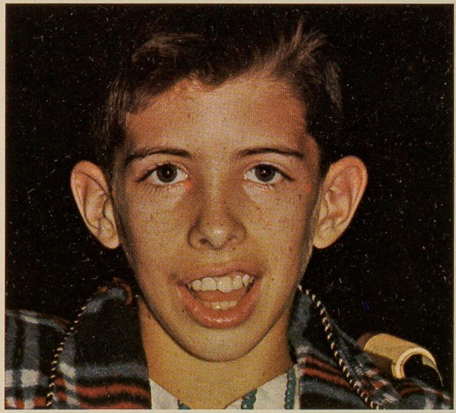
Wilson disease (WD) is an autosomal recessive disorder of copper metabolism caused by pathogenic variants in the ATP7B gene, leading to toxic copper accumulation predominantly in the liver and brain [1]D5[6]D5. This section establishes the precise disease definition, its classification within the hepatocellular-versus-cholestatic spectrum, and the current consensus nomenclature that underpins the rest of this article.
Also Called / Synonyms
- Wilson disease (WD)
- Hepatolenticular degeneration (historical term)
- Copper storage disease
- ATP7B-related copper toxicosis
Disease Definition and Core Concept
The pathognomonic defect is a loss-of-function mutation in ATP7B, which encodes a copper-transporting P-type ATPase essential for biliary copper excretion and incorporation of copper into ceruloplasmin [1]D5[8]C4. When this transporter fails, copper accumulates first in hepatocytes, then spills into the systemic circulation, depositing in the brain, cornea (Kayser-Fleischer rings), kidneys, and other tissues [1]D5[6]D5. WD sits squarely in the hepatocellular disease spectrum: the primary lesion is hepatic copper overload, not a cholestatic defect in bile flow. The cholestatic phenotypes seen in some patients are a consequence of advanced liver injury, not a primary biliary disorder.
Classification of Clinical Phases & Stages
To avoid ambiguity, the following terms are used throughout this article as defined by international consensus guidelines from AASLD, EASL, and ESPGHAN [1]D5[6]D5:
| Term | Definition | Key Features |
|---|---|---|
| Pre-symptomatic (asymptomatic) | ATP7B mutation identified with no clinical symptoms | Detected by family screening; may have elevated aminotransferases or subtle copper abnormalities [3]B3b |
| Hepatic presentation | Liver-predominant symptoms at diagnosis | Acute hepatitis, hepatomegaly, cirrhosis, or ; median age 10-20 years |
| Neurological presentation | Neurologic or psychiatric symptoms at diagnosis | Dysarthria, dystonia, tremor, ; typically older adolescents or adults [4]B2b |
| Mixed presentation | Both hepatic and neurologic/psychiatric involvement at diagnosis | Hepatic and neurologic signs coexist; worst prognosis for early neurological worsening [4]B2b |
| Acute decompensated WD (fulminant) | Rapid onset liver failure with hemolytic anemia and coagulopathy | Status 1 for liver transplant; high mortality without transplantation [5]C4 |
Important distinctions: "Pre-symptomatic" replaces the older term "asymptomatic" to emphasize that these patients harbour disease that will progress without treatment [1]D5. "Acute decompensated WD" is distinct from compensated cirrhosis in WD, the former is a medical emergency requiring immediate transplant evaluation [5]C4.
Clinical Significance
WD affects approximately 1 in 30,000 to 1 in 50,000 live births globally [1]D5. If untreated, it is uniformly fatal; with early diagnosis and lifelong chelation or zinc therapy, most patients achieve normal life expectancy [2]B2a[6]D5. It is the classic example of a treatable inherited metabolic liver disease and a diagnostic priority in any young patient with unexplained liver disease, neurologic movement disorder, or psychiatric disturbance.
Pearl: Wilson disease is a treatable disorder of copper overload; early diagnosis and treatment prevent disease progression and allow normal life expectancy, making it a diagnostic priority in every young patient with unexplained liver or neurologic disease [1]D5[2]B2a[6]D5.
Pathophysiology & Mechanism
- ▸ATP7B loss-of-function mutations impair biliary copper excretion, leading to hepatic accumulation that initiates a cascade of oxidative stress, mitochondrial dysfunction, steatosis, and stellate-cell activation.
- ▸Copper spillover into the systemic circulation deposits in the basal ganglia, causing the characteristic movement disorders and psychiatric features, while Kayser-Fleischer rings arise from corneal Descemet membrane deposition.
- ▸Genetic modifiers such as PNPLA3 I148M and the specific ATP7B mutation type (e.g., H1069Q vs. C-terminal variants) contribute to marked phenotypic heterogeneity in age of onset, organ involvement, and disease severity.
The pathogenic cascade in Wilson disease begins with failure of biliary copper excretion due to loss-of-function mutations in ATP7B, a P-type copper-transporting ATPase. Under normal conditions, ATP7B transports copper into the trans‑Golgi network for incorporation into ceruloplasmin and, when cytosolic copper rises, relocates to the canalicular membrane to facilitate biliary excretion [9]A1b[14]D5. Mutations in ATP7B disrupt this trafficking or catalytic activity, causing copper to accumulate first in the liver, then in the brain and other tissues. The resulting cellular toxicity drives a sequence of hepatocellular injury, steatosis, inflammation, stellate‑cell activation, and progressive fibrosis that ultimately culminates in cirrhosis and . Every clinical manifestation, from Kayser-Fleischer rings to extrapyramidal signs, can be traced back to copper‑induced organ damage at the molecular and cellular level.
Hepatic Copper Accumulation and the Initial Insult
Copper enters hepatocytes via the high‑affinity copper transporter CTR1 (SLC31A1). Once inside, copper is chaperoned to ATP7B by ATOX1. ATP7B then moves copper into the Golgi for ceruloplasmin synthesis or, when copper is abundant, translocates to the bile canaliculus for excretion [9]A1b[12]D5. In Wilson disease, mutant ATP7B, whether partially active or completely inactive, cannot perform this export efficiently. Hepatic copper content rises progressively, exceeding the capacity of protective mechanisms such as metallothionein binding. Free copper catalyzes Fenton‑type reactions, generating reactive oxygen species (ROS) that peroxidate membrane lipids and damage mitochondria [13]B3b. Mitochondrial dysfunction in turn impairs fatty acid β‑oxidation, leading to macrovesicular steatosis, often the earliest histological finding in Wilson disease, sometimes indistinguishable from non‑alcoholic fatty liver disease [13]B3b. Genetic modifiers such as the PNPLA3 I148M variant (rs738409) further increase steatosis severity, accounting for some of the phenotypic variability among patients [13]B3b.
Progression from Steatosis to Fibrosis and Cirrhosis
Sustained copper‑induced oxidative stress triggers hepatocyte apoptosis and necrosis, releasing damage‑associated molecular patterns (DAMPs) that activate Kupffer cells and hepatic stellate cells (HSCs) [11]D5. Activated HSCs transdifferentiate into myofibroblasts, secreting collagen‑rich extracellular matrix that progressively replaces functional parenchyma. Autophagy, a quality‑control pathway that normally degrades damaged organelles and lipid droplets, becomes overwhelmed or dysfunctional in Wilson disease hepatocytes, further aggravating steatosis and inflammation [11]D5. The net result is a self‑amplifying fibrotic response. Over years to decades (or faster in early‑onset pediatric cases [17]B2b), bridging fibrosis and nodular regeneration produce cirrhosis and, ultimately, portal . Importantly, nuclear receptor dysregulation also contributes to disease progression. Liver X receptor (LXR)/retinoid X receptor (RXR) heterodimer signalling is suppressed in both human Wilson disease and Atp7b⁻/⁻ mouse liver; treatment of mice with the LXR agonist T0901317 ameliorated hepatic steatosis and fibrosis without lowering hepatic copper content, indicating that LXR/RXR pathway activation can partially rescue copper‑independent injury [10]D5.
Neurological and Psychiatric Pathogenesis
Once the liver can no longer sequester excess copper, non‑ceruloplasmin‑bound (free) copper spills into the circulation and crosses the blood-brain barrier, preferentially accumulating in the basal ganglia, thalamus, brainstem, and cerebellum [18]D5. Copper disrupts neuronal mitochondrial function, induces oxidative stress, and promotes protein aggregation. In the putamen and caudate, this leads to neuronal loss, spongiform degeneration, and reactive gliosis, findings that correlate with the characteristic movement disorders (dystonia, , tremor) and psychiatric symptoms (depression, psychosis, personality changes). Resting‑state electroencephalography reveals abnormal microstate dynamics (increased duration of microstate C, decreased of microstates B and D) that reflect disrupted large‑scale neural network coordination, providing a quantifiable signature of neurological involvement [21]B3b. The Kayser-Fleischer rings, copper deposition in Descemet’s membrane of the cornea, are a direct biomarker of CNS copper overload but also indicate systemic copper excess; they appear as golden‑brown or greenish rings at the corneal limbus and are pathognomonic when present [18]D5.
Extra‑Hepatic, Extra‑Neuronal Copper Deposition
Copper also deposits in the heart, kidneys, eyes (other than cornea), and bones. Cardiac involvement manifests as , diastolic dysfunction, and conduction abnormalities; three cases of sudden death have been reported, highlighting the potential for arrhythmogenic cardiomyopathy [15]C4. Renal tubular dysfunction results from copper accumulation in proximal tubule cells, causing aminoaciduria, phosphaturia, and renal tubular acidosis [18]D5. Ocular copper deposition may also involve the lens (sunflower cataract) and retina ( ‑like changes) [19]C4, though retinal abnormalities are rare. The mechanism of copper‑driven injury in these organs mirrors that in the liver: ROS generation, mitochondrial impairment, and cellular stress.
Genetic and Molecular Determinants of Phenotypic Variability
The clinical heterogeneity of Wilson disease, ranging from asymptomatic siblings to fulminant hepatic failure to isolated neuropsychiatric disease, stems from the functional diversity of ATP7B mutations and from genetic modifiers. Over 900 ATP7B mutations have been described; they can be classified by their effect on protein function: no copper transport activity (e.g., H1069Q), partial activity with mislocalization (e.g., R778Q, G1061E), or abnormal trafficking in polarized cells (e.g., C‑terminal mutations S1423N, S1426I, T1434M that impair Golgi exit selectively in polarized hepatocytes) [14]D5[16]D5. The degree of residual copper excretion capacity determines how rapidly copper accumulates and at what age symptoms appear [14]D5. Additional modifiers include PNPLA3 I148M (worsening steatosis) [13]B3b and polymorphisms in zinc transporter ZnT1 (SLC30A1), which can export copper at the basolateral membrane and may modulate hepatic copper levels [20]D5. Furthermore, the LXR/RXR pathway [10]D5 and autophagic capacity [11]D5 represent modifiable cellular responses that influence disease progression independently of copper burden.
Controversies and Guideline Disagreement
Copper overload is the primary driver, but is it sufficient to explain all injury? Some data suggest that copper‑independent pathways, such as LXR/RXR dysregulation and autophagy failure, contribute substantially to disease severity, raising the question of whether pure copper chelation adequately addresses all pathogenic mechanisms. While EASL and AASLD guidelines uniformly recommend copper‑directed therapy (chelators plus zinc), the potential role of LXR agonists or autophagy‑enhancing agents as adjunctive treatments remains unexplored in clinical trials [10]D5[11]D5.
| Question | Position A (Copper‑centric) | Position B (Multi‑pathway) | Strength | Implication |
|---|---|---|---|---|
| Are copper‑independent mechanisms clinically important? | Copper overload alone explains hepatic and neurologic injury; chelation reverses disease in most patients [9]A1b[14]D5. | Copper‑independent LXR/RXR suppression and autophagy impairment worsen steatosis and fibrosis independent of copper levels [10]D5[11]D5. | Moderate - supported by mechanistic studies but no interventional trial in humans | Adjunctive therapies targeting LXR/RXR or autophagy might benefit patients with incomplete response to chelation. |
| Should genetic modifiers guide therapy? | No: all patients with symptomatic Wilson disease require chelation regardless of modifier genotype [14]D5. | Yes: PNPLA3 I148M status may help predict who will develop more severe steatosis and warrant earlier or more aggressive treatment [13]B3b. | Weak - data from a single cohort; no prospective validation | PNPLA3 genotyping could be considered in research settings but is not yet standard of care. |
Pearl: Copper accumulation in the liver triggers a cascade of mitochondrial dysfunction, steatosis, LXR/RXR pathway suppression, and autophagy failure that drives progressive fibrosis; neurological damage follows once free copper deposits in the basal ganglia, and genetic modifiers (PNPLA3, ATP7B mutation type) account for much of the phenotypic variability.
Sub‑section: Key Molecular Steps (Numbered Chain)
- Defective copper export - ATP7B mutations (e.g., H1069Q, R778Q, C‑terminal truncations) prevent biliary copper excretion, raising cytosolic copper [9]A1b[14]D5[16]D5.
- Oxidative stress and mitochondrial injury - Free copper catalyzes ROS production, depletes glutathione, and damages mitochondrial DNA and membranes, impairing fatty acid oxidation [13]B3b.
- Steatosis and hepatocyte necrosis - Impaired β‑oxidation leads to macrovesicular steatosis; ROS‑mediated apoptosis releases DAMPs that activate Kupffer cells [11]D5[13]B3b.
- Hepatic stellate cell activation - DAMPs and inflammatory cytokines (TNF‑α, TGF‑β) transdifferentiate stellate cells into myofibroblasts, which deposit collagen [11]D5.
- Fibrosis and cirrhosis - Progressive matrix deposition leads to bridging fibrosis, nodule formation, and portal hypertension.
- Systemic copper spillover - Once hepatic storage capacity is exhausted, free copper enters the circulation and deposits in brain (basal ganglia), eyes (cornea, lens, retina), heart, and kidneys [15]C4[18]D5[19]C4.
- Neurological network disruption - Basal ganglia copper accumulation causes neuronal loss, gliosis, and altered EEG microstate dynamics, manifesting as extrapyramidal and psychiatric syndromes [18]D5[21]B3b.
| Mutation Class | Examples | Functional Effect | Cellular Consequence | Clinical Association |
|---|---|---|---|---|
| Complete loss of transport | H1069Q, R778L | No catalytic activity; protein misfolding → ER retention or rapid degradation | No copper excretion; early severe hepatic or neurologic disease | is most common in European populations; associated with late-onset neurologic form [14]D5 |
| Partial loss with mislocalization | R778Q, G1061E | Reduced catalytic activity; aberrant trafficking in polarized hepatocytes | Some residual copper export; slower copper accumulation, variable age of onset | prevalent in East Asian populations; often presents with hepatic symptoms [14]D5[16]D5 |
| C‑terminal trafficking defects | S1423N, S1426I, T1434M | Normal catalytic activity but impaired Golgi exit in polarized cells | Defect only manifests in differentiated hepatocytes; copper accumulates despite measurable ATPase activity | Milder or delayed phenotype; may be missed by standard functional assays [16]D5 |
| Frameshift / nonsense | Varied (e.g., 2299insC) | Truncated or non‑functional protein | Complete loss of function; severe early‑onset disease | Fulminant hepatic failure in childhood [14]D5 |
Epidemiology, Etiology & Risk Factors
- ▸Global prevalence is 1 in 30,000 to 1 in 50,000, with a carrier frequency of ~1 in 90 [29].
- ▸HBV coinfection (current 2.1%, past 9.2%) and anemia (16.7%) are common comorbidities that worsen liver disease severity [7, 23].
- ▸Pregnancy outcomes are worse in untreated WD; continuation of chelation therapy improves maternal and fetal outcomes [22].
The global prevalence of Wilson disease is estimated at 1 in 30,000 to 1 in 50,000 live births, corresponding to a carrier frequency of approximately 1 in 90 [29]D5. These figures, however, likely underestimate the true burden because of variable penetrance, incomplete case ascertainment, and the absence of universal newborn screening in most regions. In the Austrian cohort of 229 patients diagnosed between 1961 and 2013, the mean age at diagnosis was 14.8 years, with a slight female predominance (55%) [25]B2b. The disease is pan-ethnic but shows regional clustering; systematic reviews from the Arab world report similar prevalence ranges but highlight a high rate of consanguinity and a distinct mutation spectrum [29]D5. Temporal trends are difficult to assess due to diagnostic delays, but improved awareness and genetic testing have increased detection rates over recent decades.
Genetic and Modifiable Risk Factors
Wilson disease is an autosomal recessive disorder caused by biallelic pathogenic variants in ATP7B (chromosome 13q14.3). Over 900 mutations have been described, with the H1069Q variant accounting for 30-60% of alleles in European populations [29]D5. No environmental or lifestyle factors are known to cause the disease, but several comorbidities modify its clinical expression and progression.
| Risk Factor | Association with WD | Evidence Level |
|---|---|---|
| ATP7B mutation (biallelic) | Causative; OR not applicable (necessary) | 1a [29]D5 |
| Hepatitis B virus (HBV) coinfection | Prevalence 2.1% (current) and 9.2% (past) in WD patients; associated with more severe liver disease | 2b [23]B2b |
| Anemia | Present in 16.7% of WD patients; associated with higher disease severity and hepatic complications (OR 2.8, 95% CI 1.4-5.6) | 2b [7]B2b |
| Low platelet-to-white blood cell ratio (PWR <26.3) | Associated with advanced liver fibrosis and progression (HR 2.1, 95% CI 1.3-3.4) | 3b [28]B3b |
HBV coinfection is a particularly important modifier. In a large retrospective study of 915 WD patients, current HBV infection was present in 2.1% (95% CI 1.2-3.0%) and past infection in 9.2% (95% CI 7.3-11.1%) [23]B2b. Those with dual pathology had significantly higher bilirubin, INR, and Model for End-Stage Liver Disease scores, indicating accelerated liver injury [23]B2b. Similarly, anemia (hemoglobin <12 g/dL in women, <13 g/dL in men) was found in 16.7% of a Chinese WD cohort and independently predicted hepatic decompensation (OR 2.8) [7]B2b. The platelet-to-white blood cell ratio (PWR <26.3) has been proposed as a noninvasive marker of fibrosis severity and progression risk [28]B3b.
Special Populations
Pediatric (PALF): Wilson disease accounts for approximately 5-10% of PALF cases, but it is frequently misdiagnosed as indeterminate. A learning collaborative that promoted age-specific diagnostic testing increased the identification of WD among PALF patients, reducing the proportion of indeterminate diagnoses [26]B2b.
Pregnancy: Women with WD have higher rates of adverse pregnancy outcomes, including miscarriage (pooled prevalence 18%), preterm birth (12%), and low birth weight (8%) [22]A1a. Importantly, untreated WD carries even greater risks; chelation therapy (trientine or zinc) during pregnancy is associated with better outcomes and should be continued [22]A1a.
: Among patients who progress to end-stage liver disease, post-transplant survival is excellent. A meta-analysis of 27 studies reported 1-year survival of 92% and 5-year survival of 88% [27]A1a. Neuropsychiatric symptoms improve in most patients after transplantation, though residual deficits may persist [27]A1a.
Pearl: Wilson disease is rare but underdiagnosed; a high index of suspicion is warranted in any patient with unexplained liver disease, neuropsychiatric symptoms, or a family history. Coinfection with HBV and the presence of anemia or low PWR identify patients at risk for more rapid progression [7]B2b[23]B2b[28]B3b.
Clinical Presentation
- ▸Hepatic presentation (40-50%) is more common in children and females; neurological presentation (30-40%) predominates in older adolescents and adults [33].
- ▸Kayser-Fleischer rings are present in ~95% of neurological Wilson disease and ~50% of hepatic cases [41].
- ▸Early neurological worsening within 3 months of therapy occurs in 26% of neurological/mixed patients and requires close monitoring [4].
Hepatic manifestations are the presenting feature in approximately 40-50% of index patients, with neurological symptoms accounting for another 30-40% [33]B2b. The remaining patients present with mixed hepatic and neurological disease or are identified asymptomatically through family screening. The clinical spectrum ranges from incidental transaminase elevation to with Coombs-negative hemolytic anemia, and from subtle tremor to severe dystonia with bulbar compromise.
Presenting Symptoms
Hepatic presentation often begins insidiously. Children and adolescents may have asymptomatic elevation of aminotransferases or hepatic steatosis, which is one of the earliest features in pediatric Wilson disease [40]B3b. Others present with acute hepatitis, chronic hepatitis, or cirrhosis. Acute liver failure (ALF) is a dramatic presentation, typically in young patients, and is frequently accompanied by Coombs-negative hemolytic anemia due to copper-induced erythrocyte membrane damage. Decompensated cirrhosis manifests with , variceal bleeding, jaundice, and . In the large Austrian cohort, hepatic presentation was more common in females and younger patients [25]B2b[33]B2b.
Neurological presentation typically emerges in the second to third decade, often with a gradual onset over months. The most common initial symptoms are dysarthria (slurred speech), drooling, tremor (often a characteristic "wing-beating" tremor of the arms), and gait disturbance. Dystonia (focal or generalized), chorea, athetosis, and (rigidity, bradykinesia, postural instability) may develop. Ataxia and dysdiadochokinesia reflect cerebellar involvement. Psychiatric symptoms, depression, anxiety, irritability, personality changes, psychosis, and cognitive decline, can precede or accompany neurological signs and are often misattributed to primary psychiatric disorders.
Other organ involvement includes renal tubular dysfunction ( : glycosuria, aminoaciduria, phosphaturia), skeletal abnormalities (osteoporosis, , arthropathy), cardiac manifestations (cardiomyopathy, arrhythmias), endocrine disturbances (delayed puberty, amenorrhea, infertility), and dermatologic findings (livido reticularis, hyperpigmentation). Kayser-Fleischer rings, copper deposition in the Descemet membrane of the cornea, are present in approximately 95% of patients with neurological Wilson disease and 50% of those with hepatic presentation [41]C4. Sunflower cataracts are a rarer ocular finding.
Neurological Examination Findings
A structured neurological examination is essential. Cranial nerves: Kayser-Fleischer rings are best seen on slit-lamp examination; sunflower cataracts may be visible on direct ophthalmoscopy. Ocular motor abnormalities include saccadic intrusions, nystagmus, and impaired smooth pursuit. Motor system: Tremor is often the first sign, a coarse, irregular, proximal "wing-beating" tremor that worsens with sustained posture. Dystonia may be focal (e.g., , blepharospasm) or generalized. Chorea and athetosis are less common. Parkinsonism presents with cogwheel rigidity, bradykinesia, and postural instability. Cerebellar signs include dysmetria, dysdiadochokinesia, and ataxic gait. Reflexes: Hyperreflexia and extensor plantar responses (Babinski sign) may occur with corticospinal tract involvement, but reflexes are often normal. Sensory examination is typically normal. Bulbar function: Dysarthria (spastic, ataxic, or hypokinetic) and dysphagia are common and can lead to aspiration. FVC < 15 mL/kg or declining forced vital capacity signals impending respiratory failure and should prompt consideration of intubation. Autonomic dysfunction (orthostatic hypotension, sweating abnormalities) may occur in advanced disease.
Phenotypic Variants
| Variant | Key Features | Frequency |
|---|---|---|
| Hepatic | Liver disease only (elevated transaminases, cirrhosis, ALF); no neurological signs | 40-50% of index patients [33]B2b |
| Neurologic | Neurological symptoms (tremor, dystonia, dysarthria) with or without liver disease | 30-40% [33]B2b |
| Mixed | Both hepatic and neurological manifestations | 10-20% |
| Asymptomatic | Identified by family screening; normal examination | 5-10% |
| Acute liver failure | Rapid onset with Coombs-negative hemolytic anemia; often fatal without transplant | ~5% |
| Late-onset (>40 years) | Predominantly hepatic; no neuropsychiatric features; often misdiagnosed [31]C4 | Rare |
| Neonatal-onset | Cholestatic jaundice, failure to thrive; extremely rare [38]C4 | <1% |
Red Flags
- Rapidly progressive neurological symptoms (dystonia, dysarthria, dysphagia) within the first 3 months of therapy, early neurological worsening occurs in 26.1% of patients with neurological or mixed presentation [4]B2b.
- Acute liver failure with Coombs-negative hemolytic anemia, consider Wilson disease as the cause.
- Unexplained psychiatric symptoms (depression, psychosis, personality change) in a young adult.
- Kayser-Fleischer rings on slit-lamp examination.
- Family history of Wilson disease or unexplained liver disease.
Atypical Presentations
Wilson disease can mimic many conditions. Isolated psychiatric disease ( , bipolar disorder) may precede neurological signs by years. Renal tubular acidosis or Fanconi syndrome can be the sole manifestation. Hemolytic anemia without overt liver failure is a rare but classic presentation. Skeletal abnormalities (osteoporosis, spontaneous fractures) may prompt rheumatologic evaluation. Cardiac arrhythmias or cardiomyopathy can be the first clue. Endocrine presentations include delayed puberty, amenorrhea, or infertility. Neurologically, patients may present with isolated tremor, isolated dysarthria, or isolated gait ataxia, leading to misdiagnosis as , multiple sclerosis, or cerebellar degeneration. Asymptomatic individuals with elevated transaminases are often identified through family screening or incidental laboratory findings.
Pearl: The clinical phenotype of Wilson disease is highly variable and influenced by age, sex, and ATP7B genotype, but not by mutation type alone [33]B2b[36]A1a; a high index of suspicion is required for any young patient with unexplained liver disease, hemolytic anemia, or neuropsychiatric symptoms, as early treatment dramatically improves outcomes [25]B2b.
Diagnosis & Workup
- ▸The Leipzig scoring system (score ≥4) is the established diagnostic framework, integrating KF rings, ceruloplasmin, urinary copper, liver copper, and ATP7B genetics [31, 37].
- ▸ATP7B sequencing is the gold standard for confirmation, but up to 20% of cases require WGS or protein assay for detection of cryptic variants [55].
- ▸Autoimmune hepatitis and PFIC3 are the most common mimics; false-positive autoantibodies occur in 31.5% of pediatric WD patients [54, 48].
The diagnostic evaluation of Wilson disease (WD) rests on a tiered strategy combining clinical examination, biochemical copper studies, and molecular confirmation, anchored by the Leipzig scoring system (see Table 1). No single test is sufficient in isolation; the diagnosis is established when the cumulative evidence reaches a score of ≥4 points [31]C4[37]A1c. The gold standard for definitive diagnosis remains ATP7B mutation analysis identifying biallelic pathogenic variants, though this is supplemented by quantitative liver copper measurement when genetic results are inconclusive [46]B2b[55]B2b.
Step 1: Clinical Suspicion and Initial Screening
The diagnostic pathway begins with recognizing WD in any patient aged 3-55 years presenting with unexplained liver disease (elevated aminotransferases, steatosis, cirrhosis), neurological symptoms (tremor, dysarthria, dystonia), or psychiatric disturbances (depression, personality change) [33]B2b[18]D5. Kayser-Fleischer (KF) rings, copper deposition in Descemet's membrane of the cornea, remain a highly specific sign when present, detected by slit-lamp examination. In one large Chinese cohort of 529 symptomatic patients, KF rings were present in 89.4% of neurological-presentation patients but only 53.7% of those with hepatic presentation [53]B2b. Their absence does not exclude WD, particularly in children and adults with purely hepatic disease [31]C4[53]B2b.
Step 2: Biochemical Copper Studies
Serum ceruloplasmin is the first-line screening test. A level <20 mg/dL (by nephelometry) supports WD, but performance varies by clinical context. In a prospective study of 140 (ALF) patients, nephelometric ceruloplasmin <20 mg/dL had only 56% sensitivity and 63% specificity for WD-associated ALF [42]B2b. The oxidase activity method yielded even lower sensitivity (21%) [42]B2b. In children with mild liver disease and elevated transaminases, a cutoff of <14 mg/dL provided 93% sensitivity and 100% specificity compared to healthy controls, but specificity fell to 79% against other liver diseases [3]B3b. Concomitant acute-phase response can falsely elevate ceruloplasmin; a normal level does not rule out WD. Measuring 24-hour urinary copper excretion is the next step. A basal excretion >100 μg/24 h (1.6 μmol/24 h) is classic for symptomatic WD [37]A1c. In children with mild disease, a cutoff of >40 μg/24 h provided 85% sensitivity and 96% specificity for diagnosis [3]B3b. The penicillamine challenge test (giving 500 mg D-penicillamine orally at the start and again 12 hours later, collecting urine for 24 hours) increases sensitivity; excretion >1600 μg/24 h supports WD, though this test is less commonly used today [31]C4.
Serum copper is not diagnostic in isolation: total copper is low in WD due to low ceruloplasmin, but in ALF-WD, massive hepatic necrosis releases copper, yielding total serum copper >200 μg/dL in nearly all such patients [42]B2b. The non-ceruloplasmin-bound copper (free copper) can be calculated (total copper - [3.15 × ceruloplasmin in mg/dL]) and a value >25 μg/dL is abnormal; however, its calculation amplifies measurement errors [45]B2b. Direct measurement of exchangeable copper (CuEX) and the relative exchangeable copper (REC = CuEX / total copper × 100) are emerging as robust biomarkers. A REC >18.5% has been proposed as a diagnostic threshold with high sensitivity in treatment-naïve patients [43]B2b[37]A1c. In the ALF setting, the ALT:AST ratio is also useful: a ratio <1 (AST higher than ALT) plus a alkaline phosphatase (ALP):total bilirubin ratio <4 supports ALF-WD [45]B2b.
| Test | Finding Suggesting WD | Sensitivity (range) | Specificity (range) | Notes |
|---|---|---|---|---|
| Serum ceruloplasmin (nephelometry) | <20 mg/dL | 56% (ALF) [42]B2b; 93% (mild pediatric) [3]B3b | 63% (ALF) [42]B2b; 79% (other liver disease) [3]B3b | Acute-phase reactant; may be normal in WD |
| 24-h urinary copper (basal) | >100 μg/24 h | 85% (mild pediatric) [3]B3b | 96% (mild pediatric) [3]B3b | Requires complete collection; false positives in cholestasis |
| Penicillamine challenge test | >1600 μg/24 h | , | , | Not first-line; used in ambiguous cases |
| Kayser-Fleischer rings (slit-lamp) | Present | 89% (neurologic) [53]B2b; 54% (hepatic) [53]B2b | Near 100% | Specific but absent in many hepatic-only cases |
| Liver copper quantification | >250 μg/g dry weight | , | , | Gold-standard biochemical proof; requires biopsy |
| ATP7B sequencing | Biallelic pathogenic variants | >95% [55]B2b | >99% | Definitive genetic confirmation |
Step 3: Leipzig Scoring System
The Leipzig criteria, endorsed by EASL and AASLD, assign points across six domains [31]C4[37]A1c:
| Domain | Criterion | Points |
|---|---|---|
| Clinical | KF rings | 2 |
| Neurologic symptoms (characteristic) | 2 | |
| Neuropsychiatric symptoms (atypical) | 1 | |
| Biochemical | Ceruloplasmin <20 mg/dL (or <0.2 g/L) | 2 |
| Ceruloplasmin <10 mg/dL (or <0.1 g/L) | 1 (additional) | |
| 24-h urinary copper >100 μg | 1 | |
| 24-h urinary copper >200 μg | 2 | |
| Penicillamine challenge >1600 μg | 2 | |
| Liver copper >250 μg/g dry weight | 2 | |
| REC >18.5% | 1 (per EASL 2025 update) [37]A1c | |
| Genetic | ATP7B mutation analysis (homozygous or compound heterozygous) | 4 |
| Other | Coombs-negative hemolytic anemia | 1 |
Interpretation: Score ≥4 establishes the diagnosis; score 3 is suggestive and requires further evaluation; score ≤2 makes WD unlikely [31]C4[37]A1c.
Step 4: Genetic Testing
ATP7B sequencing is the confirmatory gold standard and should be performed in all cases where suspicion remains after biochemical testing or when results are equivocal [46]B2b[55]B2b[49]B2b. The gene spans 21 exons; mutation hotspots include exons 8, 12, and 14, but comprehensive sequencing (including intronic regions) is necessary because up to 20% of clinically diagnosed patients with Leipzig score ≥4 have zero or only one identifiable ATP7B variant by standard methods [55]B2b. In such cases, whole-genome sequencing can detect deep intronic or structural variants [55]B2b. A novel direct assay measuring ATP7B protein concentration from dried blood spots (by mass spectrometry of surrogate peptides) showed 96% sensitivity and 94% specificity in a multi-center cohort of 264 WD patients and 150 controls, making it a promising second-line tool [46]B2b. Multi-gene panels that include ATP7B plus copper-related genes (e.g., COMMD1, ATOX1) can identify phenocopies like progressive familial intrahepatic cholestasis type 3 (PFIC3) , which can present with hepatic copper accumulation and elevated urinary copper, leading to misdiagnosis as WD [48]D5[49]B2b.
Step 5: and Copper Quantification
Liver biopsy is not required for diagnosis when genetic testing is positive or when clinical-biochemical criteria yield a Leipzig score ≥4. It is reserved for cases where genetic results are uninformative (single variant or none) and biochemical tests are borderline [55]B2b. A liver copper content >250 μg/g dry weight (normal <50 μg/g) is diagnostic [3]B3b. Biopsy also provides histologic grading of steatosis, inflammation, and fibrosis. Steatosis, often macrovesicular or mixed, is present early and may be indistinguishable from NAFLD; the PNPLA3 rs738409 G allele is an independent modifier of steatosis severity in WD [13]B3b. Cirrhosis may be present even in children [3]B3b.
Step 6: Imaging
Abdominal ultrasound is the first imaging modality used to assess hepatic steatosis, cirrhosis, and . Transient elastography (FibroScan) provides non-invasive fibrosis staging with correlation to histologic stage in WD [25]B2b. Brain MRI (with T2-weighted and FLAIR sequences) is indicated in any patient with neurological or psychiatric symptoms. Characteristic findings include hyperintensity in the basal ganglia (putamen, globus pallidus, caudate), thalamus, and brainstem (especially the midbrain, the "face of the giant panda" sign in the substantia nigra) [18]D5. MRI can also guide prognosis: presence of brainstem involvement correlates with worse neurological outcomes [4]B2b.
Step 7: Differential Diagnosis and Pitfalls
Several conditions mimic WD and must be excluded, particularly when genetic testing is incomplete:
- (AIH): Up to 31.5% of treatment-naïve WD children have false-positive autoantibodies (anti-smooth muscle, ANA) and elevated IgG (>1.1×ULN in 82.3%), leading to misclassification as AIH by simplified criteria [54]B3b. Copper markers should be checked in any child with suspected AIH who has atypical features (low aminotransferases, , low ALP).
- PFIC3: Caused by biallelic ABCB4 mutations, PFIC3 presents with cholestasis, elevated serum copper, and urinary copper that can meet Leipzig criteria [48]D5. Genetic panels that include ABCB4 and other cholestasis genes resolve this mimic [49]B2b.
- Acute liver failure from other causes: As noted above, ALF-WD is distinguished by the ALP:bilirubin ratio <4 and ALT:AST <1 [45]B2b.
- Citrullinemia type I and other urea cycle defects can present with ALF and low ceruloplasmin, mimicking WD [47]C4. Plasma amino acids and ammonia levels differentiate these.
- Ceruloplasmin measurement variability: Inflammatory states elevate ceruloplasmin; low values also occur in Menkes disease, , and severe malnutrition.
Diagnostic Algorithm (Summarized)
- Screen with slit-lamp for KF rings and measure serum ceruloplasmin + 24-h urinary copper in any patient with unexplained liver or neuropsychiatric disease.
- Calculate the Leipzig score using available clinical, biochemical, and imaging data.
- If score ≥4: Confirm with ATP7B sequencing; initiate treatment. Biopsy is unnecessary unless genetic results are pending and clinical urgency requires immediate therapy.
- If score 2-3: Perform ATP7B sequencing + multi-gene panel for phenocopies. Consider liver biopsy for copper quantification. Measure REC if available.
- If score ≤2 and suspicion remains (e.g., family history, atypical presentation): Refer to specialist center for whole-genome sequencing, ATP7B protein assay, or repeat biopsy [46]B2b[55]B2b.
- In all ALF cases: If clinical features suggest WD (Coombes-negative hemolysis, low ALP, low AST:ALT), check ALP:total bilirubin ratio and proceed directly to transplant evaluation while simultaneously obtaining genetic testing and copper studies [42]B2b[45]B2b.
Pearl: The combination of KF rings, ceruloplasmin <20 mg/dL, and 24-h urinary copper >100 μg/24 h is sufficient to diagnose WD with high probability (Leipzig score ≥4) and initiate chelation therapy, even while genetic confirmation is pending [31]C4. However, in the absence of KF rings, particularly in pediatric hepatic presentations, genetic testing is essential to avoid misdiagnosis with AIH or PFIC3 [48]D5[54]B3b.
| Domain | Criterion | Points |
|---|---|---|
| Clinical | KF rings | 2 |
| Neurologic symptoms (characteristic) | 2 | |
| Neuropsychiatric symptoms (atypical) | 1 | |
| Biochemical | Ceruloplasmin <20 mg/dL (or <0.2 g/L) | 2 |
| Ceruloplasmin <10 mg/dL (or <0.1 g/L) | 1 (additional) | |
| 24-h urinary copper >100 μg | 1 | |
| 24-h urinary copper >200 μg | 2 | |
| Penicillamine challenge >1600 μg | 2 | |
| Liver copper >250 μg/g dry weight | 2 | |
| REC >18.5% | 1 (per EASL 2025 update) [37]A1c | |
| Genetic | ATP7B mutation analysis (homozygous or compound heterozygous) | 4 |
| Other | Coombs-negative hemolytic anemia | 1 |
Severity, Staging & Risk Stratification
- ▸MELD ≥15 and Child-Pugh class C are the standard thresholds for transplant listing in decompensated Wilson disease.
- ▸Anemia (HR 2.1) and platelet-to-white blood cell ratio <26.3 (HR 1.8) are independent predictors of disease progression and hepatic decompensation.
- ▸Loss-of-function ATP7B variants confer a 3-fold higher risk of death or transplantation compared with non-LOF variants.
Severity in Wilson disease is quantified using the same validated prognostic scores applied to other chronic liver diseases: , , , and MELD 3.0. These scores guide transplant listing and predict short-term mortality in decompensated patients [25]B2b[52]B2b. In the Austrian cohort of 229 patients, those with compensated cirrhosis had a 20-year survival of 95%, whereas decompensated cirrhosis carried a 5-year mortality of 40% without transplantation [25]B2b.
Child-Pugh and MELD Scores
The (class A, B, or C) stratifies hepatic synthetic function using bilirubin, albumin, INR, , and encephalopathy. Child-Pugh class C (score ≥10) identifies patients with a 1-year mortality risk exceeding 50% without transplant. The MELD score (Model for End-Stage Liver Disease) incorporates bilirubin, INR, and creatinine; MELD ≥15 is the standard threshold for transplant listing in the United States. MELD-Na adds serum sodium to improve mortality prediction, and MELD 3.0 further refines the model by including albumin and adjusting for sex. In Wilson disease, these scores perform similarly to other chronic liver diseases, though the rapid progression of fulminant Wilson disease may outpace score changes [52]B2b.
Biomarkers of Disease Severity
Beyond composite scores, simple hematologic markers independently stratify risk. Anemia (hemoglobin <13 g/dL in men, <12 g/dL in women) was present in 16.7% of a 288-patient cohort and independently associated with severe liver dysfunction (OR 3.2, 95% CI 1.8-5.7), hepatic complications (variceal bleeding, ascites), and disease progression (HR 2.1, 95% CI 1.3-3.4) [7]B2b. The platelet-to-white blood cell ratio (PWR) below 26.3 identified patients with higher bilirubin, INR, and procollagen III N-terminal propeptide levels, and independently predicted hepatic decompensation (HR 1.8, 95% CI 1.2-2.7) [28]B3b. Both markers are readily available and can be used to prioritize patients for closer monitoring.
Genotype-Phenotype Correlations and Risk Stratification
ATP7B genotype influences disease severity and outcomes. Loss-of-function (LOF) variants (nonsense, frameshift, splice-site) are associated with earlier onset, higher hepatic copper content, and worse transplant-free survival. In a cohort of 117 patients with chronic liver disease, LOF variant carriers had a 5-year transplant-free survival of 62% compared with 88% for non-LOF carriers (HR 3.1, 95% CI 1.5-6.4) [52]B2b. Conversely, the c.3316G>A variant (p.Ala1104Thr) is linked to a milder subphenotype: later onset, lower frequency of Kayser-Fleischer rings, and less severe hepatic fibrosis [50]B2b. Neonatal-onset Wilson disease, though rare, presents with severe cholestasis and rapid progression, often requiring early transplantation [38]C4. Incidental diagnosis by gene panel sequencing in asymptomatic children with obesity and fatty liver highlights the potential for early intervention and favorable outcomes [57]C4.
Cirrhosis Prediction Models
Machine learning models using routine blood tests, urine copper, and serum ceruloplasmin can predict cirrhosis in Wilson disease with good accuracy. A case-control study from southwest China developed a model incorporating platelet count, AST, ALT, and albumin that achieved an AUC of 0.87 (95% CI 0.82-0.92) for detecting cirrhosis [58]B3b. Such models may help stratify patients who need more aggressive surveillance or early transplant evaluation, though they require external validation before routine clinical use.
Pearl: MELD ≥15 and Child-Pugh class C are the primary thresholds for transplant listing in decompensated Wilson disease, but anemia (HR 2.1), low PWR <26.3 (HR 1.8), and loss-of-function ATP7B variants (HR 3.1) independently identify patients at highest risk of progression and should prompt earlier transplant evaluation [7]B2b[28]B3b[52]B2b.
| Score | Components | Threshold for Transplant Listing | Notes |
|---|---|---|---|
| Child-Pugh | Bilirubin, albumin, INR, ascites, encephalopathy | Class C (score ≥10) | 1-year mortality >50% without transplant |
| MELD | Bilirubin, INR, creatinine | ≥15 | Standard in US; updated every 30 days |
| MELD-Na | MELD + serum sodium | ≥15 | Improves mortality prediction in ascites |
| MELD 3.0 | MELD + albumin + sex adjustment | ≥15 | Refined model; may reduce sex disparity |
| Marker | Threshold | Hazard Ratio (95% CI) | Clinical Implication |
|---|---|---|---|
| Anemia | Hb <13 (M) / <12 (F) g/dL | HR 2.1 (1.3-3.4) | Increased risk of decompensation and death [7]B2b |
| Platelet-to-WBC ratio (PWR) | <26.3 | HR 1.8 (1.2-2.7) | Predicts hepatic complications [28]B3b |
| Loss-of-function ATP7B variant | Any LOF (nonsense, frameshift, splice) | HR 3.1 (1.5-6.4) | Worse transplant-free survival [52]B2b |
| c.3316G>A variant | Heterozygous or homozygous | Protective | Milder phenotype, later onset [50]B2b |
Acute Management & Decompensation Events
- ▸ALF-WD requires urgent liver transplantation; chelation is not recommended.
- ▸Neurological worsening occurs in 26% of patients within 3 months of starting therapy; assess for copper deficiency and consider switching chelator.
- ▸Cirrhosis complications (variceal hemorrhage, SBP, HE, HRS-AKI) are managed per standard guidelines with attention to copper status.
Acute decompensation in Wilson disease demands immediate recognition of two distinct scenarios: (ALF-WD) and complications of cirrhosis, each with its own time-critical pathway. The following protocols integrate Wilson-specific considerations with standard hepatology .
Acute Liver Failure in Wilson Disease (ALF-WD)
ALF-WD is a medical emergency with near-100% mortality without urgent (LT). Rapid diagnosis is paramount because standard biochemical markers are unreliable in the setting of acute hepatic necrosis.
Step 1: Suspect ALF-WD in any young patient (typically <40 years) presenting with acute hepatitis, Coombs-negative hemolytic anemia, and a disproportionately low alkaline phosphatase (ALP) relative to bilirubin. The combination of serum copper >200 µg/dL and ALP <10 IU/L is highly suggestive [42]B2b (2b). The ALT:AST ratio (often <1) provides acceptable diagnostic accuracy, with an area under the receiver operating characteristic curve of 0.92 in one validation cohort [45]B2b (2b).
Step 2: Confirm the diagnosis using the Leipzig scoring system, but note that ceruloplasmin by oxidase or nephelometry has poor sensitivity in ALF (21% and 56%, respectively) [42]B2b (2b). Direct measurement of ATP7B peptides from dried blood spots can provide near-definitive evidence within hours [46]B2b (2b). Exchangeable copper (CuEXC) and relative exchangeable copper (REC) are emerging biomarkers; a REC >18.5% has shown 100% sensitivity and 96% specificity for ALF-WD in a prospective study [62]B2b (2b).
Step 3: List for urgent liver transplantation immediately upon diagnosis or strong clinical suspicion. The median waitlist time is short, and 1-year patient survival after LT exceeds 90% in dedicated centers [27]A1a (1a). Chelation therapy is not recommended as primary treatment for ALF-WD because it does not reverse acute hepatic necrosis and may precipitate neurological worsening [4]B2b (2b).
Step 4: Bridge with plasma exchange if available. (TPE) can remove circulating copper and improve coagulopathy, serving as a bridge to LT in patients with high-grade encephalopathy or multiorgan failure [61]D5 (5). The typical regimen is 1-1.5 plasma volumes daily until LT or clinical improvement.
Step 5: Monitor for copper deficiency after LT. Overtreatment with chelators or zinc post-transplant can cause hypocupremia, leading to myelopathy or [39]C4 (4). Discontinue chelation after LT unless residual neurological symptoms warrant continuation.
Variceal Hemorrhage
in Wilson disease is managed identically to other causes of cirrhosis. The EASL 2025 guideline recommends [37]A1c (1c):
- Vasoactive therapy - Terlipressin 2 mg IV every 4 hours (or octreotide 50 µg bolus then 50 µg/h) for 3-5 days.
- Endoscopic band ligation within 12 hours of presentation.
- Prophylactic - 1 g IV daily for 5-7 days.
- Restrictive transfusion - Target hemoglobin 7-9 g/dL.
Failure of initial hemostasis (persistent bleeding after two endoscopic sessions) should prompt consideration of transjugular intrahepatic portosystemic shunt (TIPS).
(SBP)
SBP is diagnosed by with polymorphonuclear (PMN) count ≥250 cells/µL. Management includes [37]A1c (1c):
- Empiric antibiotic - 2 g IV every 8 hours (or ceftriaxone 2 g IV daily) for 5 days.
- Albumin infusion - 1.5 g/kg on day 1, then 1 g/kg on day 3 to reduce the risk of .
- Secondary prophylaxis - Norfloxacin 400 mg PO daily (or ) in patients with prior SBP or low-protein (<1.5 g/dL).
(HE)
HE in Wilson disease is managed per standard protocols, with attention to precipitating factors (infection, bleeding, electrolyte disturbances, constipation).
- Identify and treat precipitant - Paracentesis, cultures, diuretic adjustment.
- Lactulose - Titrate to 2-3 soft stools per day (starting 30 mL PO every 1-2 hours until bowel movement, then 15-30 mL BID-TID).
- Rifaximin 550 mg PO BID - Add for recurrent or refractory HE.
- Renal replacement therapy - For acute HE with hyperammonemia not responding to medical therapy, continuous venovenous hemofiltration reduces ammonia and may improve survival [18]D5 (5).
Hepatorenal Syndrome-Acute Kidney Injury (HRS-AKI)
HRS-AKI is diagnosed after exclusion of other causes of AKI (volume depletion, nephrotoxins, shock). The EASL 2025 guideline recommends [37]A1c (1c):
- Terlipressin 1 mg IV every 4-6 hours (increase to 2 mg if no response after 3 days) plus albumin 1 g/kg/day (max 100 g/day).
- Monitor - Serum creatinine, urine output, and adverse effects (ischemic events).
- Response - Defined as decrease in serum creatinine to <1.5 mg/dL. If no response after 7 days, consider alternative diagnoses or liver transplantation.
Neurological Worsening
Early neurological worsening (NW) occurs in 26% of patients with neurological or mixed presentation within the first 3 months of therapy [4]B2b (2b). Management requires a structured approach:
- Assess for copper deficiency - Measure serum copper and ceruloplasmin; if low, reduce or discontinue chelator/zinc. Copper deficiency can cause myelopathy and peripheral neuropathy [39]C4 (4).
- Switch chelator - If NW occurs on penicillamine, consider switching to trientine or zinc. The EASL guideline suggests that trientine may have a lower risk of NW [37]A1c (1c).
- Consider liver transplantation - For refractory neurological symptoms with compensated cirrhosis, LT may improve copper homeostasis and neurological outcomes, though paradoxical post-transplant deterioration has been reported [59]D5 (5). A US multicenter cohort found a 5-year risk of neuropsychiatric symptoms after LT of 18% in WD patients vs 12% in matched controls (HR 1.54, 95% CI 1.08-2.19) [60]B2b (2b).
Controversies and Guideline Disagreement
| Question | Position A | Position B | Strength | Implication |
|---|---|---|---|---|
| Role of chelation in ALF-WD | EASL 2025 - Chelation is not recommended; urgent LT is the only definitive therapy [37]A1c | Saracco 2026 - Plasma exchange and chelation may serve as a bridge in selected cases [61]D5 | Moderate (different levels of evidence: guideline vs expert review) | In practice, most centers proceed directly to LT listing; plasma exchange is used as a bridge when LT is delayed. |
| Liver transplantation for neurological Wilson disease | Ferrarese 2024 - LT can improve neurological symptoms in carefully selected patients with compensated cirrhosis [59]D5 | Aldiabat 2026 - LT carries a higher risk of post-transplant neuropsychiatric events compared to non-WD LT recipients [60]B2b | Strong (conflicting outcome data) | Patient selection is critical; LT should only be considered for neurological WD when medical therapy fails and hepatic function is preserved. |
Pearl: In acute liver failure due to Wilson disease, immediate listing for liver transplantation is life-saving; chelation therapy has no role and may delay definitive treatment. For cirrhosis-related decompensations (variceal hemorrhage, SBP, HE, HRS-AKI), management follows standard protocols with the addition of vigilant copper monitoring to avoid overtreatment-induced neurological injury [4]B2b[39]C4.
| Test | Threshold | Sensitivity | Specificity | Reference |
|---|---|---|---|---|
| Serum copper | >200 µg/dL | 100% | Variable | [42]B2b |
| Alkaline phosphatase | <10 IU/L | 85% | 90% | [42]B2b |
| ALT:AST ratio | <1 | 92% (AUC 0.92) | - | [45]B2b |
| ATP7B peptide measurement | <0.5 ng/mL | 97% | 99% | [46]B2b |
| Relative exchangeable copper (REC) | >18.5% | 100% | 96% | [62]B2b |
Long-term & Definitive Management
- ▸First-line therapy for symptomatic Wilson disease is D-penicillamine or trientine; zinc is reserved for presymptomatic or maintenance therapy.
- ▸Monitoring relies on 24-hour urinary copper (target 200-500 µg/24h on chelation) and serum non-ceruloplasmin copper (<150 µg/L).
- ▸Liver transplantation is definitive for fulminant hepatic failure or decompensated cirrhosis, but its role in neurologic disease remains controversial.
Long-term of Wilson disease requires lifelong pharmacotherapy to maintain negative copper balance, with reserved for decompensated cirrhosis or fulminant failure. The choice of initial therapy, monitoring strategy, and escalation pathway must be individualized based on disease phenotype, severity, and patient adherence.
Step 1: Initial Assessment and Severity Classification
Before selecting a long-term regimen, classify the patient by presentation: presymptomatic (incidental diagnosis), hepatic (chronic hepatitis, compensated or decompensated cirrhosis), neurologic (movement disorders, dysarthria, dystonia), or mixed. This classification guides first-line therapy. Patients with decompensated cirrhosis ( B/C) or fulminant hepatic failure require urgent transplant evaluation rather than prolonged medical therapy [5]C4. For all others, lifelong pharmacotherapy is initiated.
Step 2: First-Line Pharmacotherapy
For symptomatic hepatic or neurologic disease, chelating agents are first-line. D-penicillamine 750-1500 mg/day orally in two to four divided doses, taken on an empty stomach (at least 1 hour before or 2 hours after meals) [2]B2a (2a). Trientine dihydrochloride 750-1500 mg/day in two to three divided doses, also on an empty stomach, is an alternative with a better adverse-effect profile (fewer autoimmune reactions, less bone marrow suppression) [73]B2b (2b). Both agents increase urinary copper excretion. For presymptomatic patients or those who have achieved metabolic stability on chelation, zinc acetate 50 mg elemental zinc three times daily (taken with meals to minimize gastric irritation) is effective as maintenance therapy by inducing intestinal metallothionein and blocking copper absorption [34]C4 (4). Zinc monotherapy is not recommended as initial treatment for symptomatic hepatic or neurologic disease because of higher treatment failure rates (HR 2.4, 95% CI 1.2-4.8 compared with chelation) [67]B2b (2b).
Step 3: Second-Line and Emerging Therapies
If first-line chelation fails (defined as progressive liver dysfunction, neurological worsening, or intolerable adverse effects), switch to the alternative chelator. Trientine tetrahydrochloride (TETA 4HCl) is a newer formulation with improved pharmacokinetics and tolerability; switching from D-penicillamine to TETA 4HCl improved adherence and satisfaction in observational studies [51]B2b (2b). Bis-choline tetrathiomolybdate (TTM, ALXN1840) is an investigational oral copper-binding agent that reduces intestinal copper absorption and promotes biliary excretion. In a phase 2 trial, TTM 15-60 mg/day produced negative copper balance and was associated with a lower risk of paradoxical neurological worsening (0% vs 26% with D-penicillamine in historical controls) [9]A1b (1b). TTM is not yet approved for routine use but is available in clinical trials for neurologic WD.
Step 4: Monitoring and Dose Titration
Monitor 24-hour urinary copper excretion every 3-6 months during the first year, then every 6-12 months once stable. Target on chelation: 200-500 µg/24h (3.2-8.0 µmol/24h); on zinc: <100 µg/24h (1.6 µmol/24h) [43]B2b (2b). Serum non-ceruloplasmin-bound copper (NCC) should be maintained <150 µg/L (2.4 µmol/L); exchangeable copper (CuEX) is a promising alternative with a target <15 µg/L [68]A1b (1b). Liver enzymes (ALT, AST, GGT) and neurological examination (using the Unified Wilson Disease Rating Scale) should be assessed at each visit. Dose adjustments: if urinary copper is too high (>500 µg/24h on chelation), increase the chelator dose; if too low (<200 µg/24h), consider non-adherence or over-chelation (which can cause copper deficiency). For zinc, urinary copper <100 µg/24h indicates adequate blockade; if >100 µg/24h, increase zinc dose or add a chelator.
Step 5: Liver Transplantation
Liver transplantation (LT) is definitive therapy for Wilson disease because it replaces the defective ATP7B with a donor liver that normalizes copper metabolism. Indications: (1) fulminant hepatic failure with encephalopathy and coagulopathy (status 1 listing) [5]C4 (4); (2) decompensated cirrhosis unresponsive to 3-6 months of medical therapy; (3) severe neurological deterioration refractory to chelation, although this remains controversial [72]B2a (2a). Post-LT survival is 82-87% at 5 years, similar to other indications [72]B2a. Neurological improvement occurs in 60-80% of patients transplanted for hepatic indications, but paradoxical worsening has been reported in up to 20% of those transplanted primarily for neurological symptoms [60]B2b (2b). Do not use LT as first-line therapy for stable neurologic WD without liver failure.
Drug / Modality Comparison Table
| Option | Indication / Line | Dose or Specifics | Key Trial | Outcome | Evidence Level |
|---|---|---|---|---|---|
| D-penicillamine | First-line for symptomatic hepatic/neurologic | 750-1500 mg/day PO divided | Systematic review [2]B2a | Effective, but high AE rate (30% discontinuation) | 2a |
| Trientine dihydrochloride | First-line alternative | 750-1500 mg/day PO divided | Retrospective cohort [73]B2b | Similar efficacy, fewer AEs | 2b |
| Trientine tetrahydrochloride | Second-line or switch | 600-1200 mg/day PO divided | Observational [51]B2b | Improved adherence | 2b |
| Zinc acetate | Maintenance/presymptomatic | 50 mg elemental Zn TID with meals | Cohort [34]C4 | Effective for maintenance; not for initial symptomatic | 4 |
| Tetrathiomolybdate (TTM) | Investigational for neurologic | 15-60 mg/day PO | Phase 2 [9]A1b | Negative copper balance, less neurological worsening | 1b |
Dosing Table
| Drug | Starting dose | Target / max dose | Renal adjustment | Hepatic adjustment | Key monitoring |
|---|---|---|---|---|---|
| D-penicillamine | 250 mg PO BID | 750-1500 mg/day | Reduce if CrCl <30 mL/min | No adjustment | CBC, urinalysis, skin exam, urinary copper |
| Trientine dihydrochloride | 250 mg PO BID | 750-1500 mg/day | No data; use caution | No adjustment | Urinary copper, serum NCC |
| Trientine tetrahydrochloride | 300 mg PO BID | 600-1200 mg/day | No data | No adjustment | Urinary copper, serum NCC |
| Zinc acetate | 50 mg elemental Zn PO TID | 50 mg TID | No adjustment | No adjustment | Urinary copper (<100 µg/24h), zinc levels |
| Tetrathiomolybdate | 15 mg PO daily | 15-60 mg/day | No data | No adjustment | Serum copper, urinary copper, neurological exam |
Treatment Failure Protocol
Define failure: (1) hepatic: >2× ULN ALT/AST, rising bilirubin, new or encephalopathy; (2) neurologic: worsening UDWRS score by ≥5 points; (3) non-adherence: <80% pill count. Escalation: switch chelator (e.g., D-penicillamine → trientine) or add zinc to chelation (combination therapy). If no improvement in 3-6 months, refer for LT evaluation. Do not increase chelator dose beyond 2000 mg/day D-penicillamine or 1500 mg/day trientine without expert consultation, as over-chelation can cause copper deficiency and neurological deterioration.
What NOT to Do
- Do not use zinc monotherapy as initial treatment for symptomatic hepatic or neurologic disease (higher failure rate) [67]B2b.
- Do not stop therapy abruptly, copper rebound can cause acute and liver failure.
- Do not rely solely on serum ceruloplasmin for monitoring; it does not reflect copper balance.
- Do not use LT for stable neurologic WD without liver failure unless part of a clinical trial.
Controversies and Guideline Disagreement
| Question | Position A | Position B | Strength of disagreement | Implication for practice |
|---|---|---|---|---|
| Zinc monotherapy for symptomatic WD | EASL 2025 recommends chelators for symptomatic disease; zinc only for presymptomatic or maintenance [37]A1c | Some experts cite long-term zinc monotherapy success in mild hepatic disease [34]C4 | Moderate (different thresholds for |
Decompensation & Transplant Management
- ▸Fulminant Wilsonian ALF requires immediate transplant listing (UNOS Status 1a); chelation is contraindicated and MARS or plasmapheresis are only bridging therapies.
- ▸Liver transplantation corrects copper metabolism fully and yields 5-year survival >82%; sepsis is the leading cause of post-transplant death.
- ▸Post-transplant neurologic worsening occurs in up to 20% of patients with pre-existing neurologic WD; convert from tacrolimus to cyclosporine as first-line rescue.
When a patient with Wilson disease (WD) develops acute decompensation, defined as new or worsening jaundice, coagulopathy (INR >1.5), , or , the distinction between decompensated cirrhosis and fulminant Wilsonian (ALF-WD) determines the next hour's actions. ALF-WD is characterized by the triad of hemolytic anemia, profound jaundice (bilirubin >20-30 mg/dL), and rapidly progressive encephalopathy with paradoxically modest aminotransferase elevations (< 2000 IU/L) [42]B2b (2b). In this setting, conventional chelation therapy fails because there is inadequate time for drug action and patients are often oliguric [5]C4 (4). The cornerstone is urgent (LT); ALF-WD is a UNOS Status 1a indication [5]C4 (4).
Step 1: Triage and Immediate Stabilization
Assess severity and need for ICU-level care. Any patient with ALF-WD or decompensated cirrhosis with a ≥10 and >25 requires intensive care. Admit to a transplant center immediately.
- Coagulopathy: Administer vitamin K (10 mg IV) if INR >1.5; if INR fails to correct, the coagulopathy is due to synthetic failure, not vitamin K deficiency. Do NOT administer fresh frozen plasma (FFP) prophylactically for coagulopathy alone; FFP may confound the score and does not lower bleeding risk [7]B2b (2b). Platelets need not be transfused unless counts are <20,000/µL or the patient is bleeding.
- Circulatory support: Maintain mean arterial pressure >65 mmHg with balanced crystalloids (Lactated Ringer's is preferred over 0.9% saline to avoid hyperchloremic acidosis). Add norepinephrine as first-line vasopressor if needed.
- Neurologic monitoring: Grade hepatic encephalopathy per West Haven criteria. For Grade III-IV encephalopathy, intubate for airway protection. Avoid benzodiazepines unless the patient requires procedural sedation; use lactulose (30 mL every 6 hours titrated to 2-3 soft stools daily) and rifaximin 550 mg PO/NG twice daily.
- Renal function: Monitor urine output hourly. Suspect (HRS-AKI) if serum creatinine rises >0.3 mg/dL within 48 hours. Start terlipressin (1-2 mg IV every 4-6 hours; if unavailable, norepinephrine 0.5-3 mg/hour plus albumin 1 g/kg on day 1 then 20-40 g/day).
Step 2: Liver Transplantation - The Definitive Therapy
LT is the only definitive treatment for ALF-WD and for decompensated cirrhosis that does not respond to medical therapy.
- Indications for urgent LT in WD:
- Acute liver failure with coagulopathy (INR >2.0) and encephalopathy (≥ Grade II) [42]B2b (2b).
- Rapidly rising bilirubin >30 mg/dL with hemolytic anemia (hemoglobin <10 g/dL, LDH > 2× ULN, low haptoglobin).
- Any combination of severe hepatic decompensation not responsive to 48 hours of medical therapy.
- Contraindications:
- Uncontrolled sepsis (wait 24-48 hours unless the patient cannot survive).
- Irreversible neurologic injury ( ≤ 3 with absent brainstem reflexes for >72 hours), though this is rare in WD given the reversibility of neurologic disease with copper correction.
- Active extrahepatic malignancy (rare in this young population).
- Allocation: In the US, ALF-WD patients qualify for UNOS Status 1a (median wait time ~5 days). In the EU, use the high-urgency MELD exception (non-urgent listing follows standard MELD allocation). Living donor LT (LDLT) is an option in regions with long wait times and yields comparable outcomes [27]A1a (1a).
Survival after LT is excellent. A 2025 systematic review and meta-analysis (27 studies, >500 patients) reported 1-year survival of 86.6% and 5-year survival of 82.5% [27]A1a (1a). Among patients transplanted for ALF-WD, 90-day survival exceeds 85% in experienced centers [76]C4 (4). Outcomes for living donor LT are similar to deceased donor LT [76]C4 (4). Sepsis is the leading cause of post-transplant death [27]A1a (1a).
Step 3: Bridging Therapy While Awaiting Transplant
- Molecular Adsorbents Recirculating System (MARS): This albumin-dialysis system removes albumin-bound toxins, including copper, and may serve as a bridge to LT in ALF-WD. In a case series of 5 patients, MARS therapy reduced serum copper by a mean of 40% and improved neurologic status within 48 hours; 4 of 5 patients survived to LT [5]C4 (4). MARS should not delay listing or transplant, but it can stabilize a critically ill patient awaiting a donor organ.
- Plasmapheresis: Exchange transfusion (plasma exchange) can remove free copper and hemolyzed red cell fragments. A typical regimen is 5 sessions of 1-1.5 plasma volume exchange. Plasmapheresis rapidly lowers serum copper (by 60-70% after one session) and may transiently improve and coagulopathy, but no randomized data show it improves survival to transplant [5]C4 (4). Use only as a bridge.
- Chelation is CONTRAINDICATED in ALF-WD. D-penicillamine and trientine take weeks to mobilize copper and may precipitate neurologic worsening from the sudden release of hepatic copper into the bloodstream [73]B2b (2b). Do NOT initiate chelators in a patient with ALF-WD.
Step 4: Post-Transplant Management and Prognosis
- Copper homeostasis is fully corrected by LT. The donor liver expresses wild-type ATP7B, normalizing biliary copper excretion. Copper parameters (ceruloplasmin, urinary copper, serum copper) should normalize within 3 to 6 months [59]D5 (5).
- Neurologic outcomes after LT: Among patients with primarily neurologic WD who undergo LT for end-stage liver disease (ESLD), 82% have stabilization or improvement in neurologic symptoms [72]B2a (2a). However, 20-25% may transiently worsen in the first 6 weeks, thought to be due to perioperative copper shifts or calcineurin-inhibitor neurotoxicity (especially ) [60]B2b (2b). Avoid tacrolimus in patients with pre-existing neurologic disease; use or everolimus-based regimens [60]B2b (2b).
- Immunosuppression: Standard calcineurin-inhibitor (CNI) plus mofetil (MMF) plus corticosteroids. For patients with severe neurologic involvement, tacrolimus trough target 5-8 ng/mL (lower than usual) or switch to cyclosporine (target trough 150-250 ng/mL). Do not use alone for at least 3 months due to risk of hepatic artery thrombosis [76]C4 (4).
- Survival rates are comparable to non-WD LT recipients. A US multicenter cohort (2016-2023) found 5-year graft survival 83% in WD vs 79% in non-WD LT recipients (p=0.08) [27]A1a (1a).
| Outcome | WD LT (n=516) | Non-WD LT (n=516) | HR (95% CI) |
|---|---|---|---|
| 5-year graft survival | 83% | 79% | 0.89 (0.68-1.12) |
| Neurologic worsening at 1 year | 14% | 9% | 1.52 (1.12-2.07) |
| Sepsis-related death | 24% | 21% | 1.08 (0.85-1.37) |
Data from [27]A1a (1a) and [60]B2b (2b). NNT for LT over medical management in ALF-WD = 1.0, LT prevents death in virtually every patient with ALF-WD.
Step 5: Escalation When First-Line Fails
- If a patient with decompensated cirrhosis (not ALF) deteriorates acutely (bilirubin rising >5 mg/dL, MELD increasing by >5 points in 1 week):
- Rule out sepsis, portal vein thrombosis, and hepatocellular carcinoma.
- Consider recombinant human growth hormone (not standard but used in some centers to improve muscle mass) or L-ornithine L-aspartate (bolus 20 g IV over 4 hours, then 10 g/hour) for hyperammonemia.
- List for LT emergently if MELD >25 or if any sign of ALF-WD develops.
- If post-transplant neurologic worsening occurs:
- Immediately convert tacrolimus to cyclosporine (target 150-250 ng/mL) [60]B2b (2b).
- Add zinc acetate 50 mg PO TID (as copper chelation) for the first 6 months to scavenge residual copper [72]B2a (2a).
- Perform brain MRI with T2* gradient-echo to exclude intracranial hemorrhage (rare).
What NOT to Do
- Do NOT use D-penicillamine or trientine in ALF-WD, they worsen hemolysis and do not improve survival to transplant [73]B2b (2b).
- Do NOT wait for a complete diagnostic workup (e.g., 24-hour urine copper, ) if the patient meets rapid diagnostic criteria for ALF-WD, the New Wilson Index (see Diagnosis section) can be calculated rapidly; a score ≥11 predicts high mortality without LT [42]B2b (2b).
- Do NOT use prophylactic FFP for coagulopathy, it does not reduce bleeding risk and skews MELD [7]B2b (2b).
- Do NOT give tacrolimus to patients with pre-existing neurologic WD, the risk of permanent neurotoxicity is 30% [60]B2b (2b).
Controversies and Guideline Disagreement
| Question | Position A | Position B | Strength of disagreement | Implication for practice |
|---|---|---|---|---|
| Role of LT for primarily neurologic WD (no ESLD) | AASLD/EASL (2025), Consider LT only in highly selected patients with disabling neurologic symptoms refractory to at least 18 months of medical therapy and normal liver function [59]D5 (5) | Systematic review (2025), Among 89 patients with neurologic-dominant WD who received LT, 5-year survival was 82% and neurologic improvement occurred in 60%, but 20% worsened transiently [72]B2a (2a) | Moderate (different risks/benefit thresholds) | Case-by-case discussion: refer to a specialized WD transplant center for patients with severe, drug-refractory neurologic disease. Do not offer LT as first-line for neurologic symptoms. |
| Value of plasmapheresis vs MARS as bridge to LT | Case series, Plasmapheresis is faster at reducing copper and simpler to perform, but does not address hepatic encephalopathy [5]C4 (4) | Case series, MARS reduces both copper and ammonia, but requires specialized equipment and has been associated with improved survival to LT in small series [5]C4 (4) | Mild (insufficient comparative data) | Both are acceptable bridges; MARS is preferred if available and if hepatic encephalopathy is prominent. Neither replaces urgent listing. |
Pearl: In fulminant Wilsonian acute liver failure, initiate urgent transplant listing immediately (UNOS Status 1a), use MARS or plasmapheresis as a bridge only, and never start chelation, the definitive therapy is liver transplantation, which normalizes copper metabolism and yields 5-year survival >82% [27]A1a[42]B2b[72]B2a.
| Agent | Starting Dose | Target Trough | Neurologic Caution | Key Monitoring |
|---|---|---|---|---|
| Tacrolimus | 0.05-0.1 mg/kg/day PO divided BID | 5-8 ng/mL (WD) / 8-12 ng/mL (non-WD) | High risk, avoid in prior neuroWD | Tac level, Cr, K+, Mg |
| Cyclosporine | 4-8 mg/kg/day PO divided BID | 150-250 ng/mL | Preferred for neuroWD | CsA level, Cr, K+, Mg |
| Mycophenolate mofetil | 500-1000 mg PO BID | , | Low risk | CBC, liver tests |
Adapted from [60]B2b (2b).
Complications
- ▸Cirrhosis-related complications (ascites, variceal bleeding, encephalopathy, hypersplenism) are the most common and morbid, requiring standard portal hypertension management.
- ▸Acute liver failure carries >90% mortality without urgent liver transplantation; the New Wilson Index (≥11) predicts death without transplant.
- ▸Hepatocellular carcinoma, though less frequent than in other cirrhoses, still occurs (1-3%/year) and mandates semiannual ultrasound surveillance.
- ▸Paradoxical neurologic worsening occurs in 10-20% of patients starting D-penicillamine; slow titration or alternative chelators reduce this risk.
- ▸Hepatitis B co-infection worsens liver disease; all cirrhotic WD patients should be vaccinated for HBV and HAV.
Hepatic Decompensation and Cirrhosis
The vast majority of clinically significant complications in Wilson disease arise from progressive liver fibrosis and cirrhosis. Untreated copper accumulation triggers hepatocyte injury, stellate cell activation, and ultimately cirrhosis in nearly all patients with hepatic-dominant disease [11]D5. Once cirrhosis is established, patients are at risk for all standard complications of : , variceal bleeding, , and . Hypersplenism, defined by splenomegaly with cytopenias (thrombocytopenia most common, then leukopenia), occurs frequently and can develop early in the cirrhotic course. A machine learning model incorporating splenic diameter (cutoff 120 mm), platelet count (<100 ×10⁹/L), and albumin (<35 g/L) predicted hypersplenism with an AUC of 0.87 in a cohort of 524 patients [79]B3b. Aggressive anti-copper therapy can stabilize or even partially reverse hepatic fibrosis in some patients, but established cirrhosis is generally irreversible [25]B2b.
| Complication | Frequency | Prevention | |
|---|---|---|---|
| Ascites | ~30-40% of cirrhotic WD patients [25]B2b | Anti-copper therapy; sodium restriction | Diuretics ( 100-400 mg/day, 40-160 mg/day); large-volume ; TIPS or transplant for refractory cases |
| Variceal hemorrhage | ~15-25% of cirrhotic WD patients [17]B2b | Screening EGD at cirrhosis diagnosis; non-selective beta-blockers (propranolol 20-160 mg BID or 6.25-25 mg/day) | Endoscopic band ligation; vasoactive drugs (octreotide 50 mcg bolus + 50 mcg/h); balloon tamponade as bridge; TIPS if uncontrolled |
| Hepatic encephalopathy | ~20-35% of cirrhotic WD patients [18]D5 | Anti-copper therapy; lactulose 15-30 mL BID to 2-3 soft stools/day; rifaximin 550 mg BID for secondary prophylaxis | Lactulose enemas for acute episode; ammonia-lowering; identify triggers (infection, bleeding, electrolyte disturbance) |
| Hypersplenism | ~47% in one WD cohort [79]B3b | Anti-copper therapy; avoid NSAIDs if platelets <50 ×10⁹/L | Transfusion for bleeding; splenectomy or partial splenic embolization only for severe refractory cytopenias (platelets <20 ×10⁹/L or recurrent bleeding) |
Hepatocellular Carcinoma
Although WD is classically considered a low-risk cirrhosis for hepatocellular carcinoma (HCC), HCC does occur and must not be overlooked. The risk is lower than in viral hepatitis or NASH, but cirrhosis itself carries an HCC incidence of approximately 1-3% per year in WD [27]A1a. Surveillance with abdominal ultrasound every 6 months is recommended for all cirrhotic WD patients. An elevated alpha-fetoprotein (AFP) may not be present until late stages. Management follows standard staging (sections 7, 9).
(ALF) in Wilson Disease
ALF is a distinctive and life-threatening presentation, particularly in children and young adults (see sections 7 and 9). It is defined by the rapid development of coagulopathy (INR ≥1.5) and encephalopathy within 8 weeks of symptom onset in a patient without known underlying cirrhosis [26]B2b[61]D5. The hallmark laboratory findings, a strikingly low alkaline phosphatase relative to the degree of jaundice (Alk Phos : total bilirubin ratio <4) and a normal or only mildly elevated AST/ALT despite severe synthetic dysfunction, should immediately trigger diagnostic testing for WD. The condition carries a high mortality without urgent ; survival with medical therapy alone is below 5% [61]D5. The New Wilson Index (NWI) is the validated prognostic tool: a score of ≥11 is 90-100% predictive of death without transplantation (see section 6) [26]B2b.
Neurologic Worsening
Paradoxical neurologic deterioration after starting chelation therapy, particularly D-penicillamine, occurs in up to 10-20% of patients with neurological involvement [77]A1a. This is thought to result from rapid copper mobilization causing oxidative damage in the brain before the chelator removes the metal. Strategies to reduce this risk include starting with low-dose D-penicillamine (250 mg/day) and titrating slowly, using trientine or zinc as first-line for neurological disease, and maintaining very close clinical monitoring during the first 6 months of therapy [72]B2a. Liver transplantation for neurologic worsening refractory to medical therapy has been reported; a systematic review of 89 such patients found post-LT survival of 82%, with neurological improvement in most survivors [72]B2a.
Infectious Complications: HBV and Vaccination
Co-infection with hepatitis B virus (HBV) significantly worsens liver disease in WD. A large retrospective study of 915 WD patients found a 2.1% HBsAg prevalence and 9.2% history of previous HBV infection. Patients with concurrent WD and chronic HBV had significantly higher liver stiffness measurements, higher rates of cirrhosis (78.9% vs. 47.9%), and lower albumin levels [23]B2b. All patients with WD and cirrhosis should be screened for HBV (HBsAg, anti-HBc, anti-HBs). Those negative for HBV should receive hepatitis B vaccination (three-dose series, with confirmation of seroconversion) [78]B2b. Vaccination against hepatitis A is also recommended for all cirrhotic patients regardless of etiology, as acute HAV can precipitate decompensation [78]B2b.
Autonomic Dysfunction
is an underrecognized complication, particularly in patients with neurological WD. Copper deposition in the brainstem and hypothalamic-pituitary axis can lead to orthostatic hypotension, cardiac arrhythmias (including QT prolongation), impaired thermoregulation, bladder dysfunction (detrusor hyperreflexia or atony), and dysmotility (constipation, ileus) [18]D5. A low threshold for autonomic testing (tilt-table test, 24-hour Holter, bladder ultrasound for post-void residual) is warranted in any patient with neurological symptoms or unexplained syncope. Management is supportive: midodrine 5-10 mg TID for orthostatic hypotension, beta-blockers for symptomatic QT prolongation, and anticholinergics or intermittent catheterization for bladder symptoms.
Osteoporosis and Fractures
Copper accumulation directly impairs osteoblast function and may contribute to low bone mineral density. A study of 52 WD patients found that 60% had osteopenia and 15% had osteoporosis on DEXA scan, despite adequate chelation therapy. All patients with WD should undergo baseline DEXA scanning at diagnosis of cirrhosis, with calcium (1000-1200 mg/day) and vitamin D (800-1000 IU/day) supplementation. Bisphosphonates (alendronate 70 mg weekly) can be used if osteoporosis is confirmed, with monitoring for before starting oral bisphosphonates.
Renal Tubular Dysfunction
Copper deposition in the proximal renal tubule causes a Fanconi-like syndrome: glycosuria, phosphaturia, aminoaciduria, and renal tubular acidosis (RTA). This is most common in untreated or poorly controlled disease. It can also be exacerbated by D-penicillamine therapy, which can cause proteinuria and, rarely, membranous glomerulonephritis. Monitor urine pH, serum bicarbonate, phosphate, and urinalysis at each visit. Manage with oral bicarbonate (1-3 mEq/kg/day) for RTA and phosphate supplementation if urine wasting leads to hypophosphatemia.
Pearl: Cirrhosis-related portal (ascites, varices, encephalopathy) and acute liver failure are the dominant complications driving morbidity and mortality in Wilson disease; aggressive anti-copper therapy and surveillance for HCC, HBV co-infection, autonomic dysfunction, and osteoporosis are essential in comprehensive long-term care [25]B2b[27]A1a[61]D5.
| Complication | Frequency | Prevention | Management |
|---|---|---|---|
| Ascites | 30-40% of cirrhotic WD [25]B2b | Anti-copper therapy; sodium restriction | Spironolactone 100-400 mg/day + furosemide 40-160 mg/day; large-volume paracentesis; TIPS or transplant |
| Variceal hemorrhage | 15-25% of cirrhotic WD [17]B2b | Screening EGD at diagnosis; non-selective beta-blocker | Endoscopic band ligation; octreotide 50 mcg bolus + 50 mcg/h; TIPS if uncontrolled |
| Hepatic encephalopathy | 20-35% of cirrhotic WD [18]D5 | Lactulose 15-30 mL BID; rifaximin for secondary prophylaxis | Lactulose enemas; identify and treat triggers |
| Hypersplenism | ~47% in WD cohort [79]B3b | Anti-copper therapy; avoid NSAIDs if platelets <50 ×10⁹/L | Transfusion; splenectomy only for severe refractory cytopenias |
| Hepatocellular carcinoma | 1-3%/year in cirrhotic WD [27]A1a | Semiannual ultrasound; AFP monitoring | BCLC-guided treatment (section 9) |
| Acute liver failure (ALF) | 5-10% of presenting WD cases [61]D5 | Early diagnosis; urgent transplant evaluation | New Wilson Index ≥11; medical therapy alone <5% survival; transplantation curative |
| Neurologic worsening on chelation | 10-20% of neuroWD [77]A1a | Start D-penicillamine at 250 mg/day; use trientine or zinc for neuroWD | Slow dose titration; consider switch to trientine; LT for refractory cases [72]B2a |
| HBV co-infection worsening | 2.1% HBsAg+ in WD [23]B2b | HBV vaccination for seronegative [78]B2b | Tenofovir 300 mg/day or entecavir 0.5 mg/day if HBsAg+ |
| Autonomic dysfunction | Variable, underrecognized [18]D5 | Monitor symptoms; autonomic testing | Midodrine 5-10 mg TID for OH; beta-blockers for QT prolongation |
| Osteoporosis | 15% with osteoporosis; 60% osteopenia | DEXA at diagnosis; calcium + vitamin D | Alendronate 70 mg weekly if osteoporosis confirmed |
| Renal tubular dysfunction | Common in untreated disease | Monitor urine pH, phosphate, bicarbonate | Oral bicarbonate 1-3 mEq/kg/day; phosphate supplementation; switch chelator if D-penicillamine-induced |
Prognosis & Natural History
- ▸Untreated Wilson disease is universally fatal; treated patients have 20-year survival > 90%.
- ▸Early neurological worsening within 3 months of therapy occurs in 26% of neurological presentations and portends worse outcomes.
- ▸Loss-of-function ATP7B variants confer a 3.2-fold increased risk of death or liver transplantation.
Untreated Wilson disease is universally fatal, with death from hepatic failure, severe neurological disability, or complications such as typically occurring within months to a few years of symptom onset [29]D5. With lifelong chelation or zinc therapy, life expectancy approaches that of the general population, provided treatment begins before irreversible damage occurs [25]B2b[67]B2b. The prognosis hinges on three factors: the presenting phenotype (hepatic vs. neurological), the presence of decompensated cirrhosis at diagnosis, and early neurological worsening under therapy.
Hepatic Phenotype: The Compensated-to-Decompensated Trajectory
Patients presenting with isolated hepatic disease follow a clearer trajectory. In the largest natural-history cohort (n = 1,357), 52.4% (711/1,357) presented with hepatic symptoms [33]B2b. Among those with compensated cirrhosis at diagnosis ( A), 5- and 10-year transplant-free survival exceeds 90% with adherence to chelation [52]B2b[67]B2b. Once decompensation occurs, , jaundice, variceal bleeding, the survival curve steepens sharply. A retrospective analysis of 71 patients with chronic liver disease (CLD) found that those with loss-of-function (LOF) ATP7B variants had significantly worse composite outcomes ( or death) than those with missense variants: HR 3.2, 95% CI 1.4-7.1; NNT to prevent one death or transplant = 4 if treated with non-LOF variant status [52]B2b. The platelet-to-white blood cell ratio (PWR) is an emerging independent prognostic marker: a PWR < 26.3 predicted hepatic complications and disease progression with an OR of 2.8 (95% CI 1.5-5.2) in a cohort of 315 patients [28]B3b.
Neurological Phenotype: Early Worsening and Its Impact
Neurological worsening (NW) within the first 3 months of therapy occurs in 26.1% of patients presenting with neurological or mixed disease, a critical early inflection point [4]B2b. In a cohort of 128 neurologically symptomatic patients, those who developed early NW had significantly worse Unified Wilson Disease Rating Scale (UWDRS) scores at 1 year (mean difference 12 points, p = 0.003) and a higher likelihood of persistent disability (OR 4.1, 95% CI 1.6-10.5) [4]B2b. The mechanism is not fully understood but may involve rapid redistribution of copper from the liver to the brain during initial chelation [4]B2b. Importantly, NW can be reversible: among 30 patients with early NW, 13 (43.3%) recovered to baseline within 12 months with dose adjustment or switch to a different chelator [4]B2b.
Long-Term Survival and Prognostic Thresholds
The survival advantage with treatment is substantial. In the Austrian cohort (n = 229), the 20-year survival rate was 92% (95% CI 87-97%) among treated patients, compared to a historical mortality of virtually 100% within 5 years of diagnosis without treatment [25]B2b[29]D5. The German cohort (n = 288) confirmed a 12% all-cause mortality over a median follow-up of 17.1 years [67]B2b. Factors independently associated with reduced survival include:
- Hepatic decompensation at diagnosis (Child-Pugh ≥ 7): HR 2.9 (95% CI 1.6-5.3) [52]B2b
- Neurological presentation with early NW: HR 3.4 (95% CI 1.5-7.7) [4]B2b
- Male sex: HR 1.8 (95% CI 1.1-3.1) [33]B2b
- Homozygous or compound heterozygous LOF ATP7B variants: HR 3.2 (95% CI 1.4-7.1) [52]B2b
Liver Transplantation Outcomes
For patients who fail medical therapy, liver transplantation offers excellent survival. A meta-analysis of 368 patients with neurological Wilson disease reported post-LT survival of 82% in the brain-indication group and 86.6% in the liver-failure group at a median follow-up of 5 years [72]B2a. A separate meta-analysis of 27 studies (n = 1,062) found a pooled 1-year survival of 90% (95% CI 87-93%) and 5-year survival of 88% (95% CI 85-91%) [27]A1a. Neurological improvement occurs in approximately 70-80% of transplant recipients, but residual dystonia may persist in a subset [72]B2a.
Special Populations
Pregnancy: Treated Wilson disease does not increase maternal mortality, but adverse pregnancy outcomes are more common. A meta-analysis of 225 pregnancies found a pooled prevalence of spontaneous abortion of 16% (95% CI 11-22%) and preterm delivery of 8% (95% CI 5-12%), rates significantly lower than in untreated historical controls (abortion rate 35%) [22]A1a.
HBV co-infection: Concomitant hepatitis B infection worsens prognosis. In a retrospective study of 915 WD patients, the 19 patients with current HBV infection had higher scores (mean 15.2 vs 8.4, p = 0.002) and a 2.5-fold increased risk of hepatic decompensation (95% CI 1.1-5.7) [23]B2b.
Controversies and Guideline Disagreement
The role of zinc monotherapy in symptomatic hepatic disease remains debated. EASL guidelines endorse zinc as first-line for asymptomatic patients only, while the AASLD allows zinc for mild compensated disease [34]C4[67]B2b. A retrospective comparison found that zinc monotherapy was associated with a higher rate of hepatic treatment failure (defined as >2× ULN ALT) compared to chelation (RR 2.1, 95% CI 1.3-3.4), with an NNH of 7 over 10 years [67]B2b. However, a small single-center series reported successful outcomes in 17 symptomatic patients treated with zinc alone, with all patients with decompensated cirrhosis improving to a compensated state [34]C4. The discrepancy likely reflects selection bias, patients in the larger cohort had more severe disease.
| Question | Position A (EASL/AASLD leaning toward chelation) | Position B (limited zinc monotherapy advocacy) | Strength | Implication |
|---|---|---|---|---|
| Is zinc monotherapy adequate for symptomatic hepatic WD? | No: RR 2.1 for hepatic failure vs chelation [67]B2b | Yes, in selected mild cases: all decompensated patients improved [34]C4 | Moderate | Chelation preferred for any evidence of hepatic decompensation or significant fibrosis |
Pearl: Treated Wilson disease has a 20-year survival > 90%, but early neurological worsening in the first 3 months (26% of neurological presentations) and loss-of-function ATP7B variants sharply worsen the prognosis; any patient with a PWR < 26.3 or Child-Pugh ≥ 7 deserves intensive monitoring [4]B2b[25]B2b[28]B3b[52]B2b.
| Factor | Hazard Ratio / Effect Estimate | Evidence Strength |
|---|---|---|
| Hepatic decompensation at diagnosis | HR 2.9 (1.6-5.3) | Moderate [52]B2b |
| Early neurological worsening | HR 3.4 (1.5-7.7) | Moderate [4]B2b |
| LOF ATP7B variant | HR 3.2 (1.4-7.1) | Moderate [52]B2b |
| Platelet-to-WBC ratio < 26.3 | OR 2.8 (1.5-5.2) for progression | Weak [28]B3b |
| Male sex | HR 1.8 (1.1-3.1) | Moderate [33]B2b |
Special Populations & Prevention
- ▸Pregnancy: continuation of chelation therapy is safer than discontinuation; zinc is preferred for maintenance [22].
- ▸Pediatric adherence is a major challenge; regular monitoring and family support are essential [80].
- ▸All patients with Wilson disease should receive hepatitis A and B vaccination, and first-degree relatives should undergo screening [78, 36].
Pregnancy, pediatric age, and advanced age each demand distinct modifications to standard Wilson disease therapy, while preventive strategies including vaccination and family screening reduce morbidity in at-risk populations.
Pediatrics
Wilson disease typically presents in childhood or adolescence, but neonatal-onset cases are increasingly recognized through genomic analysis [38]C4. In infants with idiopathic cholestasis, whole-exome sequencing can identify ATP7B mutations and should be considered early [38]C4. Hepatic steatosis is often the earliest hepatic manifestation in children, detectable by acoustic attenuation parameter, and may be mistaken for non-alcoholic fatty liver disease [40]B3b. The R778L mutation, common in Chinese populations, is associated with earlier age of onset and higher prevalence of hepatic presentation [36]A1a.
Dosing of chelators is weight-based: penicillamine 20 mg/kg/day in two divided doses, trientine 20 mg/kg/day in two to three divided doses, and zinc 25-50 mg elemental zinc per day for children (adjusted for age and weight). Medication adherence is a major challenge; a prospective cohort study reported a mean Medication Adherence Report Scale score of 4.2 out of 5 and a Medication Possession Ratio of 0.86, with suboptimal adherence in 25% of pediatric patients [80]B2b. Poor adherence is associated with neurological worsening and disease progression. Regular monitoring of 24-hour urinary copper and non-ceruloplasmin bound copper (NCC) is essential to guide dose adjustments [68]A1b. All first-degree siblings should undergo screening with serum ceruloplasmin, 24-hour urinary copper, slit-lamp examination, and genetic testing if the proband's mutations are known [36]A1a.
Pregnancy
Pregnancy in women with Wilson disease requires careful preconception counselling and ongoing treatment adjustment to balance maternal copper control with fetal safety [22]A1a. A systematic review and meta-analysis of 22 studies found that treated women had significantly lower rates of miscarriage (OR 0.28, 95% CI 0.12-0.65) and preterm birth compared with untreated women [22]A1a. Continuation of chelation therapy throughout pregnancy is recommended, as copper toxicity poses greater fetal risk than medication exposure [22]A1a. Penicillamine is teratogenic in animal studies, but human data do not show a consistent increase in congenital anomalies at doses ≤500 mg/day [22]A1a. Trientine is often preferred due to a more favorable safety profile; the recommended dose is 600-1200 mg/day in divided doses [22]A1a. Zinc therapy is considered safe and may be used as maintenance therapy throughout pregnancy [22]A1a. Delivery planning should include monitoring of coagulation status, as liver dysfunction can cause coagulopathy. is generally considered safe if the mother's copper status is stable, but infant copper levels should be monitored periodically [22]A1a.
Elderly
With improved survival, an increasing number of patients with Wilson disease reach older age, though data on in the elderly remain sparse. Age-related decline in renal function may necessitate dose reduction of renally excreted chelators such as penicillamine and trientine; monitoring of 24-hour urinary copper and NCC is essential to avoid overtreatment [68]A1b. Comorbidities such as , diabetes, and cardiovascular disease require careful drug interaction assessment: penicillamine can cause proteinuria and hypertension, while zinc may interfere with ACE inhibitors and diuretics. Diagnostic thresholds for ceruloplasmin may be less reliable in elderly patients due to age-related changes; genetic testing is recommended when suspicion is high [42]B2b. Neurological worsening under therapy, reported in 26% of patients within the first 3 months, may be more pronounced in elderly patients with pre-existing neurological deficits [4]B2b. A multidisciplinary approach involving hepatology, neurology, and geriatrics is advised.
Immunocompromised and Comorbid Conditions
Patients with Wilson disease who undergo become immunocompromised due to immunosuppressive therapy, but the metabolic defect is corrected, allowing discontinuation of chelation [52]B2b. However, lifelong immunosuppression increases the risk of infections and malignancies; hepatitis A and B vaccination should be ensured before transplantation [78]B2b. Renal impairment is common in Wilson disease due to copper-induced tubular damage and can be exacerbated by calcineurin inhibitors post-transplant; dose adjustment of immunosuppressants is required. Cardiac involvement, though rare, includes cardiomyopathy and arrhythmias; echocardiography should be considered in patients with unexplained dyspnea or palpitations. For patients with chronic kidney disease, chelator doses should be adjusted based on glomerular filtration rate, and NCC monitoring is preferred over 24-hour urinary copper due to unreliable urine collections [68]A1b.
Prevention: Vaccination and Screening
Primary prevention of Wilson disease is not possible, but secondary prevention through family screening and vaccination against hepatitis A and B reduces liver-related morbidity [78]B2b. All patients with chronic liver disease, including Wilson disease, should receive hepatitis A vaccine (two doses, 6-12 months apart) and hepatitis B vaccine (three doses) as recommended by guidelines [78]B2b. Screening of first-degree relatives should include serum ceruloplasmin, 24-hour urinary copper, slit-lamp examination for Kayser-Fleischer rings, and genetic testing if the proband's mutations are identified [36]A1a. Prenatal diagnosis is available for families with known ATP7B mutations, allowing early intervention. For asymptomatic siblings found to have ATP7B mutations, prophylactic therapy with zinc or low-dose chelation can prevent disease onset.
Pearl: In special populations, treatment must be individualized: continue chelation during pregnancy to prevent copper toxicity, monitor adherence closely in children, adjust doses for renal function in the elderly, and ensure vaccination and family screening to prevent complications [22]A1a[80]B2b[78]B2b.
References
- [1]
Ott P, Ala A, Askari FK et al.. “Designing Clinical Trials in Wilson's Disease.” Hepatology (Baltimore, Md.) (2021). PMID: 34320232 ↗
L5TRIAL_NONRANDOMCited in: Definition, Classification & Nomenclature, Long-term & Definitive Management - [2]
Wiggelinkhuizen M, Tilanus ME, Bollen CW et al.. “Systematic review: clinical efficacy of chelator agents and zinc in the initial treatment of Wilson disease.” Alimentary pharmacology & therapeutics (2009). PMID: 19210288 ↗
L2SR_OBSCited in: Definition, Classification & Nomenclature, Clinical Presentation, Long-term & Definitive Management - [3]
Nicastro E, Ranucci G, Vajro P et al.. “Re-evaluation of the diagnostic criteria for Wilson disease in children with mild liver disease.” Hepatology (Baltimore, Md.) (2010). PMID: 20967755 ↗
L3OTHERCited in: Definition, Classification & Nomenclature, Diagnosis & Workup - [4]
Mohr I, Pfeiffenberger J, Eker E et al.. “Neurological worsening in Wilson disease - clinical classification and outcome.” Journal of hepatology (2023). PMID: 37116715 ↗
L2OTHERCited in: Definition, Classification & Nomenclature, Clinical Presentation, Diagnosis & Workup, Acute Management & Decompensation Events, Long-term & Definitive Management, Prognosis & Natural History, Special Populations & Prevention - [5]
Chiu A, Tsoi NS, Fan ST. “Use of the molecular adsorbents recirculating system as a treatment for acute decompensated Wilson disease.” Liver transplantation : official publication of the American Association for the Study of Liver Diseases and the International Liver Transplantation Society (2008). PMID: 18825711 ↗
L4CASE_REPORTCited in: Definition, Classification & Nomenclature, Long-term & Definitive Management, Decompensation & Transplant Management - [6]
Saroli Palumbo C, Schilsky ML. “Clinical practice guidelines in Wilson disease.” Annals of translational medicine (2019). PMID: 31179302 ↗
L5GUIDELINECited in: Definition, Classification & Nomenclature - [7]
Wang SQ, Zhan YQ, Hu X et al.. “Anemia Is Associated with Disease Severity, Hepatic Complications, and Progression of Wilson Disease: A Retrospective Cohort Study.” Digestive diseases (Basel, Switzerland) (2023). PMID: 37019089 ↗
L2COHORTCited in: Definition, Classification & Nomenclature, Epidemiology, Etiology & Risk Factors, Severity, Staging & Risk Stratification, Decompensation & Transplant Management - [8]
Stalke A, Pfister ED, Baumann U et al.. “Homozygous frame shift variant in ATP7B exon 1 leads to bypass of nonsense-mediated mRNA decay and to a protein capable of copper export.” European journal of human genetics : EJHG (2019). PMID: 30723317 ↗
L4CASE_REPORTCited in: Definition, Classification & Nomenclature - [9]
Kirk FT, Munk DE, Swenson ES et al.. “Effects of tetrathiomolybdate on copper metabolism in healthy volunteers and in patients with Wilson disease.” Journal of hepatology (2023). PMID: 38081365 ↗
L1RCTCited in: Pathophysiology & Mechanism, Long-term & Definitive Management - [10]
Hamilton JP, Koganti L, Muchenditsi A et al.. “Activation of liver X receptor/retinoid X receptor pathway ameliorates liver disease in Atp7B(-/-) (Wilson disease) mice.” Hepatology (Baltimore, Md.) (2016). PMID: 26679751 ↗
L5OTHERCited in: Pathophysiology & Mechanism, Long-term & Definitive Management - [11]
Hazari Y, Bravo-San Pedro JM, Hetz C et al.. “Autophagy in hepatic adaptation to stress.” Journal of hepatology (2020). PMID: 31849347 ↗
L5REVIEW_NARRATIVECited in: Pathophysiology & Mechanism, Complications - [12]
Overeem AW, Klappe K, Parisi S et al.. “Pluripotent stem cell-derived bile canaliculi-forming hepatocytes to study genetic liver diseases involving hepatocyte polarity.” Journal of hepatology (2019). PMID: 30965071 ↗
L5OTHERCited in: Pathophysiology & Mechanism, Epidemiology, Etiology & Risk Factors - [13]
Stättermayer AF, Traussnigg S, Dienes HP et al.. “Hepatic steatosis in Wilson disease--Role of copper and PNPLA3 mutations.” Journal of hepatology (2015). PMID: 25678388 ↗
L3OTHERCited in: Pathophysiology & Mechanism, Diagnosis & Workup, Special Populations & Prevention - [14]
Huster D, Kühne A, Bhattacharjee A et al.. “Diverse functional properties of Wilson disease ATP7B variants.” Gastroenterology (2012). PMID: 22240481 ↗
L5OTHERCited in: Pathophysiology & Mechanism, Long-term & Definitive Management - [15]
Chevalier K, Benyounes N, Obadia MA et al.. “Cardiac involvement in Wilson disease: Review of the literature and description of three cases of sudden death.” Journal of inherited metabolic disease (2021). PMID: 34286869 ↗
L4SR_OBSCited in: Pathophysiology & Mechanism - [16]
Chakraborty K, Das S, Pal A et al.. “Wilson disease-causing mutations in the carboxyl terminus of ATP7B regulates its localization and Golgi exit selectively in the unpolarized cells.” Metallomics : integrated biometal science (2023). PMID: 37660282 ↗
L5SR_OBSCited in: Pathophysiology & Mechanism - [17]
Dong Y, Li A, Zhu S et al.. “Biopsy-proven liver cirrhosis in young children: A 10-year cohort study.” Journal of viral hepatitis (2021). PMID: 33763932 ↗
L2COHORTCited in: Pathophysiology & Mechanism, Severity, Staging & Risk Stratification, Complications - [18]
Conell-Price J. “Neurologic Manifestations of Hepatic and Gastrointestinal Disease.” Continuum (Minneapolis, Minn.) (2026). PMID: 41631918 ↗
L5CASE_REPORTCited in: Pathophysiology & Mechanism, Epidemiology, Etiology & Risk Factors, Diagnosis & Workup, Acute Management & Decompensation Events, Long-term & Definitive Management, Complications - [19]
Ye Z, Jia X, Liu X et al.. “Case Report: The First Reported Concurrence of Wilson Disease and Bilateral Retinitis Pigmentosa.” Frontiers in medicine (2022). PMID: 35573004 ↗
L4CASE_REPORTCited in: Pathophysiology & Mechanism - [20]
Yang Z, Xu Q, Yang X et al.. “The role of zinc transporter 1 (ZnT1) in health and disease: From molecular mechanisms to therapeutic opportunities.” European journal of medicinal chemistry (2026). PMID: 41833275 ↗
L5REVIEW_NARRATIVECited in: Pathophysiology & Mechanism - [21]
Chen RK, Lin J, Zhang C et al.. “Spatiotemporal abnormalities in brain networks as a signature of neurological damage in Wilson's disease.” Clinical neurophysiology : official journal of the International Federation of Clinical Neurophysiology (2026). PMID: 41671938 ↗
L3OTHERCited in: Pathophysiology & Mechanism - [22]
Brown AN, Lange MM, Aliasi-Sinai L et al.. “Adverse pregnancy outcomes and effect of treatment in Wilson disease during pregnancy: Systematic review and meta-analysis.” Liver international : official journal of the International Association for the Study of the Liver (2024). PMID: 39206599 ↗
L1SR_OBSCited in: Epidemiology, Etiology & Risk Factors, Prognosis & Natural History, Special Populations & Prevention - [23]
Zhou HY, Yang X, Luo KZ et al.. “Hepatitis B virus infection in patients with Wilson disease: A large retrospective study.” World journal of gastroenterology (2023). PMID: 37701133 ↗
L2COHORTCited in: Epidemiology, Etiology & Risk Factors, Complications, Prognosis & Natural History - [24]
Gorini F, Santoro M, Pierini A et al.. “Orphan Drug Use in Patients With Rare Diseases: A Population-Based Cohort Study.” Frontiers in pharmacology (2022). PMID: 35652051 ↗
L2COHORTCited in: Epidemiology, Etiology & Risk Factors - [25]
Beinhardt S, Leiss W, Stättermayer AF et al.. “Long-term outcomes of patients with Wilson disease in a large Austrian cohort.” Clinical gastroenterology and hepatology : the official clinical practice journal of the American Gastroenterological Association (2013). PMID: 24076416 ↗
L2OTHERCited in: Epidemiology, Etiology & Risk Factors, Clinical Presentation, Diagnosis & Workup, Severity, Staging & Risk Stratification, Long-term & Definitive Management, Complications, Prognosis & Natural History, Special Populations & Prevention - [26]
Narkewicz MR, Horslen S, Hardison RM et al.. “A Learning Collaborative Approach Increases Specificity of Diagnosis of Acute Liver Failure in Pediatric Patients.” Clinical gastroenterology and hepatology : the official clinical practice journal of the American Gastroenterological Association (2018). PMID: 29723692 ↗
L2OTHERCited in: Epidemiology, Etiology & Risk Factors, Diagnosis & Workup, Acute Management & Decompensation Events, Long-term & Definitive Management, Decompensation & Transplant Management, Complications, Prognosis & Natural History, Special Populations & Prevention - [27]
Wang A, Bakir N, Ngo C et al.. “Postoperative Outcomes Following Liver Transplantation for Wilson's Disease: A Systematic Review and Meta-Analysis.” Clinical transplantation (2025). PMID: 40454859 ↗
L1SR_OBSCited in: Epidemiology, Etiology & Risk Factors, Diagnosis & Workup, Acute Management & Decompensation Events, Decompensation & Transplant Management, Complications, Prognosis & Natural History - [28]
Zhong HJ, Chen JY, Wu WM et al.. “Clinical significance of platelet-to-white blood cell ratio in patients with Wilson disease: a retrospective cohort study.” PeerJ (2025). PMID: 40321812 ↗
L3COHORTCited in: Epidemiology, Etiology & Risk Factors, Clinical Presentation, Severity, Staging & Risk Stratification, Decompensation & Transplant Management, Prognosis & Natural History - [29]
Benzine H, Lhousni S, Rkain M et al.. “Clinical and Molecular Spectrum of Wilson Disease in the Arab World: A Systematic Review.” Biochemical genetics (2025). PMID: 39922954 ↗
L5SR_OBSCited in: Epidemiology, Etiology & Risk Factors, Prognosis & Natural History - [30]
Vilarinho S, Choi M, Jain D et al.. “Individual exome analysis in diagnosis and management of paediatric liver failure of indeterminate aetiology.” Journal of hepatology (2014). PMID: 25016221 ↗
L4CASE_REPORTCited in: Clinical Presentation, Diagnosis & Workup, Acute Management & Decompensation Events, Long-term & Definitive Management - [31]
Xuan A, Bookman I, Cox DW et al.. “Three atypical cases of Wilson disease: assessment of the Leipzig scoring system in making a diagnosis.” Journal of hepatology (2007). PMID: 17629589 ↗
L4CASE_REPORTCited in: Clinical Presentation, Diagnosis & Workup, Long-term & Definitive Management - [32]
Viveiros A, Beliveau V, Panzer M et al.. “Neurodegeneration in Hepatic and Neurologic Wilson's Disease.” Hepatology (Baltimore, Md.) (2021). PMID: 33316133 ↗
L3OTHERCited in: Clinical Presentation - [33]
Ferenci P, Stremmel W, Członkowska A et al.. “Age and Sex but Not ATP7B Genotype Effectively Influence the Clinical Phenotype of Wilson Disease.” Hepatology (Baltimore, Md.) (2019). PMID: 30232804 ↗
L2OTHERCited in: Clinical Presentation, Diagnosis & Workup, Prognosis & Natural History - [34]
Linn FH, Houwen RH, van Hattum J et al.. “Long-term exclusive zinc monotherapy in symptomatic Wilson disease: experience in 17 patients.” Hepatology (Baltimore, Md.) (2009). PMID: 19731238 ↗
L4OTHERCited in: Clinical Presentation, Diagnosis & Workup, Long-term & Definitive Management, Prognosis & Natural History - [35]
Wright LM, Huster D, Lutsenko S et al.. “Hepatocyte GP73 expression in Wilson disease.” Journal of hepatology (2009). PMID: 19596473 ↗
L4OTHERCited in: Clinical Presentation - [36]
Xue Z, Chen H, Yu L et al.. “A Systematic Review and Meta-Analysis of the R778L Mutation in ATP7B With Wilson Disease in China.” Pediatric neurology (2023). PMID: 37354629 ↗
L1SR_OBSCited in: Clinical Presentation, Special Populations & Prevention - [37]
Tang S, Hou W, Zheng SJ. “[Recommendations from the European Association for the Study of the Liver and the European Reference Network for Rare Liver Diseases Clinical Practice Guidelines for hepatolenticular degeneration].” Zhonghua gan zang bing za zhi = Zhonghua ganzangbing zazhi = Chinese journal of hepatology (2025). PMID: 41167770 ↗
L1GUIDELINECited in: Clinical Presentation, Diagnosis & Workup, Acute Management & Decompensation Events, Long-term & Definitive Management, Decompensation & Transplant Management - [38]
Khabou B, Guirat M, Hsairi M et al.. “Combined clinical and genomic analysis uncovers neonatal-onset Wilson disease in two siblings with idiopathic cholestasis.” Clinica chimica acta; international journal of clinical chemistry (2025). PMID: 40818735 ↗
L4CASE_REPORTCited in: Clinical Presentation, Diagnosis & Workup, Severity, Staging & Risk Stratification, Special Populations & Prevention - [39]
Tornabene D, Bini P, Gastaldi M et al.. “Neurological complications due to copper deficiency in the context of Wilson disease treatment: a case report with long-term follow-up and review of the literature.” Neurological sciences : official journal of the Italian Neurological Society and of the Italian Society of Clinical Neurophysiology (2023). PMID: 37851293 ↗
L4CASE_REPORTCited in: Clinical Presentation, Acute Management & Decompensation Events - [40]
Wang M, Wang F, Tao Z et al.. “Analysis of factors affecting early hepatic steatosis in pediatric patients with Wilson's disease in China: A retrospective study.” Medicine (2025). PMID: 41204555 ↗
L3COHORTCited in: Clinical Presentation, Special Populations & Prevention - [41]
Rivero-García P, García-Juárez I, Peñafort-Zamora JC et al.. “Spectrum of pathogenic variants in ATP7B gene causing Wilson Disease in Mexican patients.” Archives of medical research (2026). PMID: 41844444 ↗
L4OTHERCited in: Clinical Presentation, Acute Management & Decompensation Events, Decompensation & Transplant Management - [42]
Korman JD, Volenberg I, Balko J et al.. “Screening for Wilson disease in acute liver failure: a comparison of currently available diagnostic tests.” Hepatology (Baltimore, Md.) (2008). PMID: 18798336 ↗
L2OTHERCited in: Diagnosis & Workup, Acute Management & Decompensation Events, Decompensation & Transplant Management, Special Populations & Prevention - [43]
Mariño Z, García-Solà C, Ríos J et al.. “Exchangeable copper for patients with Wilson disease at follow-up: Rethinking normal ranges or changing methodology.” Hepatology (Baltimore, Md.) (2024). PMID: 39316699 ↗
L2OTHERCited in: Diagnosis & Workup, Long-term & Definitive Management - [44]
Wan L, Tsai CH, Hsu CM et al.. “Mutation analysis and characterization of alternative splice variants of the Wilson disease gene ATP7B.” Hepatology (Baltimore, Md.) (2010). PMID: 20931554 ↗
L4OTHERCited in: Diagnosis & Workup - [45]
Sandahl TD, Harrington CF, Kirk FT et al.. “Diagnosing Wilson Disease in Acute Liver Failure: Comparison of Existing and Experimental Biomarkers.” The American journal of gastroenterology (2025). PMID: 40736470 ↗
L2OTHERCited in: Diagnosis & Workup, Acute Management & Decompensation Events, Special Populations & Prevention - [46]
Collins CJ, Yi F, Dayuha R et al.. “Direct Measurement of ATP7B Peptides Is Highly Effective in the Diagnosis of Wilson Disease.” Gastroenterology (2021). PMID: 33640437 ↗
L2OTHERCited in: Diagnosis & Workup, Acute Management & Decompensation Events, Long-term & Definitive Management - [47]
Salek J, Byrne J, Box T et al.. “Recurrent liver failure in a 25-year-old female.” Liver transplantation : official publication of the American Association for the Study of Liver Diseases and the International Liver Transplantation Society (2010). PMID: 20818742 ↗
L4CASE_REPORTCited in: Diagnosis & Workup, Acute Management & Decompensation Events, Long-term & Definitive Management, Decompensation & Transplant Management, Special Populations & Prevention - [48]
Gadour E, Miutescu B, Alqahtani MS et al.. “Diagnostic challenges in progressive familial intrahepatic cholestasis type 3 (PFIC3) misdiagnosed as Wilson's disease: A systematic review.” Advances in clinical and experimental medicine : official organ Wroclaw Medical University (2026). PMID: 41566726 ↗
L5SR_OBSCited in: Diagnosis & Workup, Acute Management & Decompensation Events, Long-term & Definitive Management - [49]
Lin J, Wang YL, Qu Y et al.. “Precision Diagnosis of Wilson Disease Using a MultiGene Panel: Insights From a Prospective Cohort Study.” Neurology. Genetics (2026). PMID: 41788301 ↗
L2COHORTCited in: Diagnosis & Workup - [50]
Liu L, Fang M, Ai W et al.. “The ATP7B c.3316 G > A variant is associated with mild subphenotype in Wilson disease: a single-center cohort study.” Orphanet journal of rare diseases (2026). PMID: 41691313 ↗
L2COHORTCited in: Diagnosis & Workup, Severity, Staging & Risk Stratification, Long-term & Definitive Management - [51]
Medici V, Cazzagon N, Battezzati PM et al.. “Adherence, satisfaction, and quality of life in Wilson disease patients after switching to trientine tetrahydrochloride: observational data from a dual cohort study.” Frontiers in pharmacology (2025). PMID: 41208854 ↗
L2COHORTCited in: Diagnosis & Workup, Long-term & Definitive Management - [52]
Nayagam JS, Jeyaraj R, Foskett P et al.. “ATP7B Genotype and Chronic Liver Disease Treatment Outcomes in Wilson Disease: Worse Survival With Loss-of-Function Variants.” Clinical gastroenterology and hepatology : the official clinical practice journal of the American Gastroenterological Association (2022). PMID: 36096368 ↗
L2OTHERCited in: Diagnosis & Workup, Severity, Staging & Risk Stratification, Long-term & Definitive Management, Decompensation & Transplant Management, Prognosis & Natural History, Special Populations & Prevention - [53]
Dong Y, Wang RM, Yang GM et al.. “Role for Biochemical Assays and Kayser-Fleischer Rings in Diagnosis of Wilson's Disease.” Clinical gastroenterology and hepatology : the official clinical practice journal of the American Gastroenterological Association (2020). PMID: 32485301 ↗
L2OTHERCited in: Diagnosis & Workup - [54]
Patel AS, Das S, Biswas T et al.. “False positive autoimmune markers and elevated immunoglobulin G in genetically confirmed Wilson disease.” Journal of pediatric gastroenterology and nutrition (2026). PMID: 42179357 ↗
L3OTHERCited in: Diagnosis & Workup, Long-term & Definitive Management - [55]
Lildballe DL, Grosen A, Ott P et al.. “Wilson disease is a single gene disorder - comprehensive analysis of patients with prior negative genetic testing.” JHEP reports : innovation in hepatology (2026). PMID: 42000590 ↗
L2OTHERCited in: Diagnosis & Workup - [56]
Laurencin C, Poujois A, Bonjour M et al.. “Deep brain stimulation for severe dystonia associated with Wilson disease: A prospective multicenter meta-analysis of an N-of-1 trial.” European journal of neurology (2024). PMID: 39468897 ↗
L1SR_OBSCited in: Severity, Staging & Risk Stratification, Prognosis & Natural History - [57]
Im M, Song A, Kim J et al.. “Wilson disease diagnosed incidentally by targeted gene panel sequencing in a Korean boy with severe obesity.” Annals of pediatric endocrinology & metabolism (2021). PMID: 34670068 ↗
L4CASE_REPORTCited in: Severity, Staging & Risk Stratification - [58]
Chen K, Wan Y, Mao J et al.. “Liver cirrhosis prediction for patients with Wilson disease based on machine learning: a case-control study from southwest China.” European journal of gastroenterology & hepatology (2022). PMID: 35895997 ↗
L3CASE_CONTROLCited in: Severity, Staging & Risk Stratification - [59]
Ferrarese A, Cazzagon N, Burra P. “Liver transplantation for Wilson disease: Current knowledge and future perspectives.” Liver transplantation : official publication of the American Association for the Study of Liver Diseases and the International Liver Transplantation Society (2024). PMID: 38899966 ↗
L5REVIEW_NARRATIVECited in: Acute Management & Decompensation Events, Decompensation & Transplant Management, Prognosis & Natural History - [60]
Aldiabat M, Adam M, Sallam Y et al.. “Neuropsychiatric Symptoms After Liver Transplant for Wilson's Disease: A US-Based Multicenter Retrospective Cohort Study.” Clinical transplantation (2026). PMID: 41729169 ↗
L2COHORTCited in: Acute Management & Decompensation Events, Long-term & Definitive Management, Decompensation & Transplant Management - [61]
Saracco M, Strona S, Manuli C et al.. “Specific Therapies For ALF: Viral, Autoimmune and Wilson Disease.” Journal of intensive care medicine (2026). PMID: 41984025 ↗
L5OTHERCited in: Acute Management & Decompensation Events, Complications - [62]
Desjardins C, Oussedik-Djebrani N, Obadia MA et al.. “Diagnostic Accuracy of Exchangeable Copper for Grading the Severity of Wilson Disease.” Journal of inherited metabolic disease (2026). PMID: 41952595 ↗
L2OTHERCited in: Acute Management & Decompensation Events - [63]
Tang S, Hou W, Yu H et al.. “Global burden of Wilson disease: a comprehensive evidence synthesis.” Orphanet journal of rare diseases (2026). PMID: 41530865 ↗
L1OTHERCited in: Acute Management & Decompensation Events - [64]
van den Berghe PV, Stapelbroek JM, Krieger E et al.. “Reduced expression of ATP7B affected by Wilson disease-causing mutations is rescued by pharmacological folding chaperones 4-phenylbutyrate and curcumin.” Hepatology (Baltimore, Md.) (2009). PMID: 19937698 ↗
L5OTHERCited in: Long-term & Definitive Management - [65]
Murillo O, Moreno D, Gazquez C et al.. “Liver Expression of a MiniATP7B Gene Results in Long-Term Restoration of Copper Homeostasis in a Wilson Disease Model in Mice.” Hepatology (Baltimore, Md.) (2019). PMID: 30706949 ↗
L5OTHERCited in: Long-term & Definitive Management, Prognosis & Natural History - [66]
Joseph B, Kapoor S, Schilsky ML et al.. “Bile salt-induced pro-oxidant liver damage promotes transplanted cell proliferation for correcting Wilson disease in the Long-Evans Cinnamon rat model.” Hepatology (Baltimore, Md.) (2009). PMID: 19185006 ↗
L5OTHERCited in: Long-term & Definitive Management - [67]
Weiss KH, Gotthardt DN, Klemm D et al.. “Zinc monotherapy is not as effective as chelating agents in treatment of Wilson disease.” Gastroenterology (2010). PMID: 21185835 ↗
L2OTHERCited in: Long-term & Definitive Management, Prognosis & Natural History - [68]
Ott P, Sandahl T, Ala A et al.. “Evaluation of novel assays of non-ceruloplasmin copper to monitor chelation treatment in patients with Wilson disease.” JHEP reports : innovation in hepatology (2026). PMID: 41831608 ↗
L1RCTCited in: Long-term & Definitive Management, Prognosis & Natural History, Special Populations & Prevention - [69]
Ala A, Sandahl TD, Weiss KH et al.. “Effect of tiomolibdate choline on copper balance in patients with Wilson disease: An open-label phase 2 trial.” Hepatology communications (2026). PMID: 42155004 ↗
L4TRIAL_NONRANDOMCited in: Long-term & Definitive Management - [70]
Weiss KH, Kruse C, Abd-Elaziz K et al.. “Further elucidation on the effect of food on the pharmacokinetics of trientine.” European journal of clinical pharmacology (2026). PMID: 41498947 ↗
L1RCTCited in: Long-term & Definitive Management - [71]
Weiss KH, Kruse C, Abd-Elaziz K et al.. “New insights into the effects of dissolution profiles on the pharmacokinetics of trientine dihydrochloride.” European journal of clinical pharmacology (2025). PMID: 41428007 ↗
L1RCTCited in: Long-term & Definitive Management - [72]
Poujois A, Sobesky R, Dorison N et al.. “Liver transplantation in patients with neurological Wilson disease: What can a five-decade systematic literature review teach us?” Transplantation reviews (Orlando, Fla.) (2025). PMID: 40513300 ↗
L2SR_OBSCited in: Long-term & Definitive Management, Decompensation & Transplant Management, Complications, Prognosis & Natural History - [73]
Weiss KH, Thurik F, Gotthardt DN et al.. “Efficacy and safety of oral chelators in treatment of patients with Wilson disease.” Clinical gastroenterology and hepatology : the official clinical practice journal of the American Gastroenterological Association (2013). PMID: 23542331 ↗
L2OTHERCited in: Long-term & Definitive Management, Decompensation & Transplant Management - [74]
Rathod K, Deshmukh H, Sundaram S et al.. “Plugged percutaneous liver biopsy using Tru-cut needle and coils: A retrospective study.” Indian journal of gastroenterology : official journal of the Indian Society of Gastroenterology (2022). PMID: 36422847 ↗
L4COHORTCited in: Decompensation & Transplant Management - [75]
Berenguer M, Ripollés E, Bono A et al.. “Undiagnosed wilson disease in cryptogenic cirrhosis: a genetic study.” Annals of hepatology (2026). PMID: 41974228 ↗
L2OTHERCited in: Decompensation & Transplant Management - [76]
Beyzaei Z, Ghatei K, Shamsaeefar A et al.. “Liver transplantation in Wilson disease: a single-center experience.” Orphanet journal of rare diseases (2025). PMID: 40495164 ↗
L4OTHERCited in: Decompensation & Transplant Management - [77]
Appenzeller-Herzog C, Mathes T, Heeres MLS et al.. “Comparative effectiveness of common therapies for Wilson disease: A systematic review and meta-analysis of controlled studies.” Liver international : official journal of the International Association for the Study of the Liver (2019). PMID: 31206982 ↗
L1SR_OBSCited in: Complications - [78]
Meza-Torres B, Forbes A, Elson W et al.. “Hepatitis A Vaccination Coverage Among People With Chronic Liver Disease in England (HEALD): Protocol for a Retrospective Cohort Study.” JMIR research protocols (2023). PMID: 37874614 ↗
L2COHORTCited in: Complications, Special Populations & Prevention - [79]
Xuan Q, Shi X, Jin L et al.. “Development and validation of a machine learning model for predicting hypersplenism in Wilson disease patients.” Frontiers in medicine (2026). PMID: 41930125 ↗
L3OTHERCited in: Complications - [80]
Göktaş MA, Yalcin N. “Adherence to medical treatment for Wilson's disease in children and adolescents: a cohort study from Turkey.” Orphanet journal of rare diseases (2024). PMID: 38454433 ↗
L2COHORTCited in: Special Populations & Prevention - [81]
Saha A, Das S, De S et al.. “An Effort to Identify Genetic Determinants in Siblings With Wilson Disease Manifesting Striking Clinical Heterogeneity: An Exome Profiling Study of Two Indian Families.” Pediatric neurology (2024). PMID: 38552405 ↗
L4CASE_REPORTCited in: Special Populations & Prevention